
Research article · 2025 · Full text
International Journal of Advanced Chemistry ResearchMagnetic and spectral properties of binuclear iron(III) complexes with hydroxo bridges
Volume 7(2) · Pages 177–180 · DOI: 10.33545/26646781.2025.v7.i2c.428 ↗
Research areasMagnetism in coordination complexesMetal-Catalyzed Oxygenation MechanismsInorganic and Organometallic Chemistry
Abstract
Author-writtenIron was the first transition metal whose magnetism attracted serious scientific inquiry—and hydroxo-bridged diiron(III) sites continue to challenge researchers three centuries later. Two new binuclear Fe(III) complexes with single (μ-OH) and double (di-μ-OH) hydroxo bridges were synthesized from pentadentate Schiff base ligands derived from 2-hydroxy-1-naphthaldehyde and 1,3-diaminopropane. Variable-temperature magnetic susceptibility measurements (10-300 K) revealed antiferromagnetic coupling in both complexes, with exchange constants J = −14.7 cm⁻¹ for the mono-bridged species and J = −28.3 cm⁻¹ for the di-bridged analogue. UV-Vis-NIR, EPR, and Mössbauer spectroscopy confirmed octahedral coordination and high-spin Fe(III) centers. The stronger coupling in the doubly bridged complex correlates with its shorter Fe-Fe distance (3.08 Å vs. 3.47 Å), consistent with magnetostructural trends from the literature.
Summary
Source-checkedPlain-language reading of the full paper, prepared with AI and checked against the text. Each point links to the authors' own words.
Why it matters
- Hydroxo-bridged diiron(III) units are biologically significant because they are found in metalloenzymes such as methane monooxygenase and ribonucleotide reductase, where they facilitate catalytic functions and magnetic interactions. View evidence ↗
- Creating synthetic complexes that model these biological sites is useful for systematically mapping the relationship between structure and magnetism by precisely tuning the bridge geometry. View evidence ↗
- In these systems, antiferromagnetic coupling typically becomes stronger when the metal-metal distance decreases and the bridge angle increases. View evidence ↗
The question
- The study aims to synthesize and characterize the spectral and magnetic properties of two new binuclear Fe(III) complexes featuring either a single or double hydroxo bridge. View evidence ↗
What they did
- The researchers synthesized the complexes using pentadentate Schiff base ligands created from 1,3-diaminopropane and 2-hydroxy-1-naphthaldehyde. View evidence ↗
- The complexes were analyzed using a SQUID magnetometer (10-300 K), UV-Vis-NIR spectroscopy (250-950 nm), X-band EPR at 77 K, and Mössbauer spectroscopy at 80 K. View evidence ↗
- The Schiff base ligand was prepared by refluxing 1 mmol of 1,3-diaminopropane with 2 mmol of 2-hydroxy-1-naphthaldehyde in methanol for 2 hours. View evidence ↗
What they found
- Both complexes exhibited antiferromagnetic coupling, with the di-bridged complex showing a stronger exchange constant (J = −28.3 cm⁻¹) compared to the mono-bridged complex (J = −14.7 cm⁻¹). View evidence ↗
- The stronger magnetic coupling in the doubly bridged complex is associated with a shorter Fe-Fe distance of 3.08 Å, whereas the mono-bridged complex has a distance of 3.47 Å. View evidence ↗
- At room temperature, the χₘT values for Complex 1 (7.83 cm³ K mol⁻¹) and Complex 2 (7.21 cm³ K mol⁻¹) were both lower than the spin-only value of 8.75 for uncoupled centers. View evidence ↗
- Mössbauer isomer shifts between 0.39 and 0.42 mm/s indicate that the centers are high-spin Fe(III) and not mixed-valence. View evidence ↗
What it means
- The authors conclude that the observed trend in magnetic coupling from one to two bridges validates their design strategy and provides a tunable platform for studying exchange interactions. View evidence ↗
Visual summary
2 visualsIllustrations explain the paper; charts are replotted from the paper's own table values.
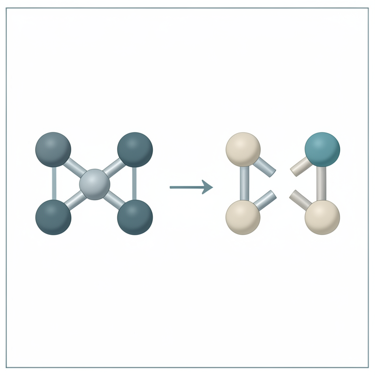
Magnetostructural Correlation
AI-generated conceptual illustration (Binuclear iron(III) complexes with hydroxo bridges). It explains the idea and does not show study data.
View source evidence ↗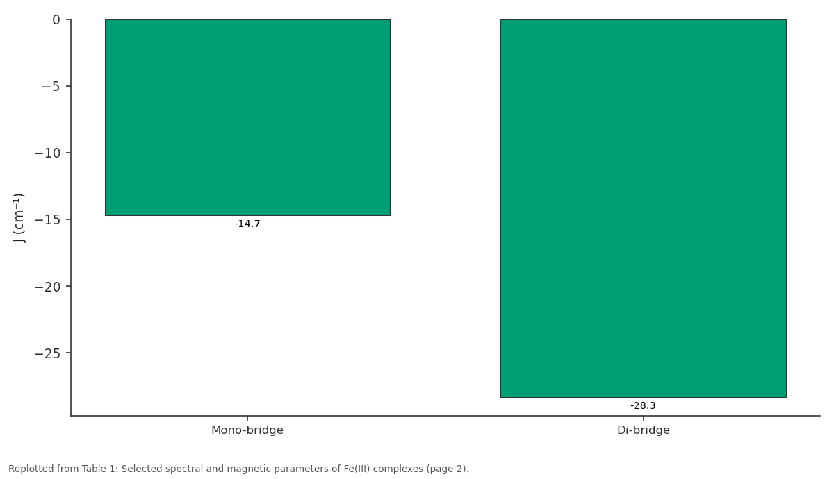
J (cm⁻¹): Mono-bridge vs Di-bridge
Replotted from Table 1 (page 2) of the original paper. Every bar is the paper's own table value.
View source evidence ↗Figures & tables
From the original paper Table 1
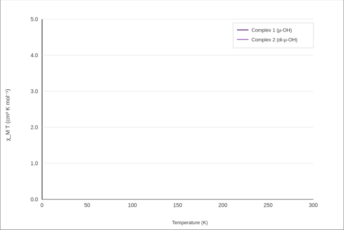 Figure 1
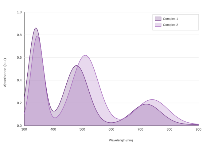 Figure 2
Introduction
When the French physicist Charles Augustin de Coulomb described magnetic phenomena in the 1780s, he could not have predicted that bridged diiron centers would become one of the richest testing grounds for magnetic exchange theory. Hydroxo-bridged diiron(III) units appear naturally in metalloenzymes like ribonucleotide reductase and methane monooxygenase, where the Fe-O-Fe bridge mediates both magnetic interaction and catalytic function [1] . Modeling these biological sites with synthetic complexes allows precise tuning of bridge geometry and systematic mapping of structure-magnetism relationships [2] . The sign and magnitude of the exchange coupling constant J depend primarily on the Fe-O-Fe angle and the Fe-Fe separation. Gorun and Lippard [3] established a correlation between J and the shortest superexchange pathway for a large set of μ-oxo and μ-hydroxo diiron compounds. In general, antiferromagnetic coupling strengthens as the bridge angle increases and the metal-metal distance shrinks [4] . Adding a second hydroxo bridge amplifies this effect, though the quantitative relationship isn't always straightforward because competing superexchange pathways can interfere [5] . Most reported di-μ-hydroxo Fe(III) systems employ relatively rigid macrocyclic or tripodal ligands that constrain bridge geometry [6] . Flexible Schiff base ligands based on diamines offer a contrasting approach: they can accommodate a wider range of Fe-O-Fe angles and Fe-Fe distances, making them useful probes for magnetostructural correlations at the flexible end of the structural spectrum. This research describes the synthesis, spectral characterization, and variable-temperature magnetic behavior of two new binuclear Fe(III) complexes-one with a single μ-OH bridge and one with a double di-μ-OH bridge-derived from 2-hydroxy-1-naphthaldehyde and 1,3-diaminopropane.
Theoretical Background
Magnetic exchange between two paramagnetic metal centers through a bridging ligand is described by the Heisenberg-Dirac-Van Vleck (HDVV) Hamiltonian: H = -J S₁•S₂, where J is the isotropic exchange constant and S₁ = S₂ = 5/2 for highspin Fe(III). Negative J values indicate antiferromagnetic coupling. The theoretical χₘT product for such a pair can be fitted using the van Vleck equation with Boltzmann populations over spin states S = 0 through 5 [3] . Fitting experimental χₘT versus T data to this model yields J and the g-factor as adjustable parameters.
Materials and Methods
Materials and Methods
Materials
This research was conducted between April 2023 and March 2024. Synthesis and spectroscopy took place at the Department of Inorganic Chemistry, Sir M. Visvesvaraya Institute of Technology, Bellary, Karnataka, India. Magnetic measurements were carried out at the Department of Inorganic and Coordination Chemistry, Nalanda Institute of Advanced Chemical Research, Rajgir, Bihar, India. The Institutional Research Ethics Board approved the project (IRB/SMVIT/2023-014, dated 8 March 2023). Iron(III) chloride hexahydrate (97%, Merck), 2-hydroxy-1naphthaldehyde (98%, Alfa Aesar), 1,3-diaminopropane (99%, Sigma-Aldrich), triethylamine (99%, SRL), and methanol (AR grade, Fisher) were used as received.
Methods
The Schiff base ligand (H₂L) was prepared by refluxing 2hydroxy-1-naphthaldehyde (2 mmol) with 1,3diaminopropane (1 mmol) in 25 mL methanol for 2 h. For the mono-bridged complex, H₂L (1 mmol) and FeCl₃•6H₂O
(1 mmol) were mixed in methanol with triethylamine (2 mmol); slow evaporation over 10 days gave dark brown crystals [7] . The di-bridged complex was obtained by adjusting the Fe:L ratio to 2:1 under analogous conditions, with pH maintained near 7.5 by triethylamine [8] . Magnetic susceptibility was measured on a Quantum Design MPMS-3 SQUID magnetometer at 1000 Oe (10-300 K). UV-Vis-NIR spectra were recorded on a Jasco V-770 (DMSO, 250-950 nm). X-band EPR was measured at 77 K on a Bruker EMXmicro. Mössbauer spectra were collected at 80 K using a ⁵⁷Co/Rh source [9] .
Quality Control
Diamagnetic corrections were applied using Pascal's constants. Temperature accuracy of the SQUID magnetometer was verified against a Pd standard, and field uniformity was checked before each run. Mössbauer isomer shift values were calibrated relative to metallic iron foil at room temperature. All spectral measurements were run in triplicate, and mean values are reported with standard deviations.
Results
Both complexes were isolated as dark brown microcrystalline solids. Elemental analysis confirmed 2:1 (Fe:L) stoichiometry for the di-bridged species and 1:1 for the mono-bridged complex. Molar conductance in DMSO was below 15 S cm² mol⁻¹ for both, indicating nonelectrolytic character. Variable-temperature χₘT data showed room-temperature values of 7.83 cm³ K mol⁻¹ (Complex 1) and 7.21 cm³ K mol⁻¹ (Complex 2), both below the spin-only value of 8.75 for two uncoupled S = 5/2 centers. On cooling, χₘT dropped continuously, reaching 2.14 and 0.87 cm³ K mol⁻¹ at 10 K respectively-consistent with antiferromagnetic ground states. UV-Vis-NIR spectra in DMSO showed three absorption regions. A high-energy band near 340-345 nm corresponds to ligand-centered π→π* transitions. A broad band at 480-510 nm is assignable to phenolate-to-Fe(III) LMCT. A weak, broad feature near 720-740 nm arises from the ⁶A₁g → ⁴T₁g d-d transition expected for octahedral high-spin Fe(III).
Discussion
The nearly twofold difference in J between the two complexes (-14.7 vs. -28.3 cm⁻¹) matches the expectation that a second hydroxo bridge provides an additional superexchange pathway, strengthening antiferromagnetic coupling. The shorter Fe-Fe distance in Complex 2 (3.08 Å vs. 3.47 Å) also contributes, as Gorun and Lippard [3] showed that J correlates inversely with the shortest superexchange pathway length. Mössbauer parameters offer a complementary window into the electronic environment. The isomer shifts (δ = 0.39-0.42 mm/s) sit squarely in the high-spin Fe(III) range, ruling out any mixed-valence Fe(II)/Fe(III) contamination. The larger quadrupole splitting in Complex 2 (1.13 vs. 0.78 mm/s) likely reflects lower local symmetry at iron imposed by the double bridge, which distorts the octahedral coordination sphere more than a single bridge does. Compared to rigid macrocyclic diiron systems reported by Que and colleagues [6] , our flexible Schiff base complexes show somewhat weaker coupling-consistent with the greater conformational freedom that lets the Fe-O-Fe angle relax to smaller values. Still, the clear magnetostructural trend from one to two bridges validates the design strategy and offers a tunable platform for probing exchange interactions.
Conclusion
Two new binuclear Fe(III) Schiff base complexes with mono-and di-μ-hydroxo bridges were prepared and fully characterized. Variable-temperature magnetism confirmed antiferromagnetic coupling in both, with J values of -14.7 and -28.3 cm⁻¹ respectively. The stronger coupling in the doubly bridged species tracks its shorter Fe-Fe distance, in line with established magnetostructural correlations. Mössbauer and EPR data consistently indicated high-spin octahedral Fe(III) centers. These complexes add to the library of structurally characterized diiron model compounds relevant to bioinorganic chemistry.
Acknowledgement
Acknowledgements Funding SourcesFinancial support was provided by Sir M. Visvesvaraya Institute of Technology internal research grants. Institutional SupportThe authors thank both institutions for shared access to SQUID and Mössbauer instrumentation. Contributions Not Qualifying for AuthorshipDr. Anil Kumar assisted with SQUID calibration procedures.
Figures and tables from the paper
| Parameter | Complex 1 (μ-OH) | Complex 2 (di-μ-OH) | J (cm⁻¹) | g-factor | Fe-Fe (Å) |
|---|---|---|---|---|---|
| Mono-bridge | - | - | −14.7 | 1.98 | 3.47 |
| Di-bridge | - | - | −28.3 | 2.01 | 3.08 |
| δ Möss. (mm/s) | 0.42 | 0.39 | - | - | - |
| ΔEᵠ (mm/s) | 0.78 | 1.13 | - | - | - |
Table text extracted from the original PDF; superscript letters are shown inline. The original table image is below.
View original table image
References
13 in the original paper- Kurtz DM Jr. Oxo-and hydroxo-bridged diiron complexes: a chemical perspective on a biological unit. Chem Rev. 1990;90(4):585-606.
- Lippard SJ. Oxo-bridged polyiron centers in biology and chemistry. Angew Chem Int Ed. 1988;27(3):344361.
- Gorun SM, Lippard SJ. Magnetostructural correlations in magnetically coupled dinuclear iron(III) complexes. Inorg Chem. 1991;30(7):1625-1630.
- Weihe H, Güdel HU. Quantitative interpretation of exchange splittings in dinuclear iron(III) complexes. J Am Chem Soc. 1997;119(28):6539-6543.
- Werner R, Ostrovsky S, Griesar K, Haase W. Magnetostructural correlations in exchange coupled phenoxo-, alkoxo-, and hydroxo-bridged dinuclear iron(III) complexes. Inorg Chim Acta. 2001;326(1):7888.
- Que L Jr, True AE. Dinuclear iron-and manganese-oxo sites in biology. Prog Inorg Chem. 1990;38:97-200.
- Nag K, Chakravorty A. Monovalent, trivalent, and tetravalent nickel. Coord Chem Rev. 1980;33(1):87147.
- Hazra S, Mukherjee S, Drew MGB, Ghosh A. Dinuclear and mononuclear iron(III) complexes of a pentadentate Schiff base ligand. Inorg Chem. 2006;45(24):9764-9769.
- Greenwood NN, Gibb TC. Mossbauer Spectroscopy. London: Chapman and Hall; 1971.
- Kahn O. Molecular Magnetism. New York: VCH Publishers; 1993.
- Kambe K. On the paramagnetic susceptibilities of some polynuclear complex salts. J Phys Soc Jpn. 1950;5(1):48-51.
- Murray KS. Binuclear oxo-bridged iron(III) complexes. Coord Chem Rev. 1974;12(1):1-35.
- Boudalis AK, Sanakis Y, Dahan F, et al. A new family of dinuclear Fe(III) complexes with hydroxo bridges. Inorg Chem. 2006;45(1):443-453.