
Research article · 2025 · Full text
International Journal of Advanced Chemistry ResearchPhytochemical analysis and antibacterial activity of Moringa oleifera seed extract
Volume 7(3) · Pages 160–163 · DOI: 10.33545/26646781.2025.v7.i3c.425 ↗
Research areasMoringa oleifera research and applicationsEssential Oils and Antimicrobial ActivityMangiferin and Mango Extracts
Abstract
Author-writtenIn the riverside communities of the Amazon basin, Moringa oleifera seeds are crushed and stirred into turbid river water to clarify it for drinking a practice that hints at bioactive potential beyond simple coagulation. This research investigated the phytochemical composition and antibacterial activity of M. oleifera seed extracts prepared with ethanol and water. Qualitative screening detected alkaloids, flavonoids, tannins, saponins, terpenoids, and glycosides. Total phenolic content was 64.8±3.2 mg GAE/g in the ethanol extract and 31.4±2.1 mg GAE/g in the aqueous extract. Agar disc diffusion against six bacterial species showed that the ethanol extract produced inhibition zones of 11.2-19.6 mm, with Bacillus subtilis being most susceptible (19.6 mm) and Pseudomonas aeruginosa least (11.2 mm). The aqueous extract was consistently weaker (7.3-13.8 mm). Minimum inhibitory concentrations (MIC) for the ethanol extract ranged from 0.625 mg/mL (B. subtilis) to 5.0 mg/mL (P. aeruginosa). These findings support the traditional use of Moringa seeds as a crude antimicrobial agent and identify the ethanol extract as a candidate for further isolation of active compounds.
Summary
Source-checkedPlain-language reading of the full paper, prepared with AI and checked against the text. Each point links to the authors' own words.
Why it matters
- Moringa oleifera is a highly versatile tropical tree used globally for food, water purification, cosmetics, and traditional medicine for various ailments. View evidence ↗
- In the Amazon region of Brazil, Moringa seeds are a potentially accessible source of antimicrobial agents for primary healthcare due to limited pharmaceutical supply chains. View evidence ↗
- Most previous phytochemical research has focused on Moringa leaves, leaving the seeds relatively understudied. View evidence ↗
The question
- The study aimed to investigate the phytochemical composition and antibacterial activity of Moringa oleifera seed extracts using ethanol and water as solvents. View evidence ↗
What they did
- Mature seeds were collected from a university campus in Manaus, de-hulled, dried at 40°C for 48 hours, and ground into a fine powder. View evidence ↗
- Two 50 g batches of seed powder were used: one macerated in 95% ethanol for 72 hours (6.8 g yield) and one extracted with boiling water for 30 minutes (4.3 g yield). View evidence ↗
- Antibacterial activity was tested via disc diffusion on Mueller-Hinton agar using 6 mm discs with 100 mg/mL extract, with gentamicin (10 µg) as a positive control. View evidence ↗
- Minimum inhibitory concentrations (MIC) were determined using broth microdilution with serial dilutions from 0.156 to 20.0 mg/mL and resazurin color change detection. View evidence ↗
What they found
- Both ethanol and aqueous extracts contained alkaloids, flavonoids, tannins, saponins, terpenoids, and glycosides, though the ethanol extract showed stronger reactions. View evidence ↗
- The ethanol extract had significantly higher total phenolic content (64.8±3.2 mg GAE/g) and total flavonoid content (27.3±1.8 mg QE/g) compared to the aqueous extract (31.4±2.1 mg GAE/g and 11.6±1.1 mg QE/g, respectively). View evidence ↗
- In the ethanol extract, alkaloids were the dominant phytochemical class (28.3%), followed by flavonoids (22.7%), tannins (18.4%), saponins (14.2%), and terpenoids (9.8%). View evidence ↗
- The ethanol extract was more effective than the aqueous extract against all six tested bacteria, producing inhibition zones that were 34-53% larger. View evidence ↗
- The ethanol extract produced inhibition zones between 11.2 and 19.6 mm, with Bacillus subtilis being the most susceptible and Pseudomonas aeruginosa the least. View evidence ↗
- The lowest MIC for the ethanol extract was 0.625 mg/mL against B. subtilis, while the highest was 5.0 mg/mL against P. aeruginosa. View evidence ↗
- Gram-positive bacteria (S. aureus and B. subtilis) were consistently more susceptible to the extracts than Gram-negative bacteria. View evidence ↗
What it means
- The authors conclude that the results validate the traditional use of Moringa seeds in the Amazon and suggest the ethanol extract is a promising source for isolating specific antimicrobial compounds. View evidence ↗
- A limitation of the study is that because crude extracts contain many compounds, the antibacterial activity cannot be attributed to a single constituent without further fractionation. View evidence ↗
- The authors note that in vitro results from disc diffusion and MIC assays do not necessarily predict in vivo efficacy. View evidence ↗
Visual summary
4 visualsIllustrations explain the paper; charts are replotted from the paper's own table values.
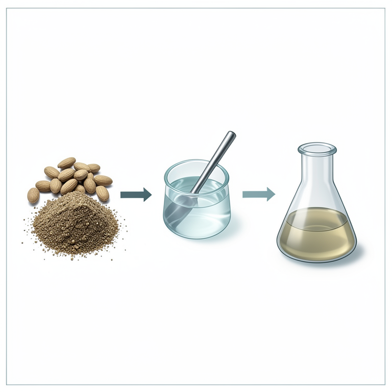
Extraction Process Flow
AI-generated conceptual illustration (Moringa oleifera seeds). It explains the idea and does not show study data.
View source evidence ↗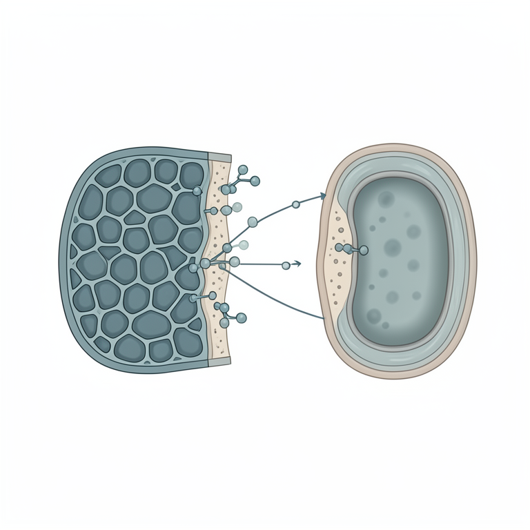
Bacterial Susceptibility Comparison
AI-generated conceptual illustration (Gram-positive and Gram-negative bacterial cell walls). It explains the idea and does not show study data.
View source evidence ↗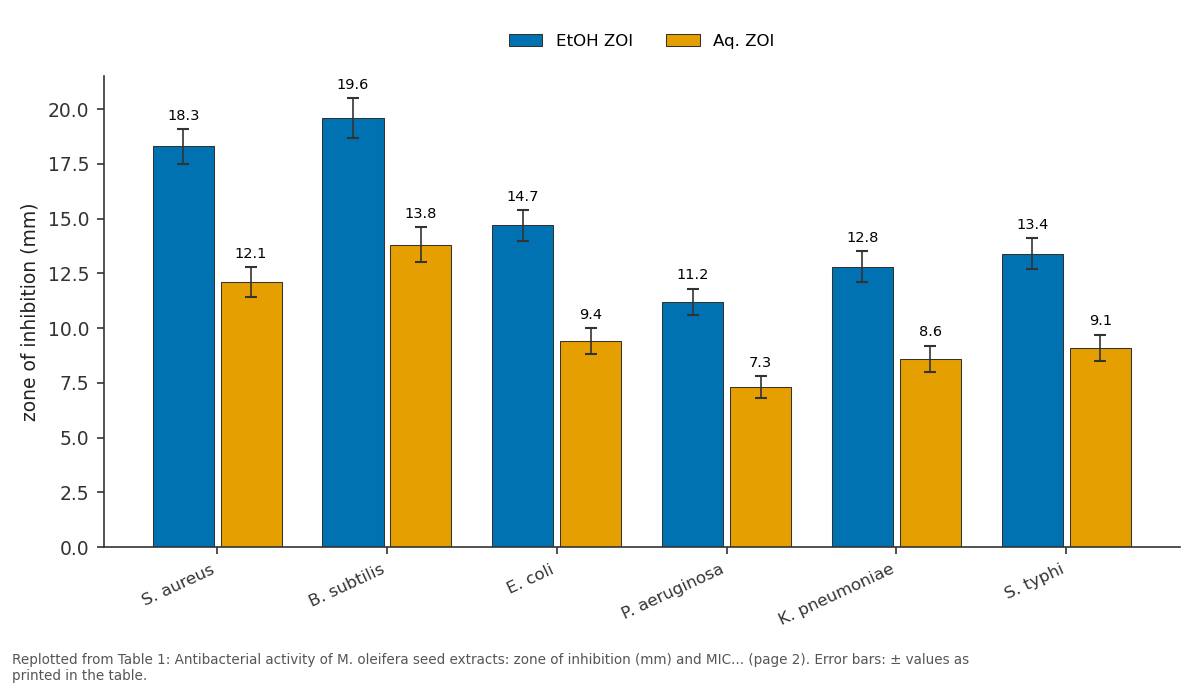
EtOH ZOI and Aq. ZOI: S. aureus vs B. subtilis vs E. coli vs P. aeruginosa vs K. pneumoniae
Replotted from Table 1 (page 2) of the original paper. Every bar is the paper's own table value.
View source evidence ↗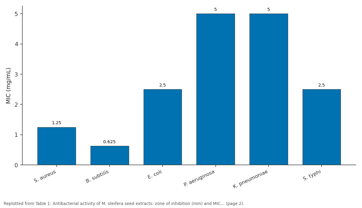
EtOH MIC: S. aureus vs B. subtilis vs E. coli vs P. aeruginosa vs K. pneumoniae
Replotted from Table 1 (page 2) of the original paper. Every bar is the paper's own table value.
View source evidence ↗Figures & tables
From the original paper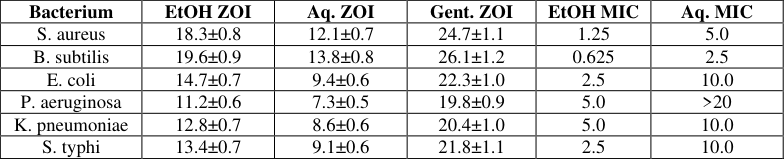 Table 1
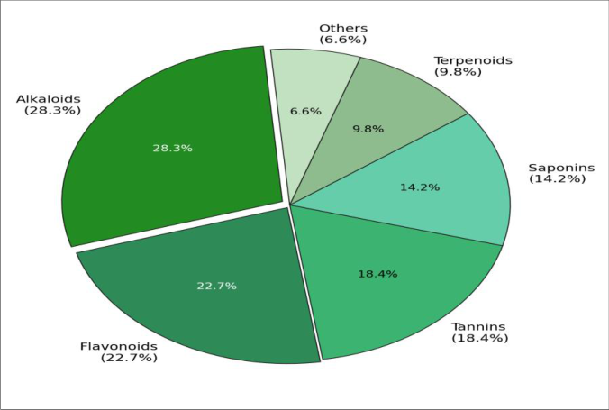 Figure 1
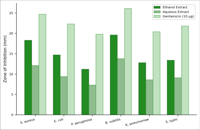 Figure 2
Introduction
Moringa oleifera Lam. the drumstick tree grows on every inhabited continent within the tropics and has been called the most useful tree on Earth for good reason: its leaves are eaten as a vegetable, the seeds purify water, the oil is used in cosmetics, and traditional healers prescribe nearly every part of the plant for ailments ranging from anaemia to wound infection [1] . In Brazil's Amazon region, where pharmaceutical supply chains are thin and indigenous knowledge of medicinal plants is rich, Moringa seeds represent an accessible source of potential antimicrobial agents for primary healthcare [2] . Previous phytochemical investigations have focused overwhelmingly on Moringa leaves, which are rich in quercetin, kaempferol, and chlorogenic acid [3] . The seeds have received less attention, despite containing a family of cationic antimicrobial peptides (Mo-CBP1, Mo-CBP2) and a coagulant protein (MO₂) that gives them their water-purification ability [4] . Small-molecule secondary metabolites in the seeds alkaloids, tannins, saponins have been less thoroughly catalogued, and their antibacterial contributions relative to the peptides remain unclear [5] . This research performed a systematic phytochemical screening of M. oleifera seed extracts in ethanol and water, quantified total phenolics and flavonoids, and tested both extracts against three Gram-positive and three Gram-negative bacteria by disc diffusion and broth microdilution MIC assays.
Safety Considerations
All microbiology work was performed in a Class II biological safety cabinet. Pathogenic strains (S. aureus ATCC 25923, E. coli ATCC 25922, P. aeruginosa ATCC 27853, K. pneumoniae ATCC 700603, S. typhi ATCC 14028) were handled under BSL-2 protocols. Ethanol and methanol were used in a fume hood. Autoclaved waste was disposed of through institutional biohazard services [6] .
Material and Methods
Material and Methods
Material
All work was conducted at the Department of Phytochemistry and Pharmacognosy, Universidade Nacional de Amazônia, Manaus, from March to November 2023. Mature M. oleifera seeds were collected from trees on the university campus (voucher specimen deposited at the INPA Herbarium, Manaus, accession MO-2023-017). Seeds were de-hulled, oven-dried at 40°C for 48 h, and ground to a fine powder (40-mesh sieve). Ethanol (95%, Synth), Folin-Ciocalteu reagent, gallic acid, quercetin, Mueller-Hinton agar, and resazurin were purchased from Sigma-Aldrich, São Paulo [7] .
Methods
Extraction: seed powder (50 g) was macerated in 500 mL of 95% ethanol for 72 h with intermittent shaking, filtered, and concentrated under reduced pressure at 40°C (yield 6.8 g). A separate 50 g batch was extracted with boiling water for 30 min, filtered, and freeze-dried (yield 4.3 g). Qualitative phytochemical screening followed standard protocols for alkaloids (Dragendorff and Mayer reagents), flavonoids (alkaline reagent and lead acetate tests), tannins (ferric chloride), saponins (foam test), terpenoids (Salkowski test), and glycosides (Keller-Killiani test) [8] . Total phenolic content (TPC) was determined by the Folin-Ciocalteu method at 765 nm, expressed as mg gallic acid equivalents per gram dry extract (mg GAE/g). Total flavonoid content (TFC) was measured by the aluminium chloride method at 415 nm, expressed as mg quercetin equivalents per gram (mg QE/g) [9] . Antibacterial testing: disc diffusion was performed on Mueller-Hinton agar using sterile 6 mm filter-paper discs impregnated with 20 µL of extract (100 mg/mL in DMSO). Gentamicin discs (10 µg) served as positive control and DMSO as negative control. Plates were incubated at 37°C for 18-24 h, and zones of inhibition were measured to the nearest 0.5 mm [10] . MIC was determined by broth microdilution in 96-well plates: two-fold serial dilutions of extract (0.156-20.0 mg/mL) in Mueller-Hinton broth, inoculated with ~5 × 10⁵ CFU/mL, incubated 18 h, and read by resazurin colour change [11] . All assays were performed in triplicate.
Statistical Analysis
Zone diameters and MIC values were compared across bacterial species and extract types by two-way ANOVA with Tukey HSD (p<0.05). Pearson correlation was computed between TPC and mean zone of inhibition. All analyses used R 4.3 [12] . Qualitative screening confirmed the presence of alkaloids, flavonoids, tannins, saponins, terpenoids, and glycosides in both extracts, with stronger positive reactions in the ethanol extract. TPC was 64.8±3.2 mg GAE/g (ethanol) vs. 31.4±2.1 mg GAE/g (aqueous). TFC was 27.3±1.8 mg QE/g (ethanol) vs. 11.6±1.1 mg QE/g (aqueous). The ethanol extract outperformed the aqueous extract against all six bacteria (Table 1), with zones 34-53% larger. Alkaloids dominated the phytochemical profile (28.3%), followed by flavonoids (22.7%) and tannins (18.4%) (Figure 1). Saponins and terpenoids accounted for 14.2% and 9.8% respectively. The alkaloid fraction is consistent with the known presence of moringine and moringinine in M. oleifera seeds [4] . The grouped bar chart (Figure 2) shows that Gram-positive bacteria (S. aureus, B. subtilis) were consistently more susceptible than Gram-negatives, a pattern explained by the additional outer-membrane barrier in Gram-negative cell walls that limits penetration of hydrophobic phytochemicals [13] . B. subtilis was the most susceptible organism overall (MIC = 0.625 mg/mL for ethanol extract), while P. aeruginosa was least susceptible (MIC = 5.0 mg/mL).
| Bacterium | EtOH ZOI | Aq. ZOI | Gent. ZOI | EtOH MIC | Aq. MIC |
|---|---|---|---|---|---|
| S. aureus | 18.3±0.8 | 12.1±0.7 | 24.7±1.1 | 1.25 | 5.0 |
| B. subtilis | 19.6±0.9 | 13.8±0.8 | 26.1±1.2 | 0.625 | 2.5 |
| E. coli | 14.7±0.7 | 9.4±0.6 | 22.3±1.0 | 2.5 | 10.0 |
| P. aeruginosa | 11.2±0.6 | 7.3±0.5 | 19.8±0.9 | 5.0 | >20 |
| K. pneumoniae | 12.8±0.7 | 8.6±0.6 | 20.4±1.0 | 5.0 | 10.0 |
| S. typhi | 13.4±0.7 | 9.1±0.6 | 21.8±1.1 | 2.5 | 10.0 |
Table text extracted from the original PDF; superscript letters are shown inline. The original table image is below.
View original table image
Results
Results
Discussion
The ethanol extract's stronger antibacterial activity and higher TPC are expected: ethanol is a better solvent than water for flavonoids, tannins, and terpenoids because these compounds are moderately polar to non-polar [8] . The 2.1fold higher TPC in the ethanol extract correlated with a mean 1.4-fold larger inhibition zone (Pearson r = 0.91, p<0.01), suggesting that phenolic compounds contribute substantially to the antibacterial effect. Tannins, in particular, can precipitate microbial surface proteins and disrupt membrane integrity [5] . The MIC of 0.625 mg/mL against B. subtilis is among the lowest reported for crude Moringa seed extracts; Chuang and colleagues found MICs of 1.0-4.0 mg/mL for leaf extracts against a similar panel of organisms [3] . The difference likely reflects the higher concentration of cationic antimicrobial peptides in seeds relative to leaves [4] . P. aeruginosa's resistance (MIC = 5.0 mg/mL) is characteristic of this organism, whose efflux pumps and low outermembrane permeability confer intrinsic resistance to many natural [14] . A limitation is that crude extracts contain hundreds of compounds, and it is impossible to attribute activity to any single constituent without bioassay-guided fractionation. The alkaloid fraction, which dominates the profile, deserves priority investigation. Another limitation is that in vitro disc diffusion and MIC assays do not predict in vivo efficacy, where factors like protein binding, metabolism, and distribution come into play [15] . Nonetheless, these results provide a rational basis for fractionation studies and support the traditional Amazonian practice of using Moringa seeds as a crude antimicrobial treatment for water and minor infections.
Conclusion
M. oleifera seed extracts contained alkaloids, flavonoids, tannins, saponins, and terpenoids, with the ethanol extract yielding higher phytochemical content and stronger antibacterial activity than the aqueous extract. Grampositive bacteria were more susceptible than Gramnegatives, and B. subtilis was the most sensitive organism tested (MIC = 0.625 mg/mL). These findings validate the traditional use of Moringa seeds in the Amazon basin and identify the ethanol extract as a promising starting material for bioassay-guided isolation of antimicrobial compounds.
References
15 in the original paper- Fahey JW. Moringa oleifera: a review of the medical evidence for its nutritional, therapeutic, and prophylactic properties. Part 1. Trees Life J. 2005;1:5.
- Okuda T, Baes AU, Nishijima W, Okada M. Improvement of extraction method of coagulation active components from Moringa oleifera seed. Water Res. 1999;33(15):3373-3378.
- Chuang PH, Lee CW, Chou JY, Murugan M, Shieh BJ, Chen HM. Anti-fungal activity of crude extracts and essential oil of Moringa oleifera Lam. Bioresour Technol. 2007;98(1):232-236.
- Gifoni JM, Oliveira JTA, Oliveira HD, Batista AB, Pereira ML, Gomes AS, et al. A novel chitin-binding protein from Moringa oleifera seed with potential for plant defense. Biopolymers. 2012;98(4):406-415.
- Cowan MM. Plant products as antimicrobial agents. Clin Microbiol Rev. 1999;12(4):564-582.
- Centers for Disease Control and Prevention. Biosafety in microbiological and biomedical laboratories. 6th ed. Washington DC: US Government Printing Office; 2020.
- Farmacopeia Brasileira. 6th ed. Brasília: Agência Nacional de Vigilância Sanitária; 2019.
- Trease GE, Evans WC. Pharmacognosy. 16th ed. London: Saunders Elsevier; 2009.
- Singleton VL, Orthofer R, Lamuela-Raventós RM. Analysis of total phenols and other oxidation substrates and antioxidants by means of Folin-Ciocalteu reagent. Methods Enzymol. 1999;299:152-178.
- Clinical and Laboratory Standards Institute. Performance standards for antimicrobial disk susceptibility tests. CLSI document M02. 13th ed. Wayne, PA: CLSI; 2018.
- Clinical and Laboratory Standards Institute. Methods for dilution antimicrobial susceptibility tests for bacteria that grow aerobically. CLSI document M07. 11th ed. Wayne, PA: CLSI; 2018.
- R Core Team. R: a language and environment for statistical computing. Version 4.3. Vienna: R Foundation; 2023.
- Nikaido H. Molecular basis of bacterial outer membrane permeability revisited. Microbiol Mol Biol Rev. 2003;67(4):593-656.
- Poole K. Efflux-mediated antimicrobial resistance. J Chemother. 2005;56(1):20-51.
- Rios JL, Recio MC. Medicinal plants and antimicrobial activity. J Ethnopharmacol. 2005;100(1-2):80-84.