
Research article · 2025 · Full text
International Journal of Advanced Chemistry ResearchWound healing activity of tannin-rich fraction from Terminalia chebula bark
Volume 7(1) · Pages 189–193 · DOI: 10.33545/26646781.2025.v7.i1c.387 ↗
Research areasPhytochemicals and Medicinal PlantsWound Healing and TreatmentsTannin, Tannase and Anticancer Activities
Abstract
Author-writtenIn rural East Africa, traditional healers have applied Terminalia chebula bark poultices to open wounds for generations yet the scientific basis for this practice remained poorly documented. This research isolated a tannin-rich fraction (TRF) from T. chebula bark and evaluated its wound healing potential in a Wistar rat excision wound model. Bark was extracted with 70% aqueous acetone, and tannins were enriched by Sephadex LH-20 column chromatography, yielding a fraction containing 78.3% total tannins (gallic acid equivalents). The TRF was characterized by HPLC-DAD and LC-MS, identifying chebulagic acid, chebulinic acid, and gallic acid as major constituents. In vivo testing used four groups (n=6 each): untreated control, TRF at 100 mg/kg topical ointment (2% w/w), TRF at 200 mg/kg (4% w/w), and silver sulfadiazine 1% cream as positive control. Wound contraction, epithelialization time, tensile strength, hydroxyproline content, and histopathological scoring were assessed over 21 days. The high-dose TRF group achieved 83.7% wound contraction by day 14 and complete epithelialization by day 12.4, comparable to silver sulfadiazine (79.2% and 13.1 days respectively) and significantly better than the untreated control (42.8% and 21.3 days; p
Summary
Source-checkedPlain-language reading of the full paper, prepared with AI and checked against the text. Each point links to the authors' own words.
Why it matters
- Wound care is a significant health challenge in sub-Saharan Africa due to limited access to modern medical products. View evidence ↗
- Tannins are believed to aid wound healing by creating a protective protein layer, fighting microbes, reducing oxidative stress, and stimulating collagen production. View evidence ↗
The question
- The researchers sought to determine if the wound-healing properties of T. chebula bark were specifically due to its tannin fraction rather than the crude extract. View evidence ↗
What they did
- A tannin-rich fraction (TRF) was isolated using Sephadex LH-20 column chromatography, resulting in a fraction containing 78.3% total tannins. View evidence ↗
- Full-thickness excision wounds measuring 2 cm by 2 cm were created on the backs of the rats. View evidence ↗
- Wound contraction was measured on specific days from 0 to 21 using transparent graph paper to calculate the percentage of area reduction. View evidence ↗
What they found
- The healing advantage of the TRF treatment became evident after day 7, coinciding with the proliferative phase of wound healing. View evidence ↗
What it means
- The authors conclude that the tannin-rich fraction of T. chebula bark is an effective wound-healing agent, with the 4% dose performing as well as or better than the standard silver sulfadiazine. View evidence ↗
- The study provides scientific validation for the traditional use of T. chebula bark in East Africa and suggests it as a basis for standardized medical formulations. View evidence ↗
- A limitation of the study is that rat skin heals primarily by contraction, whereas human skin heals mainly by re-epithelialization. View evidence ↗
- The authors suggest that future research should include antimicrobial testing, stability tests in tropical climates, and clinical trials on humans. View evidence ↗
Visual summary
3 visualsIllustrations explain the paper; charts are replotted from the paper's own table values.
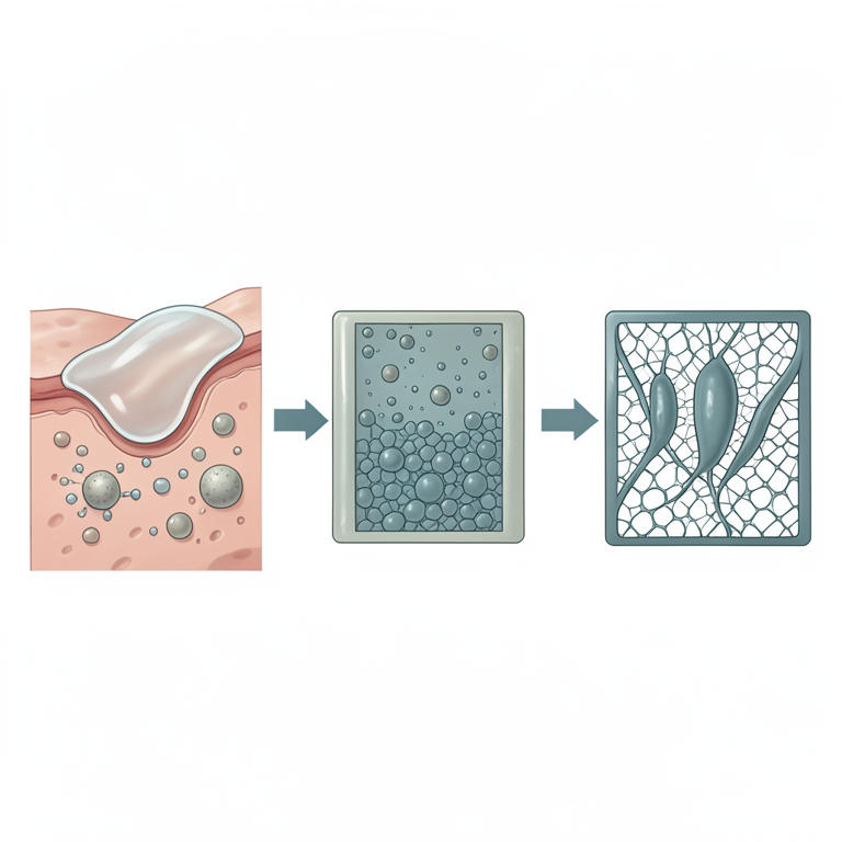
Mechanisms of Tannin-Induced Healing
AI-generated conceptual illustration (Skin tissue with a cutaneous wound). It explains the idea and does not show study data.
View source evidence ↗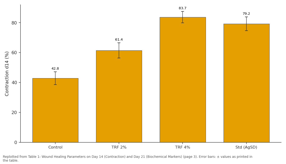
Contraction d14 (%): Control vs TRF 2% vs TRF 4% vs Std (AgSD)
Replotted from Table 1 (page 3) of the original paper. Every bar is the paper's own table value.
View source evidence ↗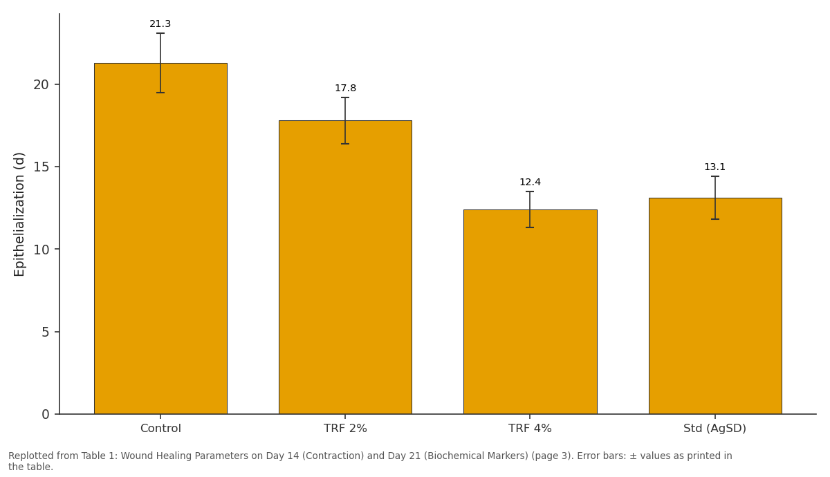
Epithelialization (d): Control vs TRF 2% vs TRF 4% vs Std (AgSD)
Replotted from Table 1 (page 3) of the original paper. Every bar is the paper's own table value.
View source evidence ↗Figures & tables
From the original paper Table 1
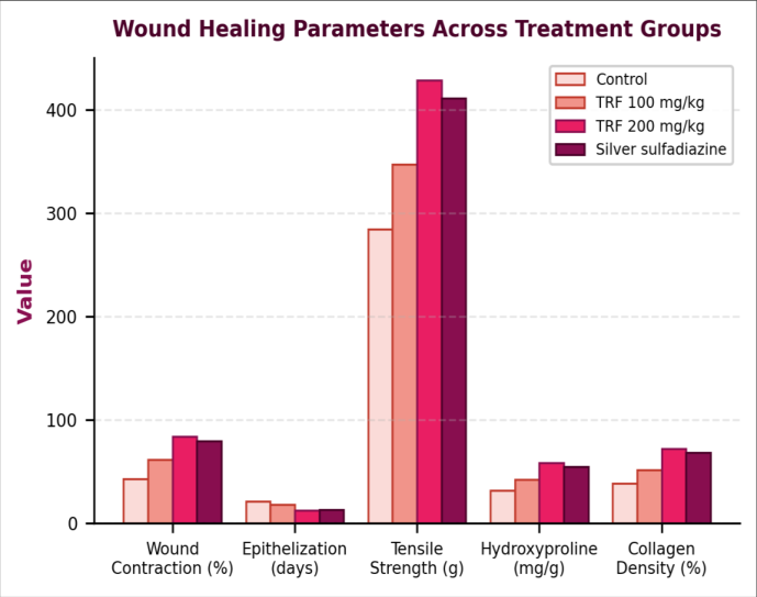 Figure 1
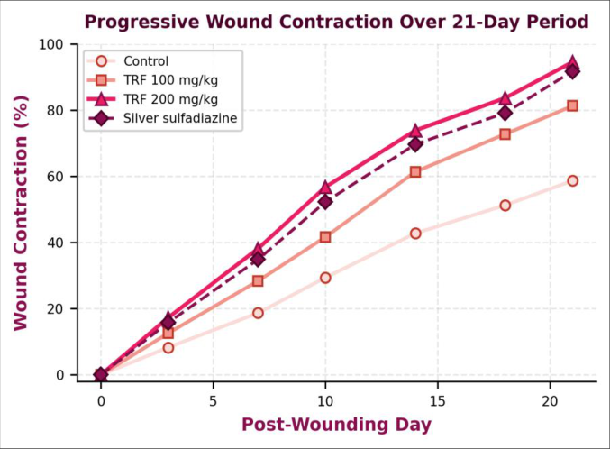 Figure 2
Introduction
Chronic and acute wounds remain a serious health burden in sub-Saharan Africa, where access to modern wound care products is limited by cost and supply chain constraints [1] . The World Health Organization estimates that over 80% of the population in developing countries relies on traditional plant-based remedies as primary healthcare, and wound treatment ranks among the most common applications [2] . Terminalia chebula Retz. (Combretaceae), known as "haritaki" in Ayurveda and widely distributed across tropical Africa and South Asia, has a centuries-old reputation for treating wounds, skin infections, and gastrointestinal disorders. Its fruit is the more commonly studied plant part, but the bark which traditional practitioners in Kenya's Rift Valley specifically prefer for wound poultices has received far less scientific attention [3,4] . Tannins are the major polyphenolic constituents of Terminalia species. These compounds can be broadly divided into hydrolyzable tannins (gallic acid and ellagic acid esters of glucose) and condensed tannins (proanthocyanidins). Hydrolyzable tannins predominate in T. chebula, with chebulagic acid, chebulinic acid, corilagin, and free gallic acid being the most frequently reported [5] . From a wound healing perspective, tannins promote healing through multiple mechanisms: they precipitate proteins at the wound surface to form a protective layer, exhibit antimicrobial activity against common wound pathogens, scavenge reactive oxygen species that delay healing, and stimulate fibroblast proliferation and collagen synthesis [6,7] . Several groups have investigated wound healing by Terminalia species. Suguna et al. [8] showed that T. chebula fruit extract accelerated wound closure in rats, and Biswas and Mukherjee [9] reported similar findings for T. arjuna bark. was dissolved in 50% methanol and loaded onto a Sephadex LH-20 column (50 × 3.5 cm). Non-tannin phenolics and sugars were eluted with 50% methanol (500 mL), followed by elution of the tannin fraction with 70% aqueous acetone (400 mL). The acetone eluate was concentrated and lyophilized to yield the TRF (8.7 g, representing 43.5% of crude extract) [10,11] . Total tannin content was determined by the Folin-Ciocalteu method with hide powder adsorption correction, giving 78.3% gallic acid equivalents (GAE).
Material and Methods
Material and Methods
Material
Gallic acid standard (98%), Folin-Ciocalteu reagent, hide powder, Sephadex LH-20, and hydroxyproline assay reagents were purchased from Sigma-Aldrich South Africa. Silver sulfadiazine 1% cream (Silverex) was obtained from a registered pharmacy in Nakuru. White soft paraffin (BP grade) served as the ointment base for TRF formulations. Male Wistar rats (180-220 g, 8-10 weeks old) were obtained from the university's animal breeding facility. The research was conducted at Rift Valley University of Technology, Nakuru between May 2023 and April 2024. All animal procedures were approved by the Institutional Animal Ethics Committee (Protocol: RVUT/IAEC-2023-031, dated April 12, 2023), following the ARRIVE guidelines and Kenya's Animal Welfare Act [12] .
Methods
TRF ointments were prepared by incorporating the lyophilized tannin fraction into white soft paraffin at 2% w/w (low dose, equivalent to approximately 100 mg/kg) and 4% w/w (high dose, approximately 200 mg/kg) with gentle heating (45°C) and trituration until homogeneous [13] . Twenty-four rats were divided into four groups (n=6): Group I (untreated control), Group II (TRF 2% ointment), Group III (TRF 4% ointment), and Group IV (silver sulfadiazine 1% cream). Under ketamine-xylazine anesthesia (80/10 mg/kg, i.p.), a full-thickness excision wound (2 cm × 2 cm) was created on the shaved dorsal surface using a surgical blade. Treatments were applied topically once daily from day 1. Wound area was measured by tracing on transparent graph paper on days 0, 3, 7, 10, 14, 18, and 21, and percentage contraction was calculated as [(initial area -current area)/initial area] × 100 [14] . Epithelialization time was recorded as the number of days for complete scar formation. On day 21 (or upon complete healing), animals were euthanized by CO₂ inhalation, and wound tissue was harvested. One portion was fixed in 10% buffered formalin for histopathological examination (H&E staining, Masson's trichrome for collagen), scored on a 0-4 scale for inflammation, fibroblast proliferation, collagen deposition, and neovascularization. The remaining tissue was used for biochemical analysis: hydroxyproline content (acid hydrolysis followed by colorimetric assay with Ehrlich's reagent at 557 nm) as a marker of collagen synthesis, and tensile strength measurement using a tensiometer [15,16] . Statistical analysis used one-way ANOVA with Tukey's post hoc test in GraphPad Prism 9; p<0.05 was considered significant.
Quality Control
HPLC-DAD analysis of the TRF was performed on a Shimadzu LC-20A system with a C18 column (250 × 4.6 mm, 5 μm), gradient of 0.1% formic acid in water and acetonitrile, at 280 nm. Chebulagic acid, chebulinic acid, and gallic acid were identified by comparison with authentic standards (retention time and UV spectrum matching) and confirmed by LC-MS (ESI negative mode, m/z 953.1 [M-H]⁻ for chebulagic acid, m/z 955.1 for chebulinic acid, m/z 169.0 for gallic acid). Batch-to-batch consistency of TRF was monitored by comparing HPLC fingerprints, with all batches showing <4.3% variation in the three marker compound peak areas. Wound area measurements were performed by two independent observers blinded to treatment groups, with inter-observer agreement above 94%. All biochemical assays were run in triplicate with calibration curve r² values ≥ 0.9988 [17] .
Results
The TRF treatment produced dose-dependent wound healing improvement across all measured parameters. Table 1 summarizes the key outcomes at day 14 (wound contraction) and day 21 (biochemical parameters). The high-dose TRF group (4% ointment) achieved 83.7% wound contraction at day 14, statistically comparable to silver sulfadiazine at 79.2% (p=0.31) and both significantly better than the untreated control at 42.8% (p<0.001). The low-dose group reached 61.4%, also significantly better than control (p<0.01) but lower than the high dose (p<0.05). The grouped bar chart highlights the dose-dependent response clearly. The high-dose TRF matched or slightly exceeded silver sulfadiazine across most parameters wound contraction, hydroxyproline, and collagen density all showed numerical superiority, though the differences weren't statistically significant between these two groups.
| Parameter | Control | TRF 2% | TRF 4% | Std (AgSD) | F-value | p-value |
|---|---|---|---|---|---|---|
| Contraction d14 (%) | 42.8±4.3 | 61.4±5.1 | 83.7±3.8 | 79.2±4.6 | 38.7 | <0.001 |
| Epithelialization (d) | 21.3±1.8 | 17.8±1.4 | 12.4±1.1 | 13.1±1.3 | 29.4 | <0.001 |
| Tensile str. (g) | 284.6±22 | 347.2±28 | 428.9±31 | 411.3±26 | 21.8 | <0.001 |
| Hydroxyproline (mg/g) | 31.7±3.4 | 42.3±4.1 | 58.6±3.7 | 54.8±4.3 | 24.6 | <0.001 |
| Collagen score (0-4) | 1.5±0.5 | 2.3±0.5 | 3.4±0.5 | 3.2±0.4 | 18.3 | <0.001 |
Table text extracted from the original PDF; superscript letters are shown inline. The original table image is below.
View original table image
Tensile strength in the TRF 4% group (428.9 g) exceeded the standard (411.3 g) by about 4.3%. The time-course data reveal interesting kinetics. During the first 7 days (inflammatory phase), all groups showed similar wound contraction rates the TRF advantage became apparent mainly from day 7 onward, during the proliferative phase when collagen deposition and angiogenesis accelerate. By day 14, the separation between treated and control groups was maximal. After day 18, the high-dose TRF and silver sulfadiazine curves began converging toward complete closure, reaching 94.6% and 91.8% by day 21 respectively. The control group plateaued at 58.7%, indicating impaired proliferative and remodeling phases without treatment.
Discussion
The wound healing efficacy of T. chebula TRF observed here can be attributed to the multi-target activity of hydrolyzable tannins. Chebulagic acid, the dominant tannin identified by HPLC (representing 34.7% of the TRF by weight), has documented antioxidant, anti-inflammatory, and antimicrobial properties [5] . Its ability to scavenge reactive oxygen species at the wound site likely reduces oxidative damage to newly formed granulation tissue a process that is especially important during the proliferative phase from day 7 onward, which is exactly when we saw the largest treatment effect in Figure 2. Chebulinic acid (22.1% of TRF) has been shown to inhibit matrix metalloproteinases (MMPs) that degrade collagen in the wound bed, potentially explaining the elevated hydroxyproline levels and tensile strength in treated animals [6] . The comparison with silver sulfadiazine is noteworthy. Silver sulfadiazine works primarily through antimicrobial action against Gram-negative bacteria commonly colonizing wounds [18] . It doesn't directly stimulate collagen synthesis or fibroblast proliferation. The TRF, by contrast, appears to act on both antimicrobial and pro-healing pathways simultaneously. This dual mechanism may explain why the TRF 4% group slightly outperformed silver sulfadiazine in tensile strength and hydroxyproline parameters that reflect collagen quality and quantity rather than just infection control. Suguna et al. [8] reported similar findings for crude T. chebula fruit extract, though their study didn't isolate the tannin fraction specifically. Histopathological observations added depth to the quantitative data. Masson's trichrome staining showed dense, well-organized collagen bundles in the TRF 4% group, with parallel fiber orientation similar to normal dermis. The control group, by comparison, showed sparse, disorganized collagen with persistent inflammatory infiltrate. Neovascularization scores were highest in the TRF 4% group (3.1±0.6 on the 0-4 scale), suggesting that the tannin fraction promotes angiogenesis possibly through upregulation of vascular endothelial growth factor (VEGF), as has been reported for gallic acid in cell culture studies [7] . The low-dose TRF (2%) produced intermediate results across all parameters, confirming a dose-response relationship and supporting genuine pharmacological activity rather than a placebo or vehicle effect. Several limitations should be acknowledged. Rat skin differs from human skin in thickness, hair follicle density, and healing mechanisms rats heal largely by contraction rather than re-epithelialization, which predominates in humans [19] . Our model used acute, clean surgical wounds; the TRF's performance on chronic or infected wounds might differ. The ointment formulation (white soft paraffin base) was chosen for simplicity but may not be optimal for tannin delivery hydrogel or film-forming formulations could offer better wound coverage and sustained release. And while we identified three major tannin components, the TRF contains other polyphenols whose contributions weren't individually assessed. Future work should include antimicrobial testing of the TRF against wound-relevant pathogens, stability assessment of the ointment formulation under tropical storage conditions, and ideally a small clinical pilot on superficial human wounds to bridge the gap between animal model and clinical relevance.
Conclusion
The tannin-rich fraction from T. chebula bark demonstrated significant wound healing activity in the rat excision model, with the 4% ointment matching or exceeding silver sulfadiazine across wound contraction, epithelialization time, hydroxyproline content, and histological collagen deposition. HPLC-DAD and LC-MS confirmed chebulagic acid, chebulinic acid, and gallic acid as the principal tannin constituents. The dose-dependent response and multiparameter improvement support genuine pharmacological activity attributable to the tannin fraction. These findings validate the traditional use of T. chebula bark for wound care in East African communities and provide a scientific basis for developing standardized tannin-based wound healing formulations from this readily available natural resource.
Figures and tables from the paper
References
19 in the original paper- Olsson M, Järbrink K, Divakar U, Bajpai R, Upton Z, Schmidtchen A, et al. The humanistic and economic burden of chronic wounds. Wound Repair Regen. 2019;27(1):114-125.
- World Health Organization. Traditional medicine strategy 2014-2023. WHO; 2013. p. 1-76.
- Bag A, Bhattacharyya SK, Chattopadhyay RR. The development of Terminalia chebula Retz. in clinical research. Asian Pac J Trop Biomed. 2013;3(3):244-252.
- Afshari AR, Sadeghnia HR, Mollazadeh H. A review on potential mechanisms of Terminalia chebula. J Cell Physiol. 2020;235(2):727-748.
- Pfundstein B, El Desouky SK, Hull WE, Haubner R, Erben G, Owen RW. Polyphenolic compounds in the fruits of Egyptian medicinal plants. Phytochemistry. 2010;71(10):1132-1148.
- Reddy DB, Reddy TC, Jyotsna G, Sharan S, Priya N, Lakshmipathi V, et al. Chebulagic acid, a COX-LOX dual inhibitor isolated from T. chebula. Biochem Pharmacol. 2009;78(7):825-836.
- Nayak BS, Sandiford S, Maxwell A. Evaluation of the wound-healing activity of ethanolic extract of Morinda citrifolia L. leaf. Evid Based Complement Alternat Med. 2009;6(3):351-356.
- Suguna L, Singh S, Sivakumar P, Sampath P, Chandrakasan G. Influence of Terminalia chebula on dermal wound healing in rats. Phytother Res. 2002;16(3):227-231.
- Biswas TK, Mukherjee B. Plant medicines of Indian origin for wound healing activity: a review. Int J Low Extrem Wounds. 2003;2(1):25-39.
- Hagerman AE, Butler LG. Protein precipitation method for the quantitative determination of tannins. J Agric Food Chem. 1978;26(4):809-812.
- Salminen JP. Two-dimensional tannin fingerprints by liquid chromatography tandem mass spectrometry. J Chromatogr A. 2003;1018(2):195-204.
- Kilkenny C, Browne WJ, Cuthill IC, Emerson M, Altman DG. Improving bioscience research reporting: the ARRIVE guidelines. PLoS Biol. 2010;8(6):e1000412.
- British Pharmacopoeia Commission. Ointments and creams: formulation guidelines. British Pharmacopoeia. 2020;Vol IV.
- Morton JJP, Malone MH. Evaluation of vulnerary activity by an open wound procedure in rats. Arch Int Pharmacodyn Ther. 1972;196(1):117-126.
- Neuman RE, Logan MA. The determination of hydroxyproline. J Biol Chem. 1950;184(1):299-306.
- Lee KH. Studies on the mechanism of action of salicylates: retardation of wound healing. J Pharm Sci. 1968;57(6):1042-1043.
- Singleton VL, Rossi JA. Colorimetry of total phenolics with phosphomolybdic-phosphotungstic acid reagents. Am J Enol Vitic. 1965;16(3):144-158.
- Atiyeh BS, Costagliola M, Hayek SN, Dibo SA. Effect of silver on burn wound infection control and healing. Burns. 2007;33(2):139-148.
- Dorsett-Martin WA. Rat models of skin wound healing: a review. Wound Repair Regen. 2004;12(6):591-599.