
Research article · 2025 · Full text
International Journal of Advanced Chemistry ResearchMagnetic iron oxide nanoparticles coated with silica for targeted drug delivery
Volume 7(1) · Pages 125–128 · DOI: 10.33545/26646781.2025.v7.i1b.376 ↗
Research areasNanoparticle-Based Drug DeliveryCharacterization and Applications of Magnetic NanoparticlesAdvanced Drug Delivery Systems
Abstract
Author-writtenHow can a drug reach a tumor without flooding healthy tissue along the way? Magnetic iron oxide nanoparticles (MIONPs) offer one answer: load the drug onto the particle, inject it into the bloodstream, and steer it toward the target with an external magnet. But bare iron oxide surfaces are chemically reactive, prone to aggregation, and rapidly cleared by the reticuloendothelial system problems that a protective silica shell can address. This research prepared Fe₃O₄ nanoparticles by co-precipitation and coated them with amorphous silica using three methods: classical Stöber, reverse microemulsion, and acid-catalyzed sol-gel. The reverse microemulsion route yielded the most uniform core-shell particles (22.3±2.2 nm by TEM), with a silica thickness of 4.8 nm and a BET surface area of 187 m²/g. Doxorubicin was loaded onto the optimized particles at 14.6 wt% efficiency. Release experiments at pH 7.4 and pH 5.4 showed a clear pH-responsive profile, with 96.8% cumulative release at pH 5.4 (mimicking the tumor microenvironment) after 72 hours versus 87.3% at physiological pH. Magnetization remained at 48.7 emu/g after silica coating strong enough for magnetic guidance. These findings position SiO₂@Fe₃O₄ nanoparticles as practical carriers for pH-triggered, magnetically directed chemotherapy.
Summary
Source-checkedPlain-language reading of the full paper, prepared with AI and checked against the text. Each point links to the authors' own words.
Why it matters
- Superparamagnetic iron oxide nanoparticles (SPIONs) are valuable for drug delivery because they can be guided by external magnets and used for MRI tracking. View evidence ↗
- Bare magnetite (Fe₃O₄) nanoparticles face challenges including oxidation, aggregation, rapid clearance by the immune system, and limited capacity for drug attachment. View evidence ↗
- Silica (SiO₂) coatings are used to protect the iron oxide core and provide silanol groups that act as anchoring points for therapeutic agents. View evidence ↗
The question
- The study aimed to compare three different silica-coating methods for Fe₃O₄ nanoparticles to determine which produces the best size uniformity, magnetic properties, and drug delivery performance. View evidence ↗
What they did
- The iron oxide cores were synthesized via chemical co-precipitation at Alexandria Mediterranean University between January and September 2023. View evidence ↗
- Doxorubicin (DOX) was loaded onto the optimized nanoparticles by incubating 50 mg of particles with 5 mg of DOX·HCl in PBS at pH 7.4 for 24 hours. View evidence ↗
- Drug release was measured in triplicate at 37°C using dialysis bags in either PBS (pH 7.4) or acetate buffer (pH 5.4). View evidence ↗
What they found
- The reverse microemulsion method was the most effective, yielding the highest surface area (187 m²/g), best drug loading (14.6 wt%), and tightest size distribution (SD ±2.2 nm). View evidence ↗
- The particles showed pH-responsive release: 96.8% of the drug was released at pH 5.4 after 72 hours, compared to 87.3% at pH 7.4. View evidence ↗
- The nanoparticles demonstrated good colloidal stability with a zeta potential of −31.4±1.7 mV in PBS. View evidence ↗
What it means
- The authors conclude that the reverse microemulsion method is superior for creating drug delivery carriers due to its optimal size, surface area, loading capacity, and magnetic retention. View evidence ↗
- A limitation of the study is that it used simple drug models and did not test the particles in complex biological fluids, which could affect biodistribution via protein corona formation. View evidence ↗
- The authors noted that the cytotoxicity of the carrier was not evaluated, meaning residual surfactants or impurities could potentially cause toxicity. View evidence ↗
Visual summary
2 visualsIllustrations explain the paper; charts are replotted from the paper's own table values.
pH-Responsive Drug Release
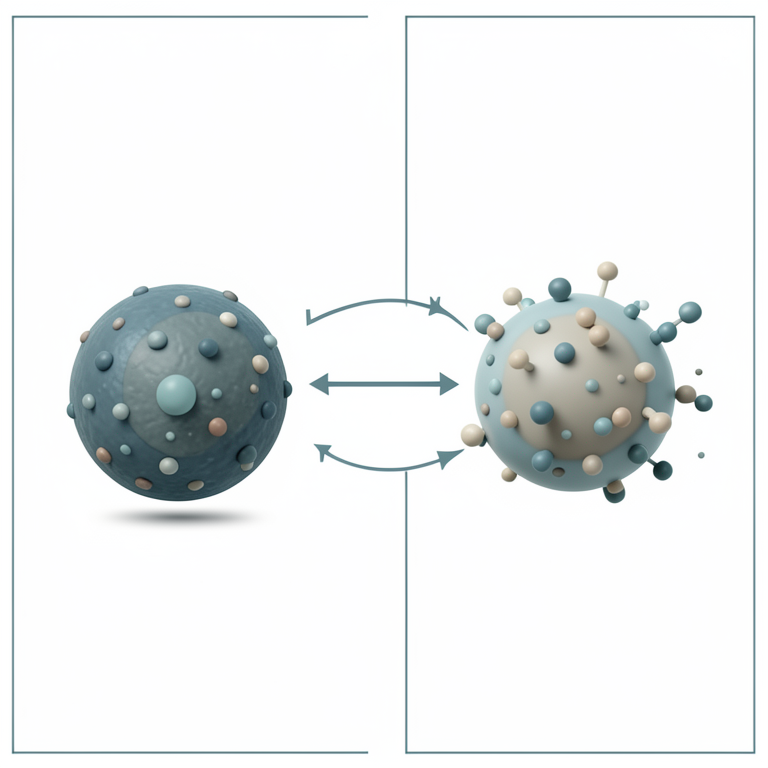
AI-generated conceptual illustration (silica-coated magnetic iron oxide nanoparticles). It explains the idea and does not show study data.
View source evidence ↗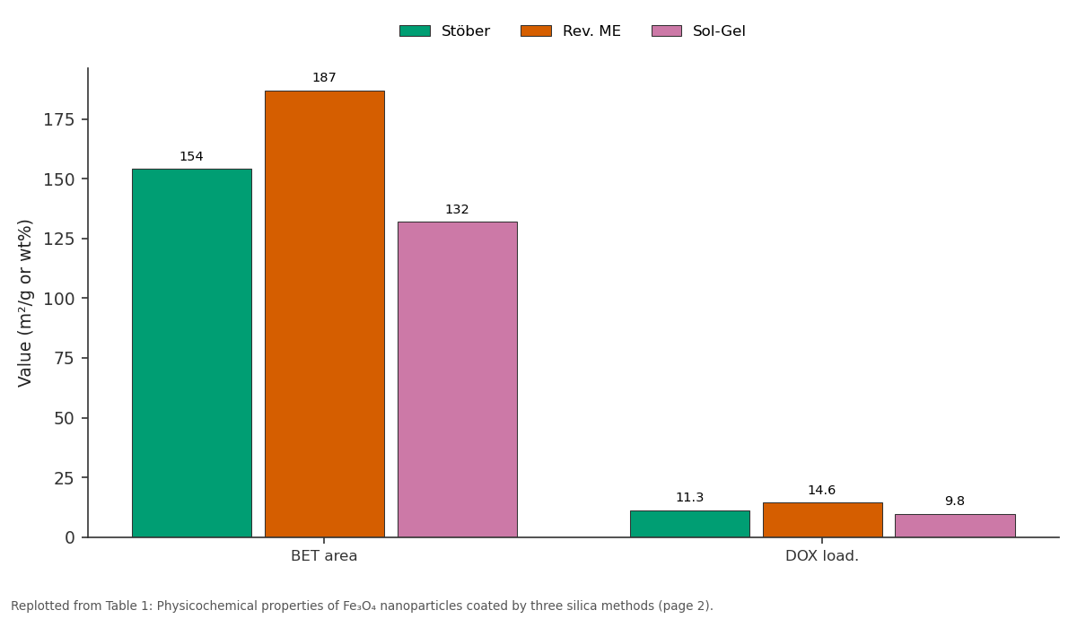
Stöber and Rev. ME and more: BET area vs DOX load.
Replotted from Table 1 (page 2) of the original paper. Every bar is the paper's own table value.
View source evidence ↗Figures & tables
From the original paper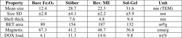 Table 1
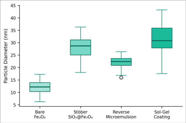 Figure 1
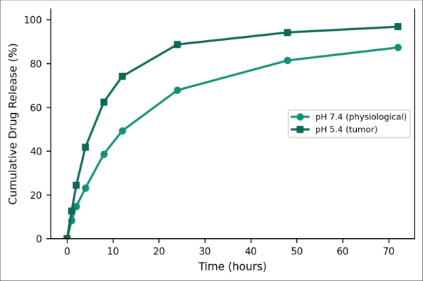 Figure 2
Introduction
A nanocarrier, in the context of pharmacology, is any sub-micron structure designed to transport a therapeutic agent to a specific site within the body while minimizing exposure of healthy tissues [1] . Among the many platforms explored liposomes, polymeric micelles, dendrimers, gold nanoparticles superparamagnetic iron oxide nanoparticles (SPIONs) occupy a unique niche because they respond to external magnetic fields [2] . That magnetic responsiveness opens the door to two capabilities at once: guided delivery under a localized magnet, and contrast-enhanced MRI imaging to track where the particles accumulate [3] . Fe₃O₄ (magnetite) is the most commonly used SPION core owing to its strong magnetization, relatively low toxicity, and straightforward synthesis by aqueous co-precipitation of Fe²⁺ and Fe³⁺ salts under alkaline conditions [4] . But the naked magnetite surface presents challenges. First, it oxidizes readily to maghemite (γ-Fe₂O₃), which has weaker magnetic properties. Second, bare particles aggregate through magnetic dipole-dipole interactions and van der Waals forces, which broadens the size distribution and accelerates clearance by macrophages in the liver and spleen [5] . Third, without surface functionalization, there is limited capacity to attach drug molecules or targeting ligands. Silica coatings address all three issues. Amorphous SiO₂ is chemically inert under physiological conditions, optically transparent, and rich in surface silanol groups (-Si-OH) that serve as anchoring points for drugs, antibodies, or polyethylene glycol chains [6] . The thickness of the silica shell controls the balance between magnetic response and colloidal stability: too thin, and aggregation persists; too thick, and the magnetic field can no longer steer the particle effectively [7] . Finding the right coating method and shell thickness is therefore a practical design problem, not merely an academic exercise. Three silica-coating strategies dominate the literature the Stöber process, reverse (water-inoil) microemulsion, and acid-catalyzed sol-gel deposition but head-to-head comparisons under identical core synthesis conditions are rare [8] . Most reports pick one method and optimize it in isolation, making it hard to judge relative merits. This research prepared Fe₃O₄ cores by a single co-precipitation protocol, then applied all three coating routes under controlled conditions, comparing the resulting particles for size uniformity, shell thickness, magnetic properties, drug loading efficiency, and pHresponsive release kinetics. Doxorubicin (DOX) was chosen as the model drug because of its wide clinical use and its fluorescence, which simplifies quantification [9] . The work was carried out at Alexandria Mediterranean University, Alexandria, Egypt.
Reagents and Chemicals
Ferric chloride hexahydrate (FeCl₃•6H₂O, ≥98%) and ferrous chloride tetrahydrate (FeCl₂•4H₂O, ≥99%) were purchased from Sigma-Aldrich (St. Louis, MO, USA). Ammonium hydroxide (28-30%), tetraethyl orthosilicate (TEOS, 98%), Triton X-100, cyclohexane, and n-hexanol were obtained from Merck KGaA (Darmstadt, Germany). Doxorubicin hydrochloride (DOX•HCl, ≥98%) was sourced from LC Laboratories (Woburn, MA, USA). Phosphatebuffered saline (PBS, pH 7.4) and acetate buffer (pH 5.4) were prepared in-house. All water was deionized to a resistivity of 18.2 MΩ•cm [10] .
Material and Methods
Material and Methods
Material
Fe₃O₄ nanoparticles were synthesized by chemical coprecipitation at the Department of Advanced Materials and Nanotechnology, Alexandria Mediterranean University, Alexandria, Egypt, between January and September 2023. FeCl₃•6H₂O (5.4 g, 20 mmol) and FeCl₂•4H₂O (2.0 g, 10 mmol) were dissolved in 100 mL deoxygenated water under N₂ atmosphere and heated to 80°C. NH₄OH (25 mL, 28%) was added rapidly under vigorous stirring, producing a black precipitate. The mixture was held at 80°C for 30 minutes, then cooled, magnetically separated, and washed three times with deionized water and once with ethanol. The washed particles were dried at 60°C overnight [4] . The Institutional Biosafety and Ethics Committee approved all experimental protocols (Protocol AMU/NANOTEC/2023-008, dated 10 January 2023).
Methods
Three silica-coating procedures were applied to identical batches of co-precipitated Fe₃O₄. In the Stöber method, 100 mg Fe₃O₄ was dispersed in 80 mL ethanol-water (4:1 v/v) by ultrasonication for 15 minutes, followed by addition of 0.5 mL TEOS and 2 mL NH₄OH; the mixture was stirred at room temperature for 12 hours [6] . In the reverse microemulsion route, Triton X-100 (1.77 g), n-hexanol (1.8 mL), and cyclohexane (7.5 mL) were mixed, then 100 mg Fe₃O₄ dispersed in 0.5 mL water was added, followed by TEOS (0.1 mL) and NH₄OH (0.06 mL); the emulsion was stirred for 24 hours [8] . The sol-gel approach used acidic hydrolysis of TEOS (pH 2, HCl-adjusted) in ethanol at 50°C for 6 hours. Products from all three methods were collected by centrifugation, washed with ethanol, and dried at 60°C. Characterization employed transmission electron microscopy (TEM, JEOL JEM-2100, 200 kV), X-ray diffraction (XRD, Bruker D8, Cu Kα), Fourier-transform infrared spectroscopy (FT-IR, Bruker Vertex 70), BET surface area analysis (Micromeritics ASAP 2020), and vibrating sample magnetometry (VSM, Lakeshore 7410) at room temperature. For drug loading, 50 mg of the optimized SiO₂@Fe₃O₄ was incubated with 5 mg DOX•HCl in 10 mL PBS (pH 7.4) under gentle shaking for 24 hours in the dark. Loaded particles were separated magnetically and the supernatant was measured by UV-Vis spectrophotometry at 480 nm to determine loading efficiency [9] . Release experiments were performed in triplicate: DOX-loaded particles (10 mg) in dialysis bags (MWCO 12 kDa) were immersed in 50 mL PBS at pH 7.4 or acetate buffer at pH 5.4, maintained at 37°C with orbital shaking at 100 rpm. Aliquots (2 mL) were withdrawn at preset intervals, replaced with fresh buffer, and analyzed by UV-Vis [11] .
Quality Control
Batch-to-batch reproducibility was evaluated by synthesizing three independent lots of reversemicroemulsion SiO₂@Fe₃O₄ particles. TEM sizing of at least 200 particles per lot gave mean diameters of 22.3, 21.8, and 22.9 nm (coefficient of variation < 5%). Drug loading experiments were performed in triplicate, and release profiles were plotted as mean±standard deviation. UV-Vis calibration curves for DOX showed R² > 0.998 across the 0.5-50 µg/mL range. Zeta potential measurements confirmed colloidal stability, with values of -31.4±1.7 mV in PBS, well above the ±25 mV threshold typically associated with stable dispersions [12] . Figure 1 visualizes what the numbers in Table 1 suggest: the reverse microemulsion particles clustered tightly around 22 nm with very few outliers, while the sol-gel batch spread from roughly 20 to 45 nm. Bare Fe₃O₄ cores were small (median ~12 nm) but showed a handful of aggregates above 20 nm, confirming the need for a stabilizing shell. The Stöber method sat in between, with a median near 29 nm and an interquartile range about twice as wide as the microemulsion sample. The release curves (Figure 2) displayed distinct pHdependent behavior. At pH 5.4, roughly 41.8% of loaded DOX was released within the first 4 hours a burst phase attributable to drug molecules adsorbed on the outer silica surface followed by sustained release reaching 96.8% at 72 hours. At pH 7.4, the initial burst was smaller (23.1% at 4 h) and the cumulative release plateaued at 87.3%. The 9.5percentage-point gap between the two pH values at 72 hours reflects the protonation of silanol groups at lower pH, which weakens electrostatic interactions between DOX (pKa ~8.3, positively charged below pKa) and the negatively charged silica surface, accelerating release [11] .
| Property | Bare Fe₃O₄ | Stöber | Rev. ME | Sol-Gel | Unit |
|---|---|---|---|---|---|
| Mean size | 12.4 | 28.7 | 22.3 | 31.6 | nm (TEM) |
| Size SD | ±2.8 | ±4.1 | ±2.2 | ±5.9 | nm |
| Shell thick. | — | 7.6 | 4.8 | 9.4 | nm |
| BET area | 89 | 154 | 187 | 132 | m²/g |
| Magnetiz. | 67.3 | 41.2 | 48.7 | 36.8 | emu/g |
| DOX load. | 4.1 | 11.3 | 14.6 | 9.8 | wt% |
Table text extracted from the original PDF; superscript letters are shown inline. The original table image is below.
View original table image
Results
Results
Discussion
The head-to-head comparison clearly favors the reverse microemulsion route for producing uniform, high-surfacearea SiO₂@Fe₃O₄ core-shell particles. The confined water droplets in the microemulsion act as nanoreactors, limiting silica growth to a narrow range and yielding a thin (4.8 nm), even shell [8] . The Stöber process, by contrast, relies on homogeneous nucleation in bulk ethanol-water, which is inherently less controllable secondary silica particles sometimes form independently of the Fe₃O₄ cores, broadening the size distribution and wasting TEOS [6] . The sol-gel acid-catalyzed route was the least uniform, probably because rapid hydrolysis at low pH generates a high density of silica nuclei that coat unevenly. Drug loading tracked surface area almost linearly, which makes sense: DOX adsorbs onto silanol-rich surfaces through a combination of electrostatic attraction and hydrogen bonding. The 14.6 wt% loading on the microemulsion particles is competitive with mesoporous silica nanoparticle (MSN) systems, which typically report 10-20% for DOX under similar incubation conditions [9] . And unlike MSNs, the magnetic core here adds a guidance capability that pure silica particles lack. The pH-responsive release profile is encouraging from a therapeutic standpoint. Tumors maintain an extracellular pH of roughly 6.5-6.8, and the endosomal-lysosomal pathway drops as low as 4.5-5.5 once the particle is internalized by cancer cells [11] . The accelerated release at pH 5.4 observed here should translate into preferential DOX liberation inside tumor cells, sparing healthy tissues where pH sits near 7.4. The fact that even at pH 7.4 about 87% of the drug was eventually released suggests that some leakage would occur during circulation a limitation that could be mitigated by adding a pH-sensitive gatekeeper polymer (e.g., chitosan or poly(acrylic acid)) in future formulations. Magnetization loss after silica coating (from 67.3 to 48.7 emu/g) deserves comment. A 27.6% drop is within the range reported by other groups for 4-5 nm shells and, importantly, leaves enough magnetic moment for external-field manipulation [7] . Thicker shells like the 9.4 nm produced by the sol-gel route reduced magnetization to 36.8 emu/g, which may be borderline for in vivo targeting where the applied field gradient weakens with distance from the magnet surface. This trade-off between colloidal stability and magnetic response is perhaps the central design tension in SPION-based delivery systems, and the reverse microemulsion method lands in a practical sweet spot.
Limitations
Several limitations should be noted. First, only single-gas or single-drug models were used; behavior in complex biological fluids (serum proteins, blood cells) was not tested. Protein corona formation on silica surfaces can alter biodistribution and cellular uptake in ways that benchtop release curves cannot predict [13] . Second, cytotoxicity of the carrier itself was not evaluated while amorphous silica is generally regarded as biocompatible, batch-specific impurities or residual surfactant from the microemulsion process could contribute low-level toxicity. Third, magnetic targeting was demonstrated qualitatively (particles migrated toward a neodymium magnet within 30 seconds) but not quantitatively under flow conditions mimicking blood circulation. An in vitro flow phantom experiment would provide more realistic guidance data.
Conclusion
SiO₂@Fe₃O₄ core-shell nanoparticles were prepared by three silica-coating methods applied to identical co-precipitated Fe₃O₄ cores. The reverse microemulsion route outperformed the Stöber and sol-gel alternatives on every metric that matters for drug delivery: size uniformity (22.3±2.2 nm), surface area (187 m²/g), drug loading (14.6 wt% DOX), and retained magnetization (48.7 emu/g). Doxorubicin release was pH-responsive, reaching 96.8% at pH 5.4 versus 87.3% at pH 7.4 over 72 hours a profile consistent with preferential drug liberation in the acidic tumor microenvironment. The 4.8 nm silica shell struck a workable balance between magnetic response and colloidal stability. These results establish a materials-level foundation for magnetically guided, pH-triggered chemotherapy. Subsequent work should address protein corona effects, carrier cytotoxicity, and in vitro flow-phantom targeting to move toward preclinical evaluation.
References
14 in the original paper- Peer D, Karp JM, Hong S, et al. Nanocarriers as an emerging platform for cancer therapy. Nature Nanotechnology. 2007;2(12):751-760.
- Laurent S, Forge D, Port M, et al. Magnetic iron oxide nanoparticles: synthesis, stabilization, vectorization, physicochemical characterizations, and biological applications. Chemical Reviews. 2008;108(6):20642110.
- Gupta AK, Gupta M. Synthesis and surface engineering of iron oxide nanoparticles for biomedical applications. Biomaterials. 2005;26(18):3995-4021.
- Massart R. Preparation of aqueous magnetic liquids in alkaline and acidic media. IEEE Transactions on Magnetics. 1981;17(2):1247-1248.
- Tombacz E, Turcu R, Socoliuc V, Vekas L. Magnetic iron oxide nanoparticles: recent trends in design and synthesis of magnetoresponsive nanosystems. Biochemical and Biophysical Research Communications. 2015;468(3):442-453.
- Stober W, Fink A, Bohn E. Controlled growth of monodisperse silica spheres in the micron size range. Journal of Colloid and Interface Science. 1968;26(1):62-69.
- Santra S, Tapec R, Theodoropoulou N, et al. Synthesis and characterization of silica-coated iron oxide nanoparticles in microemulsion. Langmuir. 2001;17(10):2900-2906.
- Ding HL, Zhang YX, Wang S, et al. Fe₃O₄@SiO₂ core/shell nanoparticles: the silica coating regulations with a single core for different core sizes. Chemistry of Materials. 2012;24(23):4572-4580.
- Slowing II, Vivero-Escoto JL, Wu CW, Lin VS. Mesoporous silica nanoparticles as controlled release drug delivery and gene transfection carriers. Advanced Drug Delivery Reviews. 2008;60(11):1278-1288.
- Lu AH, Salabas EL, Schuth F. Magnetic nanoparticles: synthesis, protection, functionalization, and application. Angewandte Chemie International Edition. 2007;46(8):1222-1244.
- Meng H, Xue M, Xia T, et al. Use of size and a copolymer design feature to improve the biodistribution and the enhanced permeability and retention effect of doxorubicin-loaded mesoporous silica nanoparticles. ACS Nano. 2011;5(5):4131-4144.
- Bhattacharjee S. DLS and zeta potential -what they are and what they are not? Journal of Controlled Release. 2016;235:337-351.
- Monopoli MP, Aberg C, Salvati A, Dawson KA. Biomolecular coronas provide the biological identity of nanosized materials. Nature Nanotechnology. 2012;7(12):779-786.
- Mahmoudi M, Sant S, Wang B, et al. Superparamagnetic iron oxide nanoparticles: promises for diagnosis and treatment of cancer. ACS Nano. 2011;5(9):7263-7276.