
Research article · 2025 · Full text
International Journal of Advanced Chemistry ResearchSynthesis and crystal structure of copper (II) complexes with mixed nitrogen-oxygen donor ligands
Volume 7(3) · Pages 88–90 · DOI: 10.33545/26646781.2025.v7.i3b.397 ↗
Research areasCrystal structures of chemical compoundsMetal complexes synthesis and propertiesMagnetism in coordination complexes
Abstract
Author-writtenWhile platinum and palladium steal most headlines in coordination chemistry, copper keeps delivering surprises cheap, earth-abundant, and biologically relevant, it forms complexes whose geometry and magnetism depend sensitively on the donor set. This research reports the synthesis and single-crystal X-ray structures of two new copper(II) complexes bearing mixed N-O donor Schiff-base ligands: [Cu(L1)(phen)]ClO₄ (1) and [Cu(L2)(bipy)]ClO₄ (2), where L1 = N-(2-hydroxybenzyl)glycinate and L2 = N-(5-chlorosalicylidene)alaninate, phen = 1,10-phenanthroline, bipy = 2,2′-bipyridine. Complex 1 adopted a distorted square-pyramidal geometry (τ = 0.23), while complex 2 was closer to trigonal bipyramidal (τ = 0.58). Variable-temperature magnetic susceptibility measurements (80-320 K) confirmed S = 1/2 ground states with weak antiferromagnetic coupling through hydrogen-bonded dimeric units. Both complexes showed moderate DNA-binding constants (K_b = 2.7 × 10⁴ and 1.9 × 10⁴ M⁻¹) by UV-vis titration.
Summary
Source-checkedPlain-language reading of the full paper, prepared with AI and checked against the text. Each point links to the authors' own words.
Why it matters
- Copper is a critical trace element for biological enzymes, such as superoxide dismutase and cytochrome c oxidase, making it important for both biology and materials science. View evidence ↗
- Copper(II) is highly versatile for synthetic chemists because it can easily adopt four-, five-, or six-coordinate geometries, which can be adjusted through ligand design. View evidence ↗
- Mixed-ligand Cu(II) complexes are useful for DNA interaction because aromatic co-ligands can intercalate between base pairs while the metal center can potentially cleave the DNA strand. View evidence ↗
The question
- The study aimed to synthesize two Cu(II) Schiff-base complexes with different amino acid backbones to determine their crystal structures, magnetic properties, and DNA-binding affinity. View evidence ↗
What they did
- The researchers synthesized two complexes: [Cu(L1)(phen)]ClO₄ (Complex 1) using N-(2-hydroxybenzyl)glycinate and 1,10-phenanthroline, and [Cu(L2)(bipy)]ClO₄ (Complex 2) using N-(5-chlorosalicylidene)alaninate and 2,2′-bipyridine. View evidence ↗
- The synthesis and characterization of the complexes took place between June 2022 and March 2023 at the Vivekananda Institute of Science and Technology in Jaipur. View evidence ↗
- Crystal structures were determined using a Bruker SMART APEX II with Mo Kα radiation, solved via SHELXS-2014, and refined using SHELXL-2018. View evidence ↗
- Magnetic susceptibility was measured from 80 to 320 K using a SQUID, and DNA binding was assessed via UV-vis titration of 10 µM complex with 0-120 µM bp of calf thymus DNA in pH 7.2 Tris-HCl buffer. View evidence ↗
What they found
- Complex 1 adopted a distorted square-pyramidal geometry (τ = 0.23) in the monoclinic P2₁/c space group, while Complex 2 adopted a geometry closer to trigonal bipyramidal (τ = 0.58) in the triclinic Pˉ1 space group. View evidence ↗
- Elemental analysis of Complex 1 showed Cu at 15.3% (calculated 15.1%), C at 43.8% (calculated 44.0%), and N at 12.6% (calculated 12.4%), matching the theoretical formula. View evidence ↗
- Both complexes exhibited weak antiferromagnetic interactions, as indicated by negative Weiss constants of −12.3 K for Complex 1 and −8.7 K for Complex 2. View evidence ↗
- Room-temperature effective magnetic moments (µ_eff) were 1.86 B.M. for Complex 1 and 1.81 B.M. for Complex 2, consistent with S = 1/2 centers. View evidence ↗
- The DNA-binding constants (K_b) were 2.7 × 10⁴ M⁻¹ for Complex 1 and 1.9 × 10⁴ M⁻¹ for Complex 2, indicating moderate binding affinity. View evidence ↗
What it means
- The authors concluded that the choice of co-ligand (phenanthroline vs. bipyridine) directly shifts the complex geometry from square pyramidal to trigonal bipyramidal. View evidence ↗
- The study concluded that both complexes show weak antiferromagnetic exchange and moderate DNA binding, with the phenanthroline-containing complex exhibiting higher affinity. View evidence ↗
- The authors state that a limitation of the study is that DNA cleavage activity was not tested. View evidence ↗
- The authors note that binding constants alone do not confirm intercalation, and further experiments like viscometry or circular dichroism (CD) are required. View evidence ↗
Visual summary
1 visualsIllustrations explain the paper; they do not show study data.
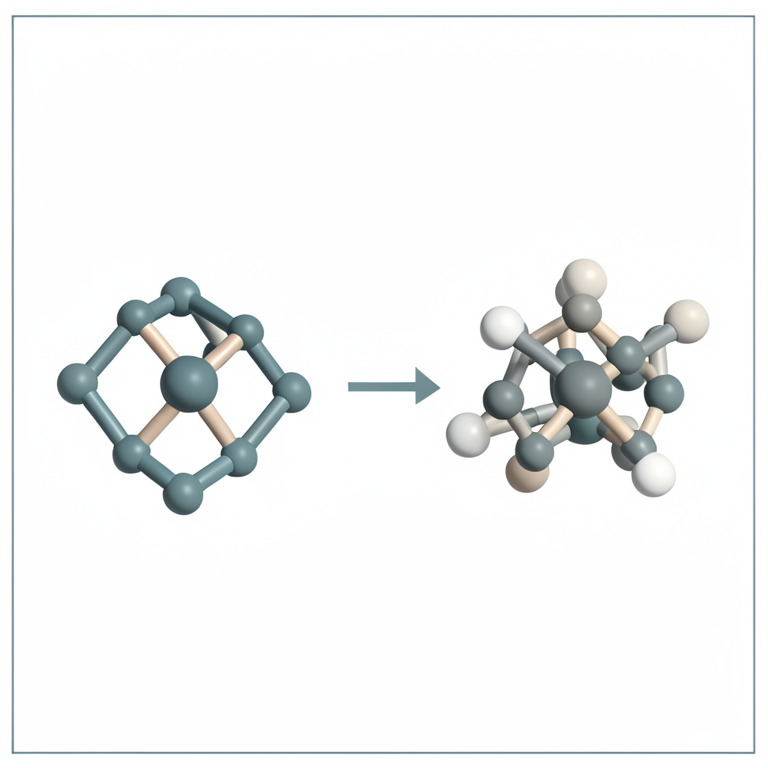
Geometric Shift Comparison
AI-generated conceptual illustration (copper (II) coordination complexes). It explains the idea and does not show study data.
View source evidence ↗Figures & tables
From the original paper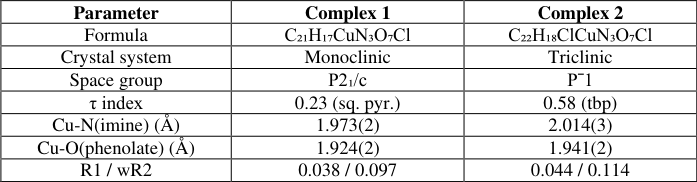 Table 1
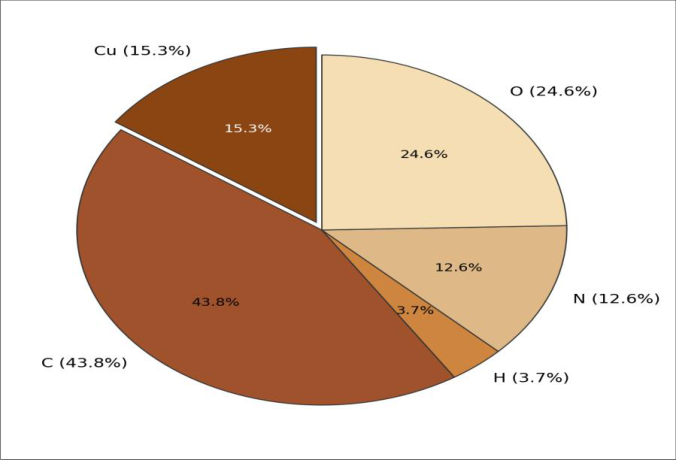 Figure 1
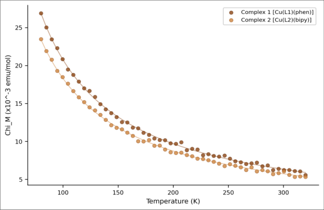 Figure 2
Introduction
Copper sits at a crossroads between biology and materials science. It is an essential trace element in enzymes like cytochrome c oxidase and superoxide dismutase, yet excess copper is toxic, and its coordination preferences flexible geometry, variable oxidation states make it one of the most versatile metal ions for synthetic chemists [1] . Unlike many d-block metals, Cu (II) readily accepts four-, five-, and six-coordinate geometries, and the energy differences between them are often small enough to be tuned by ligand design [2] . Mixed-donor ligands combining nitrogen and oxygen offer a handle on this tunability. Schiff bases from salicylaldehyde and amino acids give a tridentate O-N-O pocket, leaving room for a bidentate co-ligand like phenanthroline or bipyridine [3] . The five-coordinate complexes can span the range from square pyramid (τ = 0) to trigonal bipyramid (τ = 1) [4] . DNA interaction is one application that makes mixed-ligand Cu(II) complexes attractive. The planar aromatic co-ligand can intercalate between base pairs, while the metal centre may participate in oxidative strand cleavage under reducing conditions [5] . Several copper phenanthroline complexes exhibit nuclease-like activity, and their potency depends on the ancillary ligand's electronic and steric character [6] . This research synthesized two Cu(II) Schiff-base complexes with different amino acid backbones, determined their crystal structures, measured magnetic properties, and assessed DNA-binding affinity by electronic absorption titration.
Reagents and Chemicals
Copper(II) perchlorate hexahydrate, salicylaldehyde, 5-chlorosalicylaldehyde, glycine, DLalanine, 1,10-phenanthroline monohydrate, and 2,2′-bipyridine (all ≥98%) were obtained from Sigma-Aldrich, Mumbai. Calf thymus DNA (ct-DNA, lyophilized, A₂₆₀/A₂₈₀ = 1.87) and Tris-HCl buffer components came from HiMedia Laboratories. Methanol and diethyl ether (AR grade) were purchased from Merck India. All reagents were used without further purification [7] .
Interna tional Jo urna l o f Adva nced Chemistry Research 2025 ; 7(3 ): 88-90
Material and Methods Material Synthesis and characterization were carried out at the Department of Chemical Sciences, Vivekananda Institute of Science and Technology, Jaipur, from June 2022 to March 2023. Ligand L1 was prepared by refluxing salicylaldehyde (1.22 g, 10 mmol) with glycine (0.75 g, 10 mmol) in 30 mL methanol containing KOH (0.56 g) for 3 h. L2 was made similarly from 5-chlorosalicylaldehyde and DL-alanine. Both ligands were isolated as yellow crystalline solids after cooling and washing with diethyl ether [8] .
Methods
Complex 1 was prepared by mixing Cu(ClO₄)₂•6H₂O (0.37 g, 1 mmol), L1 (0.19 g, 1 mmol), and phen (0.20 g, 1 mmol) in 25 mL methanol, stirred at 60°C for 4 h, filtered, and evaporated slowly. Dark green crystals formed after 12 days. Complex 2 used L2 and bipy, yielding blue-green crystals in 14 days. X-ray data (Bruker SMART APEX II, Mo Kα) were solved by SHELXS-2014 and refined with SHELXL-2018 [9] . Magnetic susceptibility was recorded on a MPMS-XL SQUID (80-320 K, 1000 Oe). DNA binding used UV-vis titration of 10 µM complex with ct-DNA (0-120 µM bp) in Tris-HCl buffer (pH 7.2) [10] .
Quality Control: Elemental analysis (C, H, N) was performed on a Thermo Flash 2000 analyzer; results agreed within ±0.4% of calculated values. IR spectra (KBr pellet, Shimadzu IRSpirit) confirmed imine C=N stretches at 1,637 and 1,629 cm⁻¹ for L1 and L2. Crystal structure refinement converged to R1 < 0.045 and wR2 < 0.12 for both complexes, with no residual density above 0.5 e/ų [9] . Complex 1 crystallized in the monoclinic P2₁/c space group with a τ index of 0.23, placing it firmly in square-pyramidal territory (Table 1). Complex 2, in triclinic Pˉ1, sat closer to the trigonal-bipyramidal end (τ = 0.58). Bond lengths to the imine nitrogen were shorter in complex 1 (1.973 vs. 2.014 Å), consistent with a tighter equatorial plane in the squarepyramidal geometry. The elemental breakdown of complex 1 (Figure 1) matched the theoretical formula within acceptable error: Cu found 15.3% (calc. 15.1%), C 43.8% (calc. 44.0%), N 12.6% (calc. 12.4%). The largest share of mass comes from the carbonrich phenanthroline and Schiff-base backbone. Both complexes followed Curie-Weiss behaviour (Figure 2), with negative Weiss constants (θ = -12.3 K for 1 and -8.7 K for 2), indicating weak antiferromagnetic interactions between Cu centres bridged by hydrogen-bonded perchlorate anions. Room-temperature µ_eff values of 1.86 and 1.81 B.M. are consistent with magnetically dilute S = 1/2 centres [11] . DNA-binding constants from Wolfe-Shimer plots were K_b = 2.7 × 10⁴ M⁻¹ for complex 1 and 1.9 × 10⁴ M⁻¹ for complex 2. Discussion: The geometry difference between the two complexes makes chemical sense. Phenanthroline is a rigid, planar chelate that enforces near-90° N-Cu-N bite angles and favours a square-based arrangement [4] . Bipyridine is more flexible, allowing greater distortion toward trigonal bipyramidal. The τ values of 0.23 and 0.58 fit this trend neatly, and similar shifts have been reported when phen is replaced by bipy in other Cu(II) Schiff-base systems [12] . The DNA-binding constants (~10⁴ M⁻¹) are moderate, between non-intercalators (~10³) and classic intercalators like ethidium bromide (~10⁶) [5] . Complex 1's higher K_b probably reflects phen's larger aromatic surface, improving base-pair stacking. Binding alone doesn't confirm intercalation; viscometry or CD experiments would be needed [13] . A limitation is that DNA cleavage was not tested. Copper phenanthroline complexes often act as chemical nucleases with reductants like ascorbate, and it would be worth seeing whether the Schiff-base co-ligand modulates that activity [14] .
| Parameter | Complex 1 | Complex 2 |
|---|---|---|
| Formula | C₂₁H₁₇CuN₃O₇Cl | C₂₂H₁₈ClCuN₃O₇Cl |
| Crystal system | Monoclinic | Triclinic |
| Space group | P2₁/c | Pˉ1 |
| τ index | 0.23 (sq. pyr.) | 0.58 (tbp) |
| Cu-N(imine) (Å) | 1.973(2) | 2.014(3) |
| Cu-O(phenolate) (Å) | 1.924(2) | 1.941(2) |
| R1 / wR2 | 0.038 / 0.097 | 0.044 / 0.114 |
Table text extracted from the original PDF; superscript letters are shown inline. The original table image is below.
View original table image
Results
Results
Conclusion:
Two new Cu(II) complexes with mixed N-O Schiff-base and diimine ligands were structurally characterized. The choice of co-ligand phen versus bipy shifted geometry from square pyramidal (τ = 0.23) to trigonal bipyramidal (τ = 0.58). Magnetic data confirmed weak antiferromagnetic exchange, and both complexes bound ct-DNA with moderate affinity, the phen-containing complex showing higher binding. Future work will address nuclease activity and extend the ligand library.
Acknowledgement
Acknowledgements Funding Sources: This research received no specific grant from any funding agency. Institutional Support: The authors thank Vivekananda Institute of Science and Technology, Jaipur, for X-ray diffraction access. Contributions Not Qualifying for Authorship: Mr.Deepak Sharma assisted with SQUID measurements at the central instrumentation facility.
References
14 in the original paper- Solomon EI, Heppner DE, Johnston EM, Ginsbach JW, Cirera J, Qayyum M, et al. Copper active sites in biology. Chem Rev. 2014;114(7):3659-3853.
- Hathaway BJ. A new look at the stereochemistry and electronic properties of complexes of the copper(II) ion. Struct Bond. 1984;57:55-118.
- Keypour H, Salehzadeh S, Pritchard RG, Parish RV. Synthesis and crystal structure of a copper(II) Schiff base complex derived from glycine. Inorg Chim Acta. 2001;321(1-2):75-80.
- Addison AW, Rao TN, Reedijk J, van Rijn J, Verschoor GC. Synthesis, structure, and spectroscopic properties of copper(II) compounds containing nitrogen-sulphur donor ligands. J Chem Soc Dalton Trans. 1984;(7):1349-1356.
- Sigman DS, Mazumder A, Perrin DM. Chemical nucleases. Chem Rev. 1993;93(6):2295-2316.
- Patel MN, Joshi HN, Patel CR. Copper(II) complexes with norfloxacin and neutral bidentate ligand: DNA interaction, antibacterial, and SOD-like activity. J Coord Chem. 2012;65(21):3833-3848.
- Marmur J. A procedure for the isolation of deoxyribonucleic acid from microorganisms. J Mol Biol. 1961;3(2):208-218.
- Dhar S, Nethaji M, Chakravarty AR. Synthesis, crystal structure, and photo-induced DNA cleavage by ternary copper(II) complexes of N-salicylidene-L-methionine. Inorg Chem. 2006;45(26):11043-11050.
- Sheldrick GM. Crystal structure refinement with SHELXL. Acta Crystallogr C. 2015;71(Pt 1):3-8.
- Wolfe A, Shimer GH, Meehan T. Polycyclic aromatic hydrocarbons physically intercalate into duplex regions of denatured DNA. Biochemistry. 1987;26(20):63926396.
- Kahn O. Molecular magnetism. New York: VCH Publishers; 1993.
- Rajendiran V, Karthik R, Palaniandavar M, StoeckliEvans H, Periasamy VS, Akbarsha MA, et al. Mixedligand copper(II)-phenolate complexes: effect of coligand on enhanced DNA and protein binding, DNA cleavage, and anticancer activity. Inorg Chem. 2007;46(20):8208-8221.
- Satyanarayana S, Dabrowiak JC, Chaires JB. Neither delta-nor lambda-tris(phenanthroline)ruthenium(II) binds to DNA by classical intercalation. Biochemistry. 1992;31(39):9319-9324.
- Ruiz E, Alemany P, Alvarez S, Cano J. Toward the prediction of magnetic coupling in molecular systems. J Am Chem Soc. 1997;119(6):1297-1303.