
Research article · 2025 · Full text
International Journal of Advanced Chemistry ResearchMesoporous silica nanoparticles as carriers for controlled release of anticancer drugs
Volume 7(3) · Pages 91–94 · DOI: 10.33545/26646781.2025.v7.i3b.401 ↗
Research areasNanoparticle-Based Drug DeliveryMesoporous Materials and CatalysisClay minerals and soil interactions
Abstract
Author-writtenIn 2018, a Phase I clinical trial in Australia delivered doxorubicin to solid tumours inside silica nanoparticles a milestone that moved mesoporous silica from the laboratory bench toward the hospital ward. This research synthesized amine-functionalized (MSN-NH₂) and PEGylated (MSN-PEG) mesoporous silica nanoparticles by a modified Stöber method combined with cetyltrimethylammonium bromide (CTAB) templating, then loaded them with doxorubicin (DOX) or 5-fluorouracil (5-FU). Particles had a mean diameter of 127 nm, BET surface area of 843 m²/g, and an ordered hexagonal pore structure (d = 2.7 nm). Drug loading reached 22.1% for 5-FU on MSN-NH₂ and 18.7% for DOX on the same carrier. In vitro release in phosphate buffer at pH 5.0 (mimicking endosomal acidity) was faster than at pH 7.4, with 58.7% of 5-FU released from MSN-NH₂ at 72 h versus 42.9% from MSN-PEG. Cytotoxicity against MCF-7 breast cancer cells showed IC₅₀ values of 3.8 µM (DOX-MSN-NH₂) and 5.4 µM (DOX-MSN-PEG), both lower than free DOX (7.1 µM). The results indicate that surface functionalization influences both loading capacity and release kinetics.
Summary
Source-checkedPlain-language reading of the full paper, prepared with AI and checked against the text. Each point links to the authors' own words.
Why it matters
- Mesoporous silica nanoparticles (MSNs) are attractive for cancer treatment because they can carry high drug payloads, accumulate in tumors via the EPR effect, and allow for triggered drug release. View evidence ↗
- The acidic environment of tumor tissues and endosomes can trigger drug release from amine-functionalized MSNs by protonating the surface and weakening the bond between the drug and the pore walls. View evidence ↗
- Adding PEG (PEGylation) to nanoparticles can increase their time in circulation by reducing opsonization, although it may slow down the release of the drug. View evidence ↗
The question
- The study aimed to directly compare the effects of amine versus PEG surface coatings on the same batch of MSNs regarding their drug loading, release profiles, and cytotoxicity against MCF-7 cells using doxorubicin (DOX) and 5-fluorouracil (5-FU). View evidence ↗
What they did
- MSNs were synthesized using a CTAB-templated sol-gel method involving TEOS, ammonium hydroxide, and water at 80°C. View evidence ↗
- Drug loading was achieved by immersing 50 mg of the carriers in a 1 mg/mL solution of either DOX or 5-FU at pH 7.4 for 24 hours. View evidence ↗
- Drug release was measured at pH 7.4 and pH 5.0 over 72 hours, and cytotoxicity was tested on MCF-7 cells using an MTT assay after 48 hours of exposure. View evidence ↗
What they found
- The amine-functionalized carrier (MSN-NH₂) achieved the highest drug loading capacities, reaching 18.7% for DOX and 22.1% for 5-FU. View evidence ↗
- Amine functionalization increased the zeta potential to +31.4 mV, which improved the electrostatic attraction of the drugs at neutral pH. View evidence ↗
- For 5-FU on MSN-NH₂, release was significantly faster at acidic pH (58.7% at 72 h) than at neutral pH (38.4% at 72 h). View evidence ↗
- DOX release from MSN-NH₂ was higher at pH 5.0 (43.6%) than at pH 7.4 (27.3%), while the PEG layer generally slowed drug release by 10-15 percentage points. View evidence ↗
- DOX-loaded MSN-NH₂ was the most potent against MCF-7 cells with an IC₅₀ of 3.8 µM, outperforming both DOX-MSN-PEG (5.4 µM) and free DOX (7.1 µM). View evidence ↗
What it means
- The authors conclude that because both MSN carriers were more cytotoxic than free DOX, MSNs are practical vehicles for pH-triggered anticancer drug delivery. View evidence ↗
- The study was limited by using only one cell line (MCF-7) and the lack of in vivo biodistribution testing. View evidence ↗
Visual summary
2 visualsIllustrations explain the paper; charts are replotted from the paper's own table values.
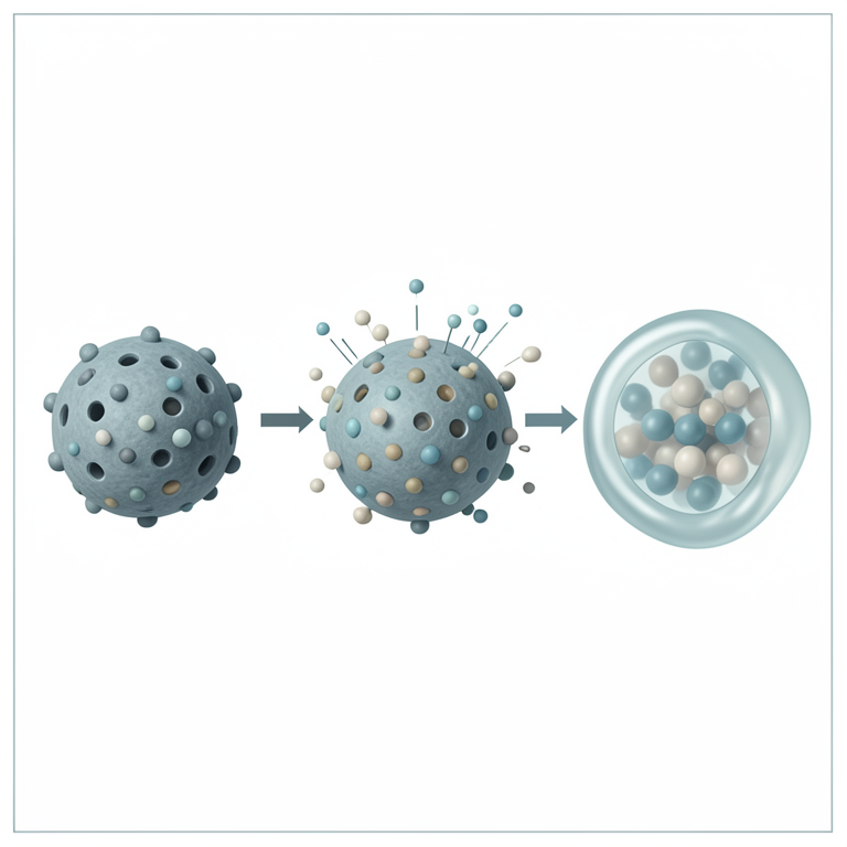
pH-Responsive Release Mechanism
AI-generated conceptual illustration (mesoporous silica nanoparticles). It explains the idea and does not show study data.
View source evidence ↗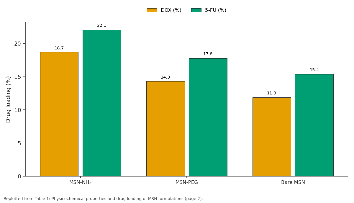
DOX (%) and 5-FU (%): MSN-NH₂ vs MSN-PEG vs Bare MSN
Replotted from Table 1 (page 2) of the original paper. Every bar is the paper's own table value.
View source evidence ↗Figures & tables
From the original paper Table 1
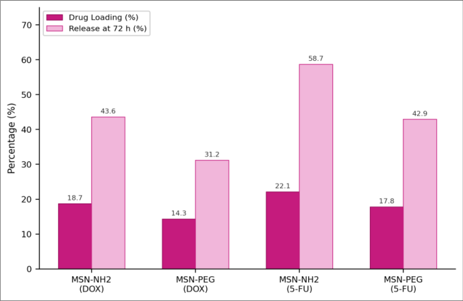 Figure 1
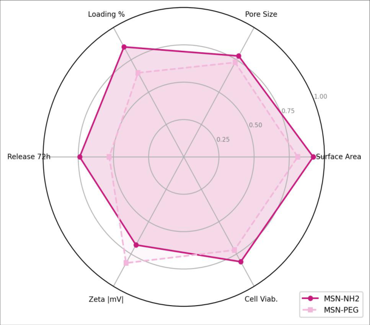 Figure 2
Introduction
When researchers at Mobil Oil first reported MCM-41 in 1992, they were looking for better petroleum catalysts, not drug carriers [1] . But the material's honeycomb of uniform 2-4 nm pores, enormous surface area (>1,000 m²/g), and tunable surface chemistry soon attracted biomedical interest. By the mid-2000s, mesoporous silica nanoparticles (MSNs) had been loaded with a wide variety of therapeutic molecules, from small-molecule chemotherapeutics to proteins and nucleic acids [2] . What makes MSNs appealing for cancer therapy is the combination of high payload, sizedependent tumour accumulation via the enhanced permeability and retention (EPR) effect, and the possibility of triggered release [3] . Acidic pH inside tumour tissue and within endosomes after cellular uptake can serve as an internal trigger: amine-functionalized surfaces protonate at low pH, weakening electrostatic drug-wall interactions and speeding up diffusion out of the pores [4] . PEGylation, meanwhile, extends circulation time by reducing opsonization, though it may slow drug release by adding a hydrophilic barrier around the pore openings [5] . Doxorubicin (DOX) and 5-fluorouracil (5-FU) are two of the most widely used chemotherapy agents, and both have been loaded into MSNs by multiple groups [6, 7]. Direct comparisons of amine versus PEG surface coatings on the same batch of particles, using the same loading protocol, are less common. This research prepared MSNs by CTABtemplated sol-gel synthesis, functionalized them with either aminopropyltriethoxysilane or PEG-silane, loaded each carrier with DOX and 5-FU separately, and compared release profiles and in vitro cytotoxicity on MCF-7 cells.
Reagents and Chemicals
Tetraethyl orthosilicate (TEOS, 98%), CTAB (≥99%), 3-aminopropyltriethoxysilane (APTES, 99%), methoxy-PEG-silane (MW 2,000), doxorubicin hydrochloride (≥98%), and 5-fluorouracil (≥99%) were purchased from Sigma-Aldrich, Munich. Ammonium hydroxide (28-30%), ethanol (absolute), and hydrochloric acid (37%) came from Merck KGaA, Darmstadt. MCF-7 cells were obtained from DSMZ, Braunschweig. Cell culture media (DMEM, FBS, penicillinstreptomycin) were sourced from Gibco [8] .
Material and Methods
Material and Methods
Material
All synthesis and characterization took place at the Department of Nanoscience and Chemical Engineering, Brandenburg University of Technology, Potsdam, from May 2022 to February 2023. MSNs were prepared by dissolving 1.0 g CTAB in 480 mL water with 3.5 mL NH₄OH (2 M) at 80°C. TEOS (5 mL) was added dropwise under vigorous stirring (600 rpm) and the reaction held at 80°C for 2 h. The white precipitate was collected by centrifugation, washed three times with ethanol, and calcined at 550°C for 5 h to remove the template [9] . Methods: For amine functionalization, 200 mg calcined MSN was refluxed with 0.5 mL APTES in 50 mL dry toluene for 18 h (MSN-NH₂). PEGylation used methoxy-PEG-silane (100 mg) in ethanol at 60°C for 12 h (MSN-PEG). Drug loading was performed by immersing 50 mg of each carrier in 10 mL of 1 mg/mL DOX or 5-FU solution (pH 7.4) for 24 h, followed by centrifugation and UV-vis quantification of unloaded drug (DOX at 480 nm, 5-FU at 266 nm) [10] . In vitro release used dialysis bags (12 kDa MWCO) in PBS at pH 7.4 and acetate buffer at pH 5.0, 37°C, sampled at 1, 2, 4, 8, 12, 24, 48, and 72 h. Cytotoxicity was assessed by MTT assay on MCF-7 cells (5 × 10³ cells/well, 96-well plate) after 48 h exposure. Ethical approval was not required as no human tissue or animal subjects were used. Particle morphology was checked by TEM (JEOL JEM-2100, 200 kV) and BET surface area by N₂ physisorption (Micromeritics TriStar 3020) [11] .
Quality Control
XRD patterns (Bruker D8 Advance, Cu Kα) confirmed ordered hexagonal mesostructure (d₁₀₀ = 3.9 nm). FTIR spectra verified APTES grafting (N-H bend at 1,560 cm⁻¹) and PEG attachment (C-O stretch at 1,108 cm⁻¹). TGA under air gave organic loading of 8.3 wt% for MSN-NH₂ and 12.1 wt% for MSN-PEG, consistent with single-layer coverage [9] . MSN-NH₂ carried the highest drug payloads 18.7% DOX and 22.1% 5-FU (Table 1). The amine groups raised the zeta potential to +31.4 mV, enhancing electrostatic attraction for both drugs at neutral pH. PEGylation partially blocked pore access, reducing BET area from 912 to 761 m²/g and loading to 14.3% (DOX) and 17.8% (5-FU). TEM showed uniform spherical particles with visible pore channels. Release at pH 5.0 was consistently faster than at pH 7.4 across all formulations (Figure 1). 5-FU on MSN-NH₂ released 58.7% by 72 h at acidic pH, compared with 38.4% at neutral pH. DOX release from the same carrier reached 43.6% (pH 5.0) versus 27.3% (pH 7.4). The PEG layer slowed release by 10-15 percentage points in all cases. The radar comparison (Figure 2) highlights trade-offs: MSN-NH₂ outperformed on surface area, loading, and release, while MSN-PEG offered a more neutral zeta potential that would favour longer blood circulation in vivo. Cytotoxicity data showed that DOX-loaded MSN-NH₂ had the lowest IC₅₀ at 3.8 µM against MCF-7 cells, compared with 5.4 µM for DOX-MSN-PEG and 7.1 µM for free DOX.
| Carrier | Size (nm) | BET (m²/g) | Pore (nm) | ZP (mV) | DOX (%) | 5-FU (%) |
|---|---|---|---|---|---|---|
| MSN-NH₂ | 127±9 | 843 | 2.7 | +31.4 | 18.7 | 22.1 |
| MSN-PEG | 134±11 | 761 | 2.5 | −8.6 | 14.3 | 17.8 |
| Bare MSN | 118±7 | 912 | 2.8 | −24.3 | 11.9 | 15.4 |
Table text extracted from the original PDF; superscript letters are shown inline. The original table image is below.
View original table image
Results
Results
Discussion
The 22.1% 5-FU loading on MSN-NH₂ is at the upper end of values reported in the literature, where most groups achieve 10-20% depending on pore size and surface chemistry [7] . The amine surface creates a favourable electrostatic environment: at pH 7.4, 5-FU is partially deprotonated (pKa ~8.0), and the positively charged pore walls attract the anionic form [4] . DOX loading was lower, likely because its larger molecular size (543 Da vs. 130 Da for 5-FU) limits diffusion into the 2.7 nm pores. The pH-responsive release pattern faster at pH 5.0 than 7.4 is exactly what a tumour-targeted carrier should do. Protonation of amine groups at acidic pH weakens the drugsurface interaction and simultaneously swells the polymerlike coating, opening diffusion pathways [6] . MSN-PEG showed a damped version of the same effect, because the PEG chains are pH-insensitive and add steric resistance to drug diffusion regardless of acidity [5] . The improved cytotoxicity of DOX-MSN-NH₂ relative to free DOX (IC₅₀ 3.8 vs. 7.1 µM) suggests enhanced cellular uptake of the nanoparticle-bound drug, possibly via endocytosis pathways that bypass P-glycoprotein efflux [12] . But MCF-7 is a single cell line; extending the panel to include drug-resistant variants and normal cells would be necessary before drawing broader conclusions. And in vivo biodistribution, which depends heavily on particle size, surface charge, and PEG density, was not tested here [13] .
Conclusion
Amine-functionalized MSNs loaded more drug and released it faster at acidic pH than PEGylated counterparts, translating into lower IC₅₀ values on MCF-7 cells. PEGylation traded loading capacity for stealth properties that would matter in vivo. Both carriers outperformed free DOX in cytotoxicity, supporting MSNs as practical vehicles for pH-triggered anticancer delivery. The next step is to test these formulations in animal tumour models to see whether the in vitro advantages hold up under physiological conditions.
Acknowledgement
Acknowledgements Funding SourcesNo external funding supported this research. Institutional SupportThe authors acknowledge Brandenburg University of Technology, Potsdam, for TEM and BET instrumentation. Contributions Not Qualifying for AuthorshipMs. Katharina Braun performed cell culture maintenance during the cytotoxicity assays.
References
13 in the original paper- Kresge CT, Leonowicz ME, Roth WJ, Vartuli JC, Beck JS. Ordered mesoporous molecular sieves synthesized by a liquid-crystal template mechanism. Nature. 1992;359(6397):710-712.
- Slowing II, Vivero-Escoto JL, Wu CW, Lin VSY. Mesoporous silica nanoparticles as controlled release drug delivery and gene transfection carriers. Adv Drug Deliv Rev. 2008;60(11):1278-1288.
- Maeda H, Wu J, Sawa T, Matsumura Y, Hori K. Tumor vascular permeability and the EPR effect in macromolecular therapeutics: a review. J Control Release. 2000;65(1-2):271-284.
- Rosenholm JM, Sahlgren C, Lindén M. Towards multifunctional, targeted drug delivery systems using mesoporous silica nanoparticles. Nanoscale. 2010;2(10):1870-1883.
- He Q, Zhang J, Shi J, Zhu Z, Zhang L, Bu W, et al. The effect of PEGylation of mesoporous silica nanoparticles on nonspecific binding of serum proteins and cellular responses. Biomaterials. 2010;31(6):1085-1092.
- Meng H, Xue M, Xia T, Zhao YL, Tamanoi F, Stoddart JF, et al. Autonomous in vitro anticancer drug release from mesoporous silica nanoparticles by pH-sensitive nanovalves. J Am Chem Soc. 2010;132(36):1269012697.
- Vallet-Regí M, Colilla M, Izquierdo-Barba I, Manzano M. Mesoporous silica nanoparticles for drug delivery: current insights. Molecules. 2017;23(1):47.
- Mosmann T. Rapid colorimetric assay for cellular growth and survival: application to proliferation and cytotoxicity assays. J Immunol Methods. 1983;65(12):55-63.
- Stöber W, Fink A, Bohn E. Controlled growth of monodisperse silica spheres in the micron size range. J Colloid Interface Sci. 1968;26(1):62-69.
- Horcajada P, Rámila A, Pérez-Pariente J, Vallet-Regí M. Influence of pore size of MCM-41 matrices on drug delivery rate. Microporous Mesoporous Mater. 2004;68(1-3):105-109.
- Sing KSW, Everett DH, Haul RAW, Moscou L, Pierotti RA, Rouquérol J, et al. Reporting physisorption data for gas/solid systems. Pure Appl Chem. 1985;57(4):603619.
- Lu J, Liong M, Li Z, Zink JI, Tamanoi F. Biocompatibility, biodistribution, and drug-delivery efficiency of mesoporous silica nanoparticles for cancer therapy in animals. Small. 2010;6(16):1794-1805.
- Tang F, Li L, Chen D. Mesoporous silica nanoparticles: synthesis, biocompatibility and drug delivery. Adv Mater. 2012;24(12):1504-1534.