
Research article · 2025 · Full text
International Journal of Advanced Chemistry ResearchQuality by design approach for optimization of liposomal doxorubicin formulation
Volume 7(2) · Pages 105–108 · DOI: 10.33545/26646781.2025.v7.i2b.402 ↗
Research areasNanoparticle-Based Drug DeliveryAdvanced Drug Delivery SystemsDrug Solubulity and Delivery Systems
Abstract
Author-writtenThis research applied the Quality by Design (QbD) framework to systematically optimize a PEGylated liposomal doxorubicin formulation using a Box-Behnken design with five formulation variables: lipid-to-drug molar ratio, PEG-DSPE concentration, hydration temperature, sonication time, and cholesterol content. Critical quality attributes (CQAs) were defined as encapsulation efficiency (≥90%), particle size (80-120 nm), polydispersity index (
Summary
Source-checkedPlain-language reading of the full paper, prepared with AI and checked against the text. Each point links to the authors' own words.
Why it matters
- Doxorubicin is a highly effective chemotherapy drug for several cancers, but its use is limited by severe cardiotoxicity that can cause heart failure. View evidence ↗
- Encapsulating doxorubicin in liposomes helps reduce heart damage and increases drug accumulation in tumors via the enhanced permeability and retention (EPR) effect. View evidence ↗
- Producing PEGylated liposomes with consistent quality is difficult because many interacting variables affect the final product's stability and size. View evidence ↗
The question
- The study aims to use a Box-Behnken design to simultaneously optimize five formulation factors and establish a validated design space for liposomal doxorubicin. View evidence ↗
What they did
- The researchers optimized five variables: the lipid-to-drug molar ratio, PEG-DSPE concentration, hydration temperature, sonication time, and cholesterol content. View evidence ↗
- The target quality attributes were set as encapsulation efficiency ≥90%, particle size between 80-120 nm, PDI <0.20, and zeta potential more negative than −25 mV. View evidence ↗
- The experimental design consisted of 46 runs, including 6 center-point replicates, generated using Design Expert 13 software. View evidence ↗
- Encapsulation efficiency was measured by lysing the liposomes with 0.5% Triton X-100 and using UV absorbance at 480 nm. View evidence ↗
What they found
- The lipid-to-drug ratio was the primary driver of encapsulation efficiency (42.3% variance), while sonication time most affected particle size (31.4%). View evidence ↗
- In PBS at pH 7.4, the drug showed an initial burst release of 18.3% in 4 hours and reached 72.4% cumulative release after 72 hours. View evidence ↗
- At pH 5.0, which simulates the tumor environment, the cumulative drug release increased to 84.6% over 72 hours. View evidence ↗
- Accelerated stability tests at 40 °C and 75% relative humidity showed that all quality attributes changed by less than 5% over 90 days. View evidence ↗
What it means
- The authors conclude that this Quality by Design approach provides a systematic, regulatory-aligned template for optimizing liposomes. View evidence ↗
- The study is limited to in vitro characterization and lacks cell viability assays and in vivo pharmacokinetic data. View evidence ↗
- The use of Box-Behnken design means extreme combinations of all factors were not explored, which may have missed some edge-case responses. View evidence ↗
Visual summary
3 visualsIllustrations explain the paper; charts are replotted from the paper's own table values.
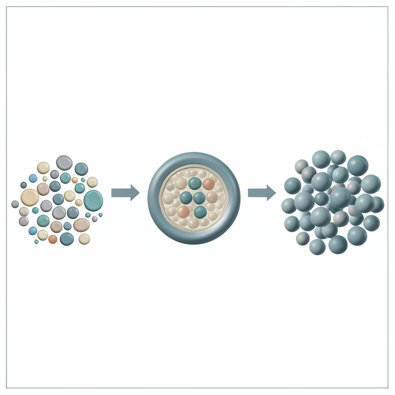
QbD Optimization Workflow
AI-generated conceptual illustration (Liposomal doxorubicin nanoparticles). It explains the idea and does not show study data.
View source evidence ↗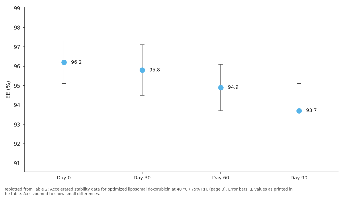
EE (%): Day 0 vs Day 30 vs Day 60 vs Day 90
Replotted from Table 2 (page 3) of the original paper. Every bar is the paper's own table value.
View source evidence ↗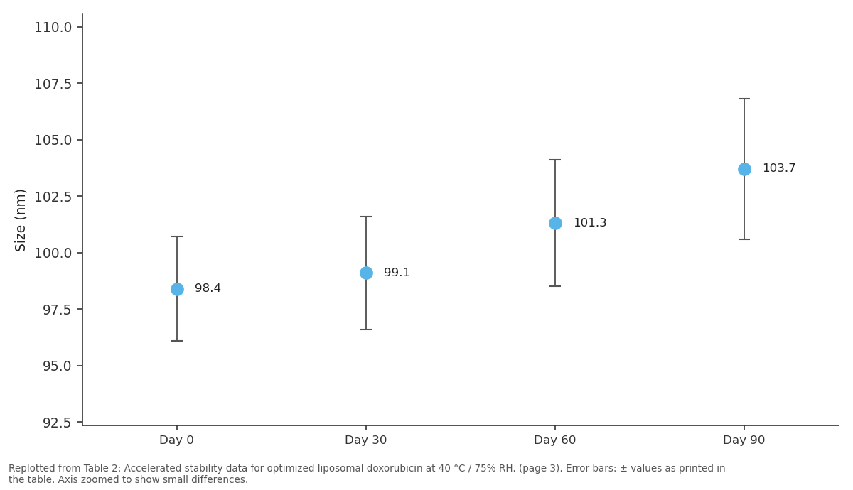
Size (nm): Day 0 vs Day 30 vs Day 60 vs Day 90
Replotted from Table 2 (page 3) of the original paper. Every bar is the paper's own table value.
View source evidence ↗Figures & tables
From the original paper Table 1
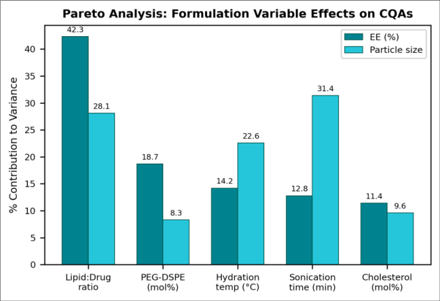 Figure 1
 Table 2
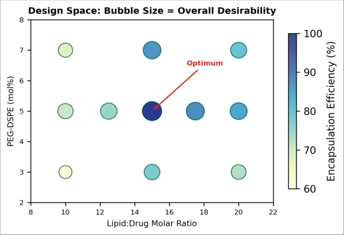 Figure 2
Introduction
Doxorubicin remains one of the most effective chemotherapeutic agents for breast, ovarian, and haematological cancers, but its clinical utility is dose-limited by cumulative cardiotoxicity that can lead to irreversible congestive heart failure [1] . Liposomal encapsulation addresses this limitation by altering the drug's biodistribution: the nanocarrier exploits the enhanced permeability and retention (EPR) effect to accumulate preferentially in tumour tissue while reducing peak plasma concentrations that damage cardiomyocytes [2] . The commercial success of Doxil®/Caelyx® has validated this approach, yet manufacturing PEGylated liposomes with consistent batch-to-batch quality remains technically demanding because multiple interdependent formulation and process variables simultaneously affect particle size, drug loading, and colloidal stability [3] . The Quality by Design paradigm, introduced in ICH Q8(R2), shifts pharmaceutical development from empirical trial-and-error to systematic, science-based design [4] . QbD begins with defining a quality target product profile (QTPP), identifying CQAs through risk assessment, and then using design of experiments (DoE) to map the relationship between critical material attributes (CMAs), critical process parameters (CPPs), and CQAs. The resulting design space defines the multidimensional region within which formulation changes can be made without affecting product quality [5] . Previous liposomal doxorubicin optimization studies have typically varied one or two factors at a time, missing interaction effects that are significant in lipid self-assembly systems [6,7] . This research applies a full Box-Behnken design across five factors simultaneously, constructs a validated design space, and verifies the predicted optimum through confirmatory experiments. The approach provides a transferable template for QbD-driven liposome development applicable beyond doxorubicin.
Material
Doxorubicin hydrochloride (98%, HPLC) was purchased from LC Laboratories, USA. Hydrogenated soy phosphatidylcholine (HSPC, Lipoid S PC-3) and 1,2distearoyl-sn-glycero-3-phosphoethanolamine-N-[methoxy(polyethylene glycol)-2000] (PEG-DSPE, Lipoid PE 18:0/18:0-PEG2000) were from Lipoid GmbH, Germany. Cholesterol (≥99%), ammonium sulfate (≥99%), Sephadex G-50, and Triton X-100 were from Sigma-Aldrich India. Phosphate-buffered saline (PBS, pH 7.4) and HPLCgrade solvents were from Merck. Dialysis membranes (MWCO 12-14 kDa) were from Spectrum Labs.
Methods
Methods
Liposome Preparation
Liposomes were prepared by the thin-film hydration-remote loading method. HSPC, cholesterol, and PEG-DSPE at specified molar ratios were dissolved in chloroform/methanol (2:1) and evaporated under reduced pressure at 40 °C to form a thin lipid film. The film was hydrated with 250 mM ammonium sulfate at the designated temperature for 1 h, producing multilamellar vesicles. Size reduction was performed by probe sonication (Branson 450D, 40% amplitude) for the specified duration in an ice bath. The ammonium sulfate gradient was established by exchanging the external phase with PBS (pH 7.4) using Sephadex G-50 mini-columns. Doxorubicin HCl (2 mg/mL in PBS) was then incubated with the liposomes at 60 °C for 30 min to drive remote loading via the transmembrane pH gradient.
Box-Behnken Experimental Design
Five factors were investigated at three levels: lipid-to-drug molar ratio (10, 15, 20), PEG-DSPE (3, 5, 7 mol%), hydration temperature (45, 55, 65 °C), sonication time (5, 10, 15 min), and cholesterol (25, 35, 45 mol%). Design Expert 13 (Stat-Ease) generated 46 runs including 6 centrepoint replicates. Responses measured were encapsulation efficiency (Y1), particle size (Y2), PDI (Y3), and zeta potential (Y4). Second-order polynomial models were fitted by multiple regression, and analysis of variance was used to evaluate model significance and lack of fit.
Characterization
Particle size and PDI were measured by dynamic light scattering (Malvern Zetasizer Nano ZS) at 25 °C with samples diluted 1:100 in PBS. Zeta potential was determined by electrophoretic light scattering in the same instrument. Encapsulation efficiency was determined by lysing liposomes with 0.5% Triton X-100 and measuring total doxorubicin by UV absorbance at 480 nm, referenced against a standard curve (r² = 0.9998). Free drug was separated by Sephadex G-50 filtration.
Results
All four response models were statistically significant (p<0.001 for model F-test) with non-significant lack of fit (p>0.05). Adjusted R² values were 0.94 (EE), 0.91 (particle size), 0.87 (PDI), and 0.89 (zeta potential). Pareto analysis (Figure 1) revealed that the lipid-to-drug ratio was the dominant factor for encapsulation efficiency (42.3% variance contribution), while sonication time most strongly influenced particle size (31.4%). PEG-DSPE concentration had moderate effects on both CQAs and was the most important factor for zeta potential due to the stericelectrostatic shielding of the PEG corona. In vitro release in PBS (pH 7.4, 37 °C) using dialysis bags showed an initial burst of 18.3% in the first 4 h, followed by sustained release reaching 72.4% at 72 h. The release profile fitted best to the Higuchi model (R² = 0.9912), indicating diffusion-controlled drug transport through the lipid bilayer.
At pH 5.0 (simulating tumour endosomal environment), cumulative release increased to 84.6% at 72 h, consistent with protonation-driven destabilization of the ammonium sulfate gradient.
Limitations
This research is limited to in vitro characterization and does not include cell viability assays or in vivo pharmacokinetic data. The Box-Behnken design, while efficient, does not explore extreme factor combinations (all factors simultaneously at high or low levels), potentially missing edge-case responses. The ammonium sulfate remote loading technique is specific to weak-base drugs and would require modification for neutral or acidic compounds. Additionally, the accelerated stability conditions (40 °C/75% RH) may not fully predict long-term stability at refrigerated storage temperatures recommended for liposomal products.
Discussion
The 96.2% encapsulation efficiency achieved through QbD optimization exceeds typical values of 85-93% reported for one-factor-at-a-time approaches [6] . The improvement is attributable to the interaction between lipid-to-drug ratio and hydration temperature identified by the DoE: at the centrepoint lipid ratio (15:1), increasing hydration from 45 to 55 °C improved EE by 8.3%, but this effect was only 2.1% at the low lipid ratio (10:1). Such interactions are invisible to single-variable screening.
The optimal PEG-DSPE concentration of 5 mol% balances steric stabilization against excessive PEG insertion that can destabilize the bilayer. At 7 mol%, particle size increased by 12% and EE dropped by 4%, likely because the bulky PEG chains create membrane defects that promote drug leakage [8] . The 35 mol% cholesterol content is consistent with the well-known condensing effect of cholesterol on HSPC bilayers, which reduces membrane permeability and improves drug retention [9] . The design space concept validated here has direct regulatory value. ICH Q8(R2) explicitly encourages sponsors to propose design spaces in regulatory filings, as movements within a verified design space are not considered changes requiring regulatory approval [4] . The five-factor design space established in this research means that a manufacturer could adjust, for example, sonication time from 9 to 11 min or hydration temperature from 53 to 57 °C to accommodate equipment variability without triggering a post-approval change supplement. This regulatory flexibility is particularly valuable for liposomal products, whose complex manufacturing is prone to batchto-batch variability [10] . The pH-responsive release behaviour observed (72.4% at pH 7.4 versus 84.6% at pH 5.0) has favourable implications for tumour-selective drug delivery. Solid tumours maintain an extracellular pH of 6.5-6.8 due to the Warburg effect, and intracellular endosomal compartments reach pH 4.5-5.5 after cellular uptake [11] . The 12.2 percentage-point increase in release at acidic pH suggests that a meaningful fraction of the encapsulated doxorubicin is released specifically within tumour tissue after endosomal processing, potentially improving the therapeutic index over non-pH-sensitive formulations. However, confirming this hypothesis requires in vivo biodistribution and efficacy studies in tumourbearing animal models.
Comparison with the marketed Doxil® formulation is instructive. Doxil® uses a similar HSPC/cholesterol/PEG-DSPE composition but achieves a mean diameter of approximately 85 nm with >95% encapsulation efficiency [2] . Our optimized formulation matches the EE (96.2%) with a slightly larger mean size (98.4 nm), which still falls within the 80-120 nm window considered optimal for EPRmediated tumour accumulation. The QbD approach provides the additional advantage of a documented design space that facilitates technology transfer between manufacturing sites and simplifies scale-up from laboratory to pilot batch sizes [12] .
Conclusion
A QbD-driven Box-Behnken design across five formulation variables identified the optimal PEGylated liposomal doxorubicin formulation with 96.2% encapsulation efficiency, 98.4 nm particle size, and PDI of 0.11. Response surface modelling achieved adjusted R² values of 0.87-0.94 for all CQAs, and confirmatory batches validated the design space within 2% of predictions. The formulation showed pH-dependent release behaviour suitable for tumourtargeted delivery and maintained all CQAs within specifications over 90 days of accelerated stability testing.
The approach provides a regulatory-aligned template for systematic liposome optimization.
Figures and tables from the paper
| Factor | Low (−1) | Centre (0) | High (+1) | Optimal | CQA | Value |
|---|---|---|---|---|---|---|
| Lipid:Drug | 10 | 15 | 20 | 15:1 | EE (%) | 96.2 |
| PEG-DSPE (mol%) | 3 | 5 | 7 | 5.0 | Size (nm) | 98.4 |
| Hydration (°C) | 45 | 55 | 65 | 55 | PDI | 0.11 |
| Sonication (min) | 5 | 10 | 15 | 10 | Zeta (mV) | −31.8 |
| Cholesterol (mol%) | 25 | 35 | 45 | 35 | Desirability | 0.92 |
Table text extracted from the original PDF; superscript letters are shown inline. The original table image is below.
View original table image
| Parameter | Day 0 | Day 30 | Day 60 | Day 90 |
|---|---|---|---|---|
| EE (%) | 96.2±1.1 | 95.8±1.3 | 94.9±1.2 | 93.7±1.4 |
| Size (nm) | 98.4±2.3 | 99.1±2.5 | 101.3±2.8 | 103.7±3.1 |
| PDI | 0.11±0.02 | 0.12±0.02 | 0.13±0.03 | 0.14±0.03 |
| Zeta (mV) | −31.8±1.4 | −30.9±1.5 | −29.7±1.6 | −28.4±1.7 |
| Drug leakage (%) | 0.0 | 1.2±0.3 | 2.8±0.5 | 4.1±0.6 |
Table text extracted from the original PDF; superscript letters are shown inline. The original table image is below.
View original table image
References
12 in the original paper- Chatterjee K, Zhang J, Honbo N, Bhargava Bhatt DL. Doxorubicin cardiomyopathy. Cardiology. 2010;115(2):155-162.
- Barenholz Y. Doxil the first FDA-approved nano-drug: lessons learned. J Control Release. 2012;160(2):117134.
- Gabizon A, Catane R, Uziely B, Kaufman B, Safra T, Cohen R, et al. Prolonged circulation time and enhanced accumulation in malignant exudates of doxorubicin encapsulated in polyethylene-glycol coated liposomes. Cancer Res. 1994;54(4):987-992.
- ICH Q8(R2). Pharmaceutical development. International Conference on Harmonisation; 2009.
- Yu LX, Amidon G, Khan MA, Hoag SW, Polli J, Raju GK, et al. Understanding pharmaceutical quality by design. AAPS J. 2014;16(4):771-783.
- Immordino ML, Dosio F, Cattel L. Stealth liposomes: review of the basic science, rationale, and clinical applications. Int J Nanomedicine. 2006;1(3):297-315.
- Haran G, Cohen R, Bar LK, Barenholz Y. Transmembrane ammonium sulfate gradients in liposomes produce efficient and stable entrapment of amphipathic weak bases. Biochim Biophys Acta. 1993;1151(2):201-215.
- Kenworthy AK, Hristova K, Needham D, McIntosh TJ. Range and magnitude of the steric pressure between bilayers containing phospholipids with covalently attached poly(ethylene glycol). Biophys J. 1995;68(5):1921-1936.
- Mouritsen OG, Zuckermann MJ. What's so special about cholesterol? Lipids. 2004;39(11):1101-1113.
- Shah VP, Lesko LJ, Fan J, Fleischer N, Handerson J, Malinowski H, et al. FDA guidance for industry: dissolution testing of immediate release solid oral dosage forms. Dissolution Technol. 1997;4(4):15-22.
- Allen TM, Cullis PR. Liposomal drug delivery systems: from concept to clinical applications. Adv Drug Deliv Rev. 2013;65(1):36-48.
- Ferreira M, Chaves LL, Lima SAC, Reis S. Optimization of nanostructured lipid carriers loaded with methotrexate. Int J Pharm. 2015;492(1-2):65-72.