
Research article · 2026 · Full text
International Journal of Biology SciencesConcurrent canine parvoviral enteritis and Isospora infection in a German Shepherd puppy: Clinical, coprological and molecular diagnosis
Volume 8(4) · Pages 08–10 · DOI: 10.33545/26649926.2026.v8.i4a.612 ↗
Research areasAnimal Virus Infections StudiesViral gastroenteritis research and epidemiologyVeterinary Medicine and Surgery
Abstract
Author-writtenA 3-month-old male German Shepherd puppy was presented with inappetance, vomiting and acute onset of bloody, foul-smelling diarrhoea. Clinical examination revealed dullness, pale conjunctival mucous membranes, severe dehydration, pyrexia and tensed abdomen. Faecal antigen rapid test was positive for canine parvoviral enteritis, while coprological examination revealed Isospora spp. oocysts. Molecular confirmation by PCR amplified a 400 bp fragment specific for canine parvovirus. The puppy was treated with intensive supportive therapy including intravenous fluids, antibiotics, antiemetics, gastric protectants and anticoccidial drugs for five days. Gradual clinical and haematological improvement was observed with resolution of vomiting and diarrhoea. The case highlights the importance of early diagnosis, molecular confirmation and aggressive supportive management in canine parvoviral enteritis complicated by concurrent Isospora infection.
Summary
Source-checkedPlain-language reading of the full paper, prepared with AI and checked against the text. Each point links to the authors' own words.
Why it matters
- Canine parvoviral enteritis (CPE) is a highly contagious disease caused by canine parvovirus type 2 (CPV-2) that often leads to significant mortality in young, unvaccinated puppies. View evidence ↗
- The virus primarily targets rapidly dividing cells in the bone marrow and intestinal crypts, causing systemic compromise and severe pathology in the intestines. View evidence ↗
- Co-infections with intestinal parasites like Isospora spp. can worsen gastrointestinal damage and make it harder for affected puppies to recover. View evidence ↗
The question
- The study aims to document a case of concurrent parvoviral enteritis and Isosporosis in a German Shepherd puppy. View evidence ↗
What they did
- The subject was a 3-month-old male German Shepherd puppy weighing 5.3 kg treated at the Veterinary Clinical Complex in Orathanadu. View evidence ↗
- Initial screening for parvovirus was conducted using a rapid antigen test on a fecal sample. View evidence ↗
- Coprological examination was used to identify the presence of Isospora spp. oocysts in the feces. View evidence ↗
- Molecular confirmation of the parvovirus was performed via PCR analysis of extracted fecal DNA, targeting a specific 400 bp fragment. View evidence ↗
- The puppy received an intensive supportive and symptomatic treatment regimen for five consecutive days. View evidence ↗
What they found
- The puppy was diagnosed with concurrent canine parvoviral enteritis and Isospora infection based on a positive rapid antigen test, PCR amplification of a 400 bp fragment, and the detection of oocysts in feces. View evidence ↗
- Treatment led to the cessation of vomiting by the second day and a progressive reduction in diarrhea, with fecal consistency improving significantly by day four. View evidence ↗
- The puppy's white blood cell count decreased from a state of leukocytosis on day one (17 × 10³/µL) to near-normal levels by day four (9.5 × 10³/µL). View evidence ↗
What it means
- The authors conclude that early diagnosis, molecular confirmation, and aggressive supportive care are critical for managing parvoviral enteritis when complicated by Isospora infection. View evidence ↗
- The authors recommend comprehensive fecal screening for all diarrheic puppies, especially in areas with high environmental contamination. View evidence ↗
Figures & tables
From the original paper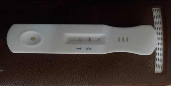 Figure 1
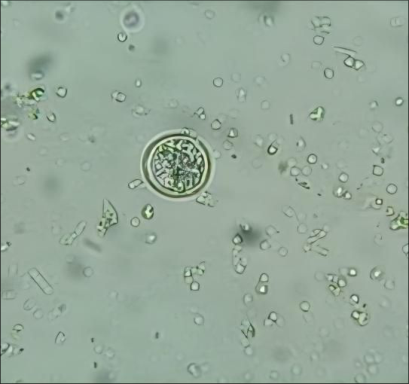 Figure 2
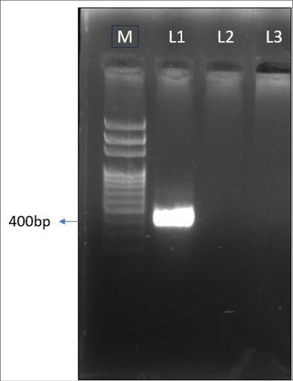 Figure 3
Introduction
Canine parvoviral enteritis (CPE) is a highly contagious viral gastroenteritis of domestic dogs caused by canine parvovirus type 2 (CPV-2), characterized by acute haemorrhagic diarrhoea, vomiting, dehydration, leukopenia and significant mortality in young and unvaccinated puppies. CPV predominantly affects the rapidly dividing cells of the intestinal crypts and bone marrow, resulting in severe enteric pathology and systemic compromise (Abhiram et al., 2023) [1] . In India, CPV continues to be a major cause of canine morbidity and mortality. Epidemiological surveys report high prevalence of CPV infection in diarrhoeic dogs, with PCR incidence rates commonly above 30-40% in clinically suspected cases (Abhiram et al., 2023) [2] . In a PCR-based study from Kolkata, nearly one-third of faecal samples from diarrhoeic dogs tested positive for CPV-2 with the highest occurrence in puppies aged 1-6 months. Likewise, a regional study around Tirupati detected CPV infection in approximately 40.25% of diarrhoeic dogs, with the youngest age groups (<3 months) most frequently affected (Mohan et al., 2025) [2] . Concurrent intestinal parasitism, such as coccidial infections (Isospora spp.) may further exacerbate gastrointestinal pathology and hinder recovery in affected puppies, yet reports on mixed infections in Indian canine populations remain limited. This case report details the concurrent infection of parvoviral enteritis and Isosporosis in a German Shepherd puppy.
Case history and observation
A 3-month-old male German Shepherd puppy weighing 5.3 kg was presented to the Veterinary Clinical Complex, Veterinary College and Research Institute, Orathanadu with a history of inappetance, repeated vomiting and acute onset of bloody, foul-smelling diarrhoea for the past two days. The owner reported progressive weakness and recumbency prior to presentation. On clinical examination, the animal appeared poor in general condition and was dull and depressed. The conjunctival mucous membranes were pale, indicating compromised perfusion. Severe dehydration was evident, as assessed by prolonged capillary refill time (>4 seconds) and delayed skin tenting (>3 seconds). Physiological parameters revealed a heart rate of 120 beats/min, respiratory rate of 50 breaths/min and rectal temperature of 39.6 °C. Abdominal palpation revealed a tensed abdome, and tenesmus was observed during defecation. The faeces were bloody, foul smelling and watery in consistency. For laboratory confirmation, blood and faecal samples were collected aseptically prior to initiation of therapy. Whole blood was collected from the cephalic vein into EDTA vacutainers for haematological examination. Fresh faecal material was collected directly from the rectum using sterile gloves and containers for coprological examination and further diagnostic testing. Initial screening using a canine parvovirus enteritis (CPE) antigen rapid test performed on the faecal sample yielded a positive result indicating active parvoviral infection (Fig. 1). Coprological examination of the faecal sample revealed the presence of Isospora spp. oocysts (Fig. 2) suggestive of concurrent coccidial infection. For molecular confirmation, DNA was extracted from the faecal sample and subjected to polymerase chain reaction (PCR) analysis. The PCR assay amplified a specific 400 bp fragment (Fig. 3) confirming the presence of canine parvovirus (parvoviral enteritis). These findings established a diagnosis of canine parvoviral enteritis complicated by concurrent Isospora infection.
Treatment and result
Based on the clinical severity, positive antigen and molecular confirmation of canine parvoviral enteritis and concurrent Isospora infection an intensive supportive and symptomatic treatment regimen was instituted and continued for five consecutive days. Fluid therapy was initiated using dextrose normal saline (DNS) and Ringer's lactate (RL) administered intravenously at 50 ml each to correct severe dehydration and electrolyte imbalance. Broad-spectrum antimicrobial coverage was provided with ceftriaxone @ 20 mg/kg IV to prevent secondary bacterial septicaemia. Metronidazole @ 25 mg/kg IV was administered to control anaerobic bacterial overgrowth. To manage vomiting and gastric irritation, ondansetron @ 0.2 mg/kg IV and ranitidine @ 2 mg/kg IM were administered. Considering the detection of Isospora spp. oocysts, sulphonamide-trimethoprim combination @ 15 mg/kg IM was included for anticoccidial therapy. Tribivet® (vitamin B1, B6 and B12) @ 2 ml IV was given as supportive therapy to improve appetite and metabolic recovery. The puppy showed a gradual and consistent clinical improvement following initiation of therapy. Vomiting subsided by the second day of treatment and the frequency and severity of diarrhoea reduced progressively, with marked improvement in faecal consistency by day four. Appetite improved gradually and the animal regained alertness and normal posture. Serial haematological evaluation revealed a progressive improvement in haemoglobin, packed cell volume, and erythrocyte count indicating correction of dehydration and haemoconcentration. Leukocytosis observed on day one (17 × 10³/µL) gradually returned towards normal limits by day four (9.5 × 10³/µL). Differential leukocyte counts showed partial recovery of neutrophil counts with a relative decrease in lymphocytosis over the observation period.
Discussion
Canine parvoviral enteritis (CPE) is recognized as one of the most significant causes of acute haemorrhagic gastroenteritis in young dogs across India, particularly in unvaccinated pups under six months of age. Several Indian studies have consistently demonstrated high prevalence of CPE in this age group with clinical presentations (Mohan et al., 2025) [2] similar to those in the present case. The hallmark features of CPE include haemorrhagic, foul-smelling diarrhoea, severe dehydration, vomiting and profound depression which were well documented in Indian veterinary scenario (Sharma, 2025) [4] . In a field study using rapid antigen kits in Central India, vomiting and severe dehydration were reported (Mandawat et al., 2025) [3] in the majority of confirmed CPV cases reinforcing the typical clinical picture seen in this case. Haematological alterations in CPE commonly include leukocytosis or leukopenia depending on disease stage and secondary complications. Studies from India have documented significant changes in total leukocyte and differential counts in parvoviral cases (Zope et al., 2023) [5] , similar to the elevated WBC with lymphocytosis and depressed neutrophil proportions observed early in treatment in the present report. Such patterns reflect the immune response to viral enteritis and secondary bacterial translocation and septicaemia which often complicates clinical recovery. While parvovirus alone can produce severe gastroenteritis, concurrent infections with intestinal parasites such as Isospora are not uncommon in field settings, especially in young, unvaccinated animals exposed to environmental contamination (Baghel et al., 2024) [6] . Therapeutic management emphasizing aggressive fluid therapy, antiemetics, broad-spectrum antimicrobials and correction of electrolyte imbalance remain the cornerstone of CPE treatment, as reported in multiple Indian clinical studies (Shruthi and Ajay, 2023; Sharma, 2025) [4,7] . The gradual clinical and hematologic improvement seen over the treatment period in this case mirrors findings which correlate early supportive care with higher survival rates in infected pups. In summary, the clinical course, diagnostic results and therapeutic response in this case are consistent with canine parvoviral enteritis. The additional detection of Isospora spp. highlights the importance of comprehensive fecal screening in diarrhoeic puppies, particularly in regions where environmental contamination is a significant risk factor.
References
7 in the original paper- Abhiram S, Mondal T, Samanta S, Batabyal K, Joardar SN, Samanta I, et al. Occurrence of canine parvovirus type 2c in diarrhoeic faeces of dogs in Kolkata, India. Virusdisease. 2023;34(2):339-344. DOI: 10.1007/s13337-023-00817-y.
- Mohan A, Goud KS, Shobhamani B, Reddy TN. Incidence of canine parvovirus infection in and around Tirupati. Indian J Vet Med. 2025;45(1):54-56.
- Mandawat S, Singh R, Mohammed N, Singh Meena D, Jyoti, Chaudhary W, et al. Epidemiological study of canine parvoviral enteritis in Jaipur, Rajasthan. Indian J Vet Sci Biotechnol. 2025;21(6):168-170.
- Sharma A. Diagnostic and therapeutic approaches in canine parvovirus: a clinical study. Int J Anim Vet Fish Allied Sci. 2025;11(4).
- Zope PS, Waghmare SP, Pajai KS, Dighe DG, Hajare SW, Kamdi BP. Molecular diagnosis and haematobiochemical alteration in canine parvoviral infection in dogs. Indian J Vet Sci Biotechnol. 2023;19(5):93-96.
- Baghel KR, Raj IL, Bisen S, Ratre HK. Successful therapeutic management of CystoIsospora infection in dog. Indian Vet J. 2024;101(12):25-27.
- Shruti G, Ajay K. Canine parvo viral enteritis in dogs: diagnostic and therapeutic evaluation. J Vet Anim Sci. 2023;54(1):71-78.