
Research article · 2026 · Full text
International Journal of Biology SciencesCross transmission study and pathogenesis of Trichomonas gallinae in different hosts in Assam, India
Volume 8(4) · Pages 11–18 · DOI: 10.33545/26649926.2026.v8.i4a.613 ↗
Research areasBird parasitology and diseasesParasitic Infections and DiagnosticsHelminth infection and control
Abstract
Author-writtenTo determine the virulent nature of the Trichomonas gallinae and its transmissibility to different hosts, experiment was performed with or without immunosuppressive drugs in chicken, duck, quails and mice taking pigeon as its natural host. Pigeon strain of T. gallinae orally inoculated at concentration of 4x10⁴ trophozoites in birds and intra peritoneal inoculation in mice revealed presence of parasites in all bird species while mice developed abscess which is an indicator of T. gallinae infection.
Summary
Source-checkedPlain-language reading of the full paper, prepared with AI and checked against the text. Each point links to the authors' own words.
Why it matters
- Avian trichomoniasis, commonly called canker, is a potentially fatal disease that causes significant economic and health impacts on the poultry industry and can lead to declines in bird populations. View evidence ↗
- While Trichomonas gallinae primarily affects the upper digestive tract, it can also involve the respiratory system and other internal organs. View evidence ↗
The question
- The study aimed to determine the virulence of T. gallinae and its ability to transmit to chickens, ducks, quails, and mice, both with and without the use of immunosuppressive drugs. View evidence ↗
What they did
- Groups consisted of a healthy control, a healthy inoculated group receiving 4x10^4 trophozoites orally, and an immunosuppressed inoculated group treated with a single 0.2 ml I/M dose of dexamethasone sodium. View evidence ↗
What they found
- In immunosuppressed pigeons (Group III), three birds died naturally by day 13 due to oropharyngeal blockages, and parasite concentrations reached 'very high' (++++) levels. View evidence ↗
- Healthy inoculated pigeons (Group II) developed symptoms starting on day 3, progressing to cachectic conditions and heavy oral lesions by day 11, though no mortality occurred. View evidence ↗
- Immunosuppressed quails (Group III) showed no changes until day 7, developed diarrhea and weakness by day 9, and tested positive for parasites (+) from day 13, with no deaths recorded. View evidence ↗
- Immunosuppressed mice (Group III) remained healthy until day 7, after which they developed weakness and rough hair, resulting in one death on day 13. View evidence ↗
- Immunosuppressed chickens (Group III) developed drowsiness and diarrhea by day 3, progressing to severe diarrhea and high parasite levels (+++) from day 7 to 15, without mortality. View evidence ↗
What it means
- The authors conclude that they have successfully detailed the cross-transmission and pathogenesis of T. gallinae from pigeons to chickens, ducks, quails, and mice, marking the first such report from Assam and India. View evidence ↗
Visual summary
1 visualsIllustrations explain the paper; they do not show study data.
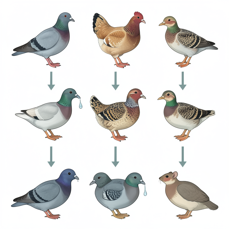
Experimental Grouping Flowchart
AI-generated conceptual illustration (pigeons, chickens, ducks, quails, and mice). It explains the idea and does not show study data.
View source evidence ↗Figures & tables
From the original paper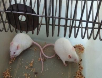 Figure 1
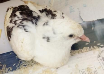 Figure 2
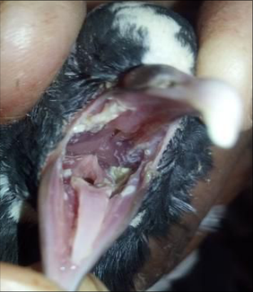 Figure 2
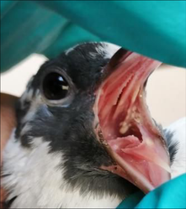 Figure 2
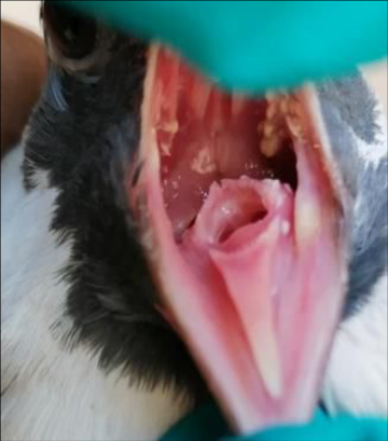 Figure 3
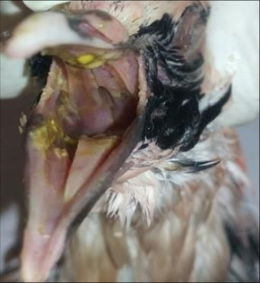 Figure 3
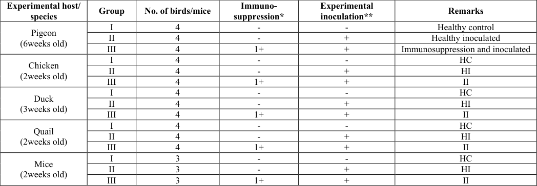 Table 1
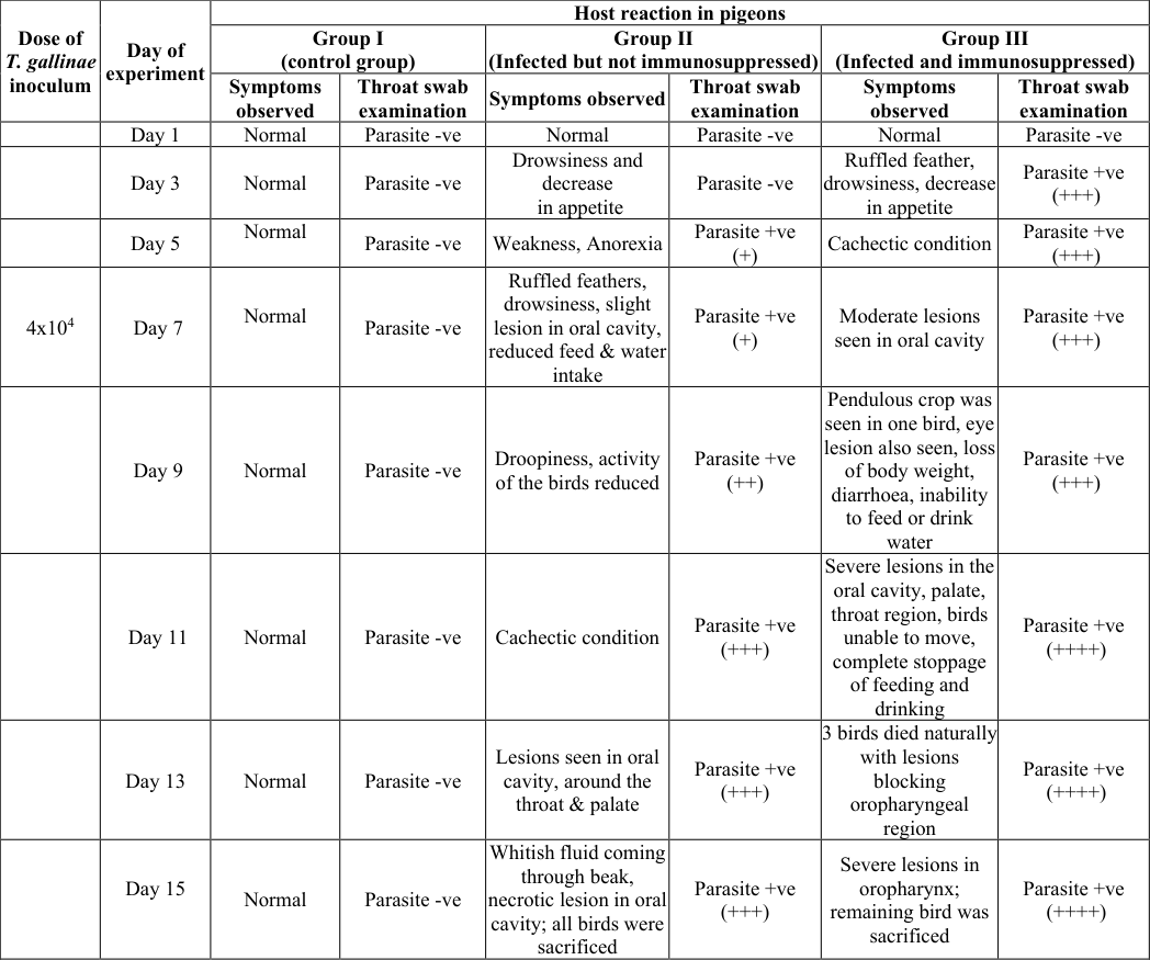 Table 2
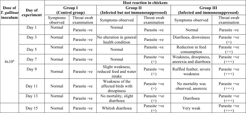 Table 3
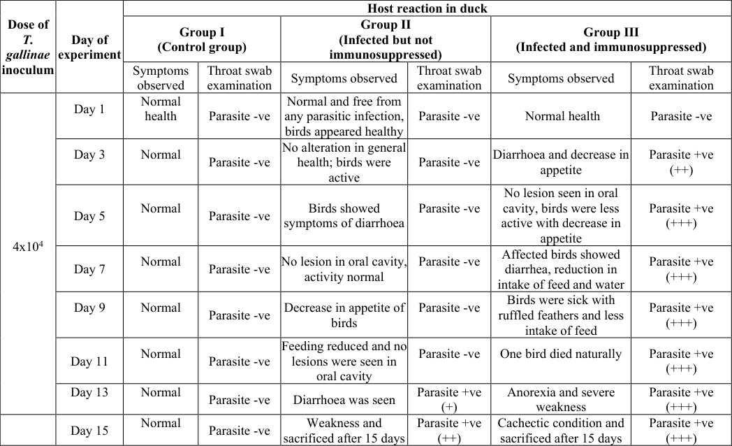 Table 4
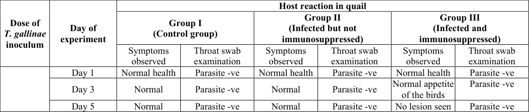 Table 5
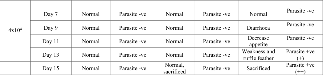 Table 6
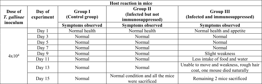 Table 6
Introduction
Trichomonas gallinae, the causative agent of Avian trichomoniasis, is a flagellate protozoan parasite under class Zoomastigophorea and order Trichomonadida. In natural condition, this parasite occur mainly in the columbiformes birds (pigeon and mourning dove) although several
Grouping of the birds and mice
Twelve birds from each species (pigeon, chicken, duck and quail) were put into three groups (I, II, III) each with four birds (Fig 1 ,Table 1). Group I birds was considered as healthy control; group II were healthy without immunosuppression but experimentally inoculated (orally) with 4X10 4 trophozoites of T. gallinae; group III birds were both immunosuppressed and inoculated with T. gallinae. In the case of mice experimentation, 9 mice were divided into three equal groups (I, II, III). However, T. gallinae was inoculated peritoneally. Immunosuppression was done with injection of dexamethasone sodium as per experimental design @ 0.2 ml I/M in a single dose. Oral inoculation of T. gallinae was also done with 410 4 trophozoites.
| Experimental host/ species | Group | No. of birds/mice | Immuno- suppression* | Experimental inoculation** | Remarks |
|---|---|---|---|---|---|
| Pigeon (6weeks old) | I | 4 | - | - | Healthy control |
| II | 4 | - | + | Healthy inoculated | |
| III | 4 | 1+ | + | Immunosuppression and inoculated | |
| Chicken (2weeks old) | I | 4 | - | - | HC |
| II | 4 | - | + | HI | |
| III | 4 | 1+ | + | II | |
| Duck (3weeks old) | I | 4 | - | - | HC |
| II | 4 | - | + | HI | |
| III | 4 | 1+ | + | II | |
| Quail (2weeks old) | I | 4 | - | - | HC |
| II | 4 | - | + | HI | |
| III | 4 | 1+ | + | II | |
| Mice (2weeks old) | I | 3 | - | - | HC |
| II | 3 | - | + | HI | |
| III | 3 | 1+ | + | II |
Table text extracted from the original PDF; superscript letters are shown inline. The original table image is below.
View original table image
Monitoring of infection
Experimental birds and mice of each group were kept under careful observation up to 15 days for appearance of clinical symptoms and mortality. Throat swabs were also examined on every alternate day from day 3 for detection of parasites and grading was done as very high (++++), high (+++), moderate (++), mild (+) and negative (-). Experimentally inoculated surviving birds were sacrificed after 15 days of observation for parasitological and pathological study.
Results and Discussion
Cross transmissibility studies in pigeon Host reaction due to experimental inoculation of T. gallinae is presented in Table 2. The birds of group I neither showed any symptoms of illness nor parasites were detected on throat swab examination. The group II birds developed clinical symptoms from 3 rd day post inoculation such as drowsiness, decrease in appetite and from 5 th day onwards exhibited ruffled feathers (Fig 2 i), slight lesions in oral cavity and from 9 th day, the birds showed droopiness, and on 11 th day were in cachectic condition with heavy lesions in oral cavity, around the throat and palate (Fig 3 ) Parasites could be detected on throat swab examination from 5 th day of infection while high concentrations were seen from 11 th -15 th days post inoculation. No mortality was seen in group II birds and on 15 th day, whitish fluid drooled out through beak along with necrotic lesions in the oral cavity. The group III birds showed ruffled feather, drowsiness, decreased appetite on 3 rd day post inoculation and high concentration of parasites (+++) on throat swab examination. On 7 th day, moderate lesions were seen in oral cavity and on 9 th day, the birds had pendulous crop (Fig 2 ii), lesion in eye, diarrhoea, inability to feed or drink water. On 11 th day onwards severe lesions were grossly seen in the oral cavity, palate, throat, inability to move (Fig 2 iii), complete stoppage of feeding and drinking, with very high concentration of parasites (++++). On 13 th day of infection, 3 birds died naturally due to lesions blocking the oropharyngeal region. The remaining bird was sacrificed on 15 th day. Transmission of T. gallinae by experimental means has been performed earlier in avian and mammalian hosts by different workers. In columbiform birds, experimental transmission by oral intubation of cultured or fresh parasite suspensions was used (Cauthen, 1936; Conti et al., 1985; Powell and Hollander, 1982; Stabler and Kihara, 1954) [3,4,15,20] . Many of the previous papers reported simple cross-transmission experiments to establish whether or not T. gallinae would produce disease. Powell and Hollander (1982) [15] transmitted T. gallinae from pigeons to ring doves. Stabler and Herman (1951) [19] also transmitted T. gallinae from mourning doves to domestic pigeons. Intrahepatic, intramuscular, intraperitoneal and subcutaneous injections of T. gallinae have been used to produce trichomonad pathogenicity. In line with the present observations, Neimanis et al. (2010) [14] also reported similar clinical signs such as lethargy; gaping beaks which may be covered in food remains; laboured breathing; drooling; closed eyes; matted and wet feathers around the beak; head and chest and dishevelled feathers held away from the body exhibited by birds. Mohamed et al. (2009) [13] reported that natural and experimental affected squabs with trichomoniasis showed depression, off-food and dyspnea, in addition to offensiveodor-fluid, drooling from the opened peak, due to lesions in the oral cavity. Moreover, a large caseated mass was present in the buccal cavity. Sometimes greenish watery diarrhea was noticed. Similar signs along with occlusion of the crop with caseated materials were also reported by Abd-ElMotelib and Galal (1993) [1] . It was reported that when Trichomonas-free, non-immune pigeons were infected with a known avirulent T. gallinae isolate, they did not elicit an immune response or develop any lesion even after being inoculated with 1 x 10 6 organisms. In contrast, when infected with a known virulent strain, Trichomonas-free, non-immune pigeons died within six to nine days following inoculation of a single trichomonad.
| Dose of T. gallinae inoculum | Day of experiment | Host reaction in pigeons | — | — | — | — | — |
|---|---|---|---|---|---|---|---|
| Group I (control group) | Group II (Infected but not immunosuppressed) | Group III (Infected and immunosuppressed) | |||||
| Symptoms observed | Throat swab examination | Symptoms observed | Throat swab examination | Symptoms observed | Throat swab examination | ||
| Day 1 | Normal | Parasite -ve | Normal | Parasite -ve | Normal | Parasite -ve | |
| Day 3 | Normal | Parasite -ve | Drowsiness and decrease in appetite | Parasite -ve | Ruffled feather, drowsiness, decrease in appetite | Parasite +ve (+++) | |
| Day 5 | Normal | Parasite -ve | Weakness, Anorexia | Parasite +ve (+) | Cachectic condition | Parasite +ve (+++) | |
| 4x10⁴ | Day 7 | Normal | Parasite -ve | Ruffled feathers, drowsiness, slight lesion in oral cavity, reduced feed & water intake | Parasite +ve (+) | Moderate lesions seen in oral cavity | Parasite +ve (+++) |
| Day 9 | Normal | Parasite -ve | Droopiness, activity of the birds reduced | Parasite +ve (++) | Pendulous crop was seen in one bird, eye lesion also seen, loss of body weight, diarrhoea, inability to feed or drink water | Parasite +ve (+++) | |
| Day 11 | Normal | Parasite -ve | Cachectic condition | Parasite +ve (+++) | Severe lesions in the oral cavity, palate, throat region, birds unable to move, complete stoppage of feeding and drinking | Parasite +ve (++++) | |
| Day 13 | Normal | Parasite -ve | Lesions seen in oral cavity, around the throat & palate | Parasite +ve (+++) | 3 birds died naturally with lesions blocking oropharyngeal region | Parasite +ve (++++) | |
| Day 15 | Normal | Parasite -ve | Whitish fluid coming through beak, necrotic lesion in oral cavity; all birds were sacrificed | Parasite +ve (+++) | Severe lesions in oropharynx; remaining bird was sacrificed | Parasite +ve (++++) |
Table text extracted from the original PDF; superscript letters are shown inline. The original table image is below.
View original table image
Cross transmissibility studies in chicken
Host reaction following experimental inoculation of T. gallinae in chicken is presented in Table 3.
| Dose of T. gallinae inoculum | Day of experiment | Host reaction in chickens | — | — | — | — | — |
|---|---|---|---|---|---|---|---|
| Group I (Control group) | Group II (Infected but not immunosuppressed) | Group III (Infected and immunosuppressed) | |||||
| Symptoms observed | Throat swab examination | Symptoms observed | Throat swab examination | Symptoms observed | Throat swab examination | ||
| 4x10⁴ | Day 1 | Normal | Parasite -ve | Normal | Parasite -ve | Normal | Parasite -ve |
| Day 3 | Normal | Parasite -ve | No alteration in general health condition | Parasite -ve | Diarrhoea, drowsiness | Parasite +ve (+) | |
| Day 5 | Normal | Parasite -ve | Normal | Parasite -ve | Reduction in feed consumption | Parasite +ve (++) | |
| Day 7 | Normal | Parasite -ve | Normal | Parasite +ve (+) | Weakness, droopiness, anorexia and diarrhoea | Parasite +ve (+++) | |
| Day 9 | Normal | Parasite -ve | Slight weakness, reduced feed and water intake | Parasite +ve (+) | Ruffled feather, severe weakness | Parasite +ve (+++) | |
| Day 11 | Normal | Parasite -ve | Weakness of the affected birds with droopiness | Parasite +ve (+) | No mortality was observed, anorexic | Parasite +ve (+++) | |
| Day 13 | Normal | Parasite -ve | No mortality, slight diarrhoea | Parasite +ve (+) | Diarrhoea | Parasite +ve (+++) | |
| Day 15 | Normal | Parasite -ve | Whitish diarrhoea | Parasite +ve (+) | Very weak | Parasite +ve (+++) |
Table text extracted from the original PDF; superscript letters are shown inline. The original table image is below.
View original table image
The birds of group I neither showed any symptoms of illness nor parasites were detected on throat swab examination. The group II birds did not show any clinical symptoms till 6 th day of post infection but parasites were detected on day 7. Symptoms like weakness, reduced feed and water intake etc. were observed from 9 th day onwards. On 11 th day of infection, the birds showed droopiness, diarrhoea and mild parasites were detected (+). No mortality was recorded till 15 day and all the birds were sacrificed. Group III birds had diarrhoea, became drowsy with low parasites on throat swab examination from 3 th day onwards. Droopiness, anorexia, severe diarrhoea with high concentration of parasites (+++) were seen from 7 th day till 15 th day of observation but no mortality was seen. On15 th day of infection, birds were sacrificed for detailed pathological studies.
Chickens and turkeys have been orally intubated with T. gallinae from pigeons (Levine et al., 1941; Levine and Brandly, 1939) [10,11] . Gabaldon and Andrews (1935) [7] also used chickens for experimental transmission. Levine (1985) [12] reported that infections in chickens are rare. There is paucity of literature to compare the pathogenesis and pathology of T. gallinae infection in chicken as observed in our present study. However, Sasikala et al. (2017) [17] reported only natural case of Trichomoniasis in 2 months old Desi chicken in Orathanadu and finding Trichomonas sp. trophozoites by cytological examination of the scrapings from crop mucosa.
Cross transmissibility studies in duck
Host reaction following experimental inoculation of T. gallinae in duck is presented in Table 4.
| Dose of T. gallinae inoculum | Day of experiment | Host reaction in duck | — | — | — | — | — |
|---|---|---|---|---|---|---|---|
| Group I (Control group) | Group II (Infected but not immunosuppressed) | Group III (Infected and immunosuppressed) | |||||
| Symptoms observed | Throat swab examination | Symptoms observed | Throat swab examination | Symptoms observed | Throat swab examination | ||
| 4x10⁴ | Day 1 | Normal health | Parasite -ve | Normal and free from any parasitic infection, birds appeared healthy | Parasite -ve | Normal health | Parasite -ve |
| Day 3 | Normal | Parasite -ve | No alteration in general health; birds were active | Parasite -ve | Diarrhoea and decrease in appetite | Parasite +ve (++) | |
| Day 5 | Normal | Parasite -ve | Birds showed symptoms of diarrhoea | Parasite -ve | No lesion seen in oral cavity, birds were less active with decrease in appetite | Parasite +ve (+++) | |
| Day 7 | Normal | Parasite -ve | No lesion in oral cavity, activity normal | Parasite -ve | Affected birds showed diarrhea, reduction in intake of feed and water | Parasite +ve (+++) | |
| Day 9 | Normal | Parasite -ve | Decrease in appetite of birds | Parasite -ve | Birds were sick with ruffled feathers and less intake of feed | Parasite +ve (+++) | |
| Day 11 | Normal | Parasite -ve | Feeding reduced and no lesions were seen in oral cavity | Parasite -ve | One bird died naturally | Parasite +ve (+++) | |
| Day 13 | Normal | Parasite -ve | Diarrhoea was seen | Parasite +ve (+) | Anorexia and severe weakness | Parasite +ve (+++) | |
| Day 15 | Normal | Parasite -ve | Weakness and sacrificed after 15 days | Parasite +ve (++) | Cachectic condition and sacrificed after 15 days | Parasite +ve (+++) |
Table text extracted from the original PDF; superscript letters are shown inline. The original table image is below.
View original table image
The birds of group I neither showed any symptoms of illness nor parasites were detected on throat swab examination. From 5 th day onwards, group II birds showed diarrhoea, decreased appetite and absence of gross oral lesions. Very less parasites were detected (+) in throat swab examination from 13 th day onwards. No mortality was noticed in this group. Group III birds had diarrhoea, decreased appetite, moderate concentration of parasite (++) from 3 rd day onwards and gradually becoming high (+++) till 15 th day. On 11 th day, one bird died naturally of infection while the remaining 3 birds in cachectic condition were sacrificed.
There is not much literature available to compare the experimental design, transmission, pathogenesis and pathology of T. gallinae infection in duck as carried out in the present study. However, Tsai et al. (1997) [22] recorded natural outbreak of trichomonosis due to T. gallinae in farmed ducks showing signs and symptoms of respiratory and intestinal form.
Cross transmissibility studies in quail
Host reaction following experimental inoculation of T. gallinae in quail is presented in Table 5.
| Dose of T. gallinae inoculum | Day of experiment | Host reaction in quail | — | — | — | — | — |
|---|---|---|---|---|---|---|---|
| Group I (Control group) | Group II (Infected but not immunosuppressed) | Group III (Infected and immunosuppressed) | |||||
| Symptoms observed | Throat swab examination | Symptoms observed | Throat swab examination | Symptoms observed | Throat swab examination | ||
| Day 1 | Normal health | Parasite -ve | Normal health | Parasite -ve | Normal health | Parasite -ve | |
| Day 3 | Normal | Parasite -ve | Normal | Parasite -ve | Normal appetite of the birds | Parasite -ve | |
| Day 5 | Normal | Parasite -ve | Normal | Parasite -ve | No lesion seen | Parasite -ve |
Table text extracted from the original PDF; superscript letters are shown inline. The original table image is below.
View original table image
In birds of group I (uninfected control) no clinical symptoms/ lesions were seen and parasites were also not detected on throat swab examination. In group II birds no gross lesion or symptoms was noticed till 15 th day and all birds were sacrificed. In group III birds till 7 th day of infection, no alteration was observed. On 9 th day birds having diarrhoea gradually showed signs of weakness and ruffled feathers. Parasite were detected from 13 th day onwards (+) but no mortality was seen and all birds were sacrificed on 15 th day of infection.
In line with our present findings, Levine et al. (1941) [11] reported the results of feeding experiments on the transmission of Trichomonas to Bobwhite quails. No lesions were found in the trachea, liver or other abdominal organs of any of the quail and trichomonads were not found in the intestinal tract posterior to the gizzard thus agreeing to our report. Numerous protozoa were demonstrable in the necrotic areas and in the yellowish fluid seen in the crop and proventriculus of some of the infected quails.
Cross transmissibility studies in mice
Host reaction following experimental inoculation of T. gallinae in mice is presented in Table 6. Group I mice (uninfected control) were normal in health and activity. In group II mice no mortality was noticed till 15 days of experiment and all mice were sacrificed. In group III, all mice appeared normal and apparently healthy till 7 th day. Gradually they started showing symptoms of weakness, decreased appetite, inability to move, rough body hair coat and one mouse died on day 13 th while the remaining 2 mice were sacrificed on 15 th day of infection.
| Host reaction in mice | — | — | — | — |
|---|---|---|---|---|
| Dose of T. gallinae inoculum | Day of experiment | Group I (Control group) | Group II (Infected but not immunosuppressed) | Group III (Infected and immunosuppressed) |
| Symptoms observed | Symptoms observed | Symptoms observed | ||
| 4x10⁴ | Day 1 | Normal health | Normal health | Normal health and appetite |
| Day 3 | Normal | Normal | Normal | |
| Day 5 | Normal | Normal | Normal | |
| Day 7 | Normal | Normal | Normal | |
| Day 9 | Normal | Normal | Slight weakness | |
| Day 11 | Normal | Normal | Less intake of food and water | |
| Day 13 | Normal | Normal | Unable to move and weakness, rough hair coat, one mouse died naturally | |
| Day 15 | Normal | Normal condition and all the mice were sacrificed | Remaining 2 mice sacrificed |
Table text extracted from the original PDF; superscript letters are shown inline. The original table image is below.
View original table image
| 4x10⁴ | — | — | — | — | — | — | — |
|---|---|---|---|---|---|---|---|
| Day 7 | Normal | Parasite -ve | Normal | Parasite -ve | Normal | Parasite -ve | |
| Day 9 | Normal | Parasite -ve | Normal | Parasite -ve | Diarrhoea | Parasite -ve | |
| Day 11 | Normal | Parasite -ve | Normal | Parasite -ve | Decrease appetite | Parasite -ve | |
| Day 13 | Normal | Parasite -ve | Normal | Parasite -ve | Weakness and ruffle feather | Parasite +ve (+) | |
| Day 15 | Normal | Parasite -ve | Normal, sacrificed | Parasite -ve | Sacrificed | Parasite +ve (++) |
Table text extracted from the original PDF; superscript letters are shown inline. The original table image is below.
View original table image
Acknowledgement
AcknowledgmentsThe authors are grateful to the Dean, College of Veterinary Science, Assam Veterinary and Fishery University for providing necessary facilities to carry out the research work. They also declare that there is no conflict of interest related to the work.
References
22 in the original paper- Abdel El-Motelib TY, Galal BG. Some studies on Trichomonas gallinae infection in pigeons. Assiut Vet Med J. 1993;30(59):277-288.
- Borji H, Razmi GH, Movassaghi AH. Prevalence and pathological lesion of Trichomonas gallinae in pigeons of Iran. J Parasit Dis. 2011;35(2):186-189.
- Cauthen G. Studies on Trichomonas columbae, a flagellate parasitic in pigeons and doves. Am J Hyg. 1936;23(1):132-242.
- Conti JAR, Frohlich K, Forrester DJ. Experimental transfer of Trichomonas gallinae (Rivolta, 1878) from white-winged doves to mourning doves. J Wildl Dis. 1985;21(3):229-232.
- Frost JK, McLure MT. Intracellular Trichomonas vaginalis and Trichomonas gallinae in natural and experimental infections. J Parasitol. 1961;47(2):302303.
- Frost JK, Honigberg BM. Comparative pathogenicity of Trichomonas vaginalis and Trichomonas gallinae to mice. II: Histopathology of subcutaneous lesions. J Parasitol. 1962;48(6):898-918.
- Gabaldon A, Andrews J. Experimental lesions caused by Trichomonas columbae in the chicken. J Parasitol. 1935;21(6):451.
- Girard YA, Rogers KH, Woods LW, Chouicha N, Miller WA, Johnson CK. Dualpathogen etiology of avian trichomonosis in a declining band-tailed pigeon population. Infect Genet Evol. 2014;24:146-156.
- Honigberg BM. Comparative pathogenicity of Trichomonas vaginalis and Trichomonas gallinae to mice. I. Gross pathology, quantitative evaluation of virulence, and some factors affecting pathogenicity. J Parasitol. 1961;47(4):545-671.
- Levine ND, Brandly A. A pathogenic Trichomonas from the upper digestive tract of chickens. J Am Vet Med Assoc. 1939;95:77-78.
- Levine ND, Boley LE, Hester HR. Experimental transmission of Trichomonas gallinae from the chicken to other birds. Am J Hyg. 1941;33(Sec. C):23-32.
- Levine ND. Veterinary Protozoology. Ames (IA): Iowa State University Press; 1985. p. 414.
- Mohamed EI, Gehan H, El-Sakkar, Magda MM Moursi. Pathological studies on pigeon trichomoniasis with reference to the associated bacteria. Egypt J Comp Path Clin Path. 2009;22(2):67-87.
- Neimanis AS, Handeland K, Isomursu M, Agren E, Mattsson R, Hamnes IS. First report of epizootic trichomoniasis in wild finches (Family Fringillidae) in southern Fennoscandia. Avian Dis. 2010;54(1):136-141.
- Powell EC, Hollander WF. Trichomonas gallinae infections in the ring dove (Streptopelia risoria). J Wildl Dis. 1982;18(1):89-90.
- Robinson RA, Lawson B, Toms MP, Peck KM, Kirkwood JK, Chantrey J. Emerging infectious disease leads to rapid population declines of common British birds. PLoS ONE. 2010;5(8):e12245.
- Sasikala M, Selvaraj J, Prasath NB, Ahamad DB. Trichomoniasis in a desi chicken. Indian Vet J. 2017;94(12):85-86.
- Stabler RM. Variations in virulence of strains of Trichomonas gallinae in pigeons. J Parasitol. 1948;34(2):147-149.
- Stabler RM, Herman CM. Upper digestive tract trichomoniasis in mourning doves and other birds. Trans N Am Wildl Conf. 1951;16:145-162.
- Stabler RM, Kihara JT. Infection and death in the pigeon resulting from the oral implantation of single individuals of Trichomonas gallinae. J Parasitol. 1954;40(6):706.
- Stockdale JE, Dunn JC, Goodman SJ, Morris AJ, Sheehan DK, Grice PV, et al. The protozoan parasite Trichomonas gallinae causes adult and nestling mortality in a declining population of European turtle doves, Streptopelia turtur. Parasitology. 2015;142(3):490-498.
- Tsai SS, Chang TC, Kuo M, Itakura C. Respiratory and intestinal trichomoniasis in mule ducks. Avian Pathol. 1997;26(3):651-656.