
Research article · 2026 · Full text
International Journal of Biology SciencesSurgical management of teat fistula in a crossbred jersey cows: A case study
Volume 8(4) · Pages 19–21 · DOI: 10.33545/26649926.2026.v8.i4a.614 ↗
Research areasVeterinary Medicine and SurgeryMilk Quality and Mastitis in Dairy CowsVeterinary Orthopedics and Neurology
Abstract
Author-writtenA study was conducted in the department of veterinary surgery and radiology, VCC, RIVER in cows (6) having history of teat injury due to trauma. Clinical examination revealed deep lacerated wound on the left hind teat (3), right fore teat (2) and left for teat (1) with fistula at the mid teat level. All the physiological parameters were within the normal range. The animals were sedated using inj. xylazine at the dose rate of 0.1mg/kg intravenously and ring block was employed using 2% lignocaine hydrochloride. Under aseptic condition the fistulus tract was debrided and closed by three-layer closure using 3-0 polyglactin 910 and skin was apposed by sterile disposable skin staples. The teat canal patency was maintained by infant feeding tube 10 placed insitu. Post operatively inj. streptopenicillin 2.5g administered I/M for 5 days and intra-mammary infusion with inj. Gentamicin 5ml mixed with inj. Metronidazole 15ml for 3 days. All the animals showed complete recovery on 14th day without any complication (eventfully). Pre and postoperative ultrasound scanning confirmed the healing of the teat walls.
Summary
Source-checkedPlain-language reading of the full paper, prepared with AI and checked against the text. Each point links to the authors' own words.
Why it matters
- Maintaining udder and teat health is critical for dairy farmers because diseases in these areas reduce productivity and lead to significant economic losses. View evidence ↗
- Teat lacerations that result in fistulas are medical emergencies, as delays in repair can cause teat necrosis or mastitis. View evidence ↗
- Teat fistulas can be congenital or acquired through full-thickness injuries that penetrate the teat cistern. View evidence ↗
The question
- The study aims to describe the successful surgical management of teat fistulas in crossbred Jersey cows. View evidence ↗
What they did
- The study involved six pleuriparous crossbred Jersey cows weighing between 290 kg and 360 kg, all of whom had experienced traumatic teat injuries. View evidence ↗
- Physical exams showed lacerated wounds approximately 2.5 cm by 1 cm that created a 50% vertical cut extending into the teat canal, causing milk to leak from the mid-teat. View evidence ↗
- Surgical preparation included intravenous sedation with xylazine at 0.1 mg/kg and local anesthesia via a 2% lignocaine hydrochloride ring block. View evidence ↗
- The surgical repair involved a three-layer closure: the muscular layer was closed with a double-layer simple continuous suture using 3-0 polyglactin 910, and the skin was closed with disposable stainless steel staples. View evidence ↗
- To ensure the teat canal remained open, a size 10 infant feeding tube made of modified polyvinyl was inserted and secured with a stay suture. View evidence ↗
- Post-operative care included intramuscular Meloxicam (0.5 mg/kg for 2 days) and Streptopenicillin (2.5 g for 7 days), along with intramammary infusions of Gentamicin (5 ml) and Metronidazole (15 ml). View evidence ↗
What they found
- All six cows achieved complete recovery by the 14th post-operative day without experiencing any complications. View evidence ↗
- The healing process resulted in minimal scarring and the restoration of normal milk yield and milking ability. View evidence ↗
- Post-operative ultrasound scans confirmed healing, showing a hyperechoic area of mucosa at the teat wall. View evidence ↗
What it means
- The authors recommend the use of disposable skin staples for wound closure as they provide good edge apposition and minimize scar formation. View evidence ↗
Visual summary
1 visualsIllustrations explain the paper; they do not show study data.
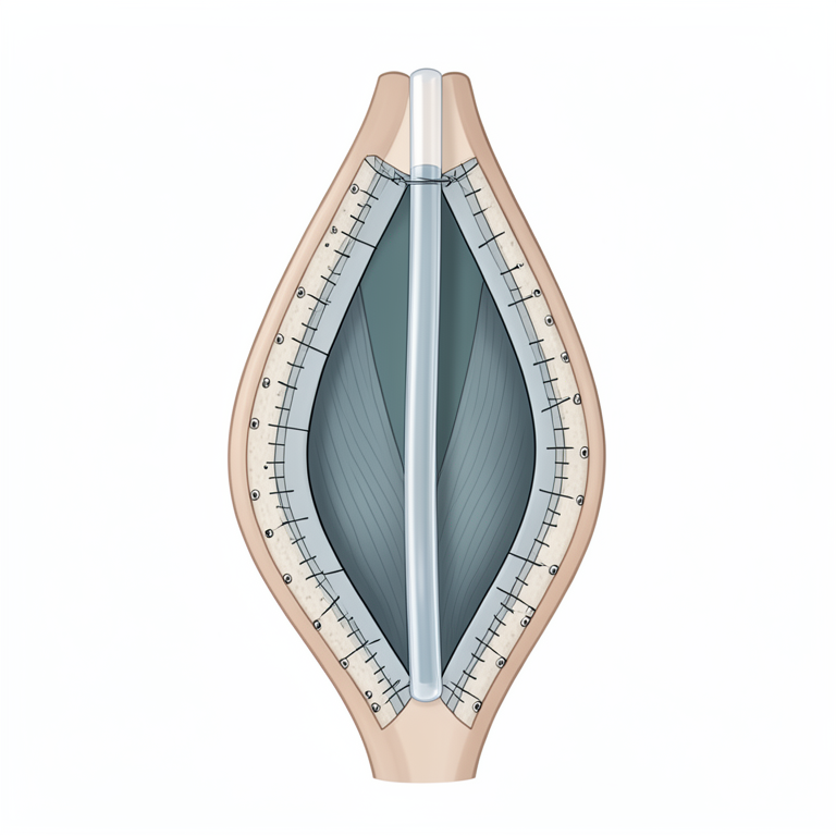
Surgical Closure Concept
AI-generated conceptual illustration (crossbred jersey cow teat). It explains the idea and does not show study data.
View source evidence ↗Figures & tables
From the original paper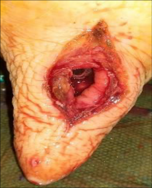 Figure 1
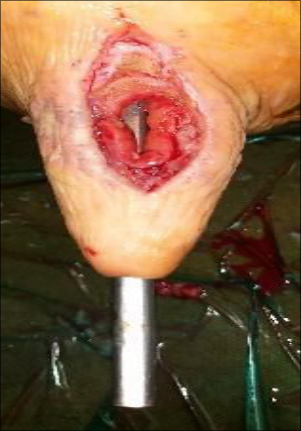 Figure 2
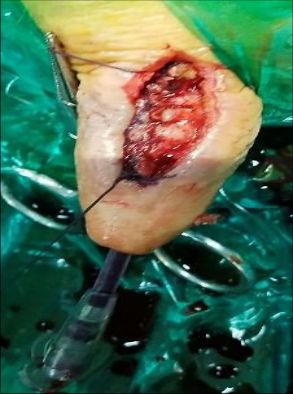 Figure 3
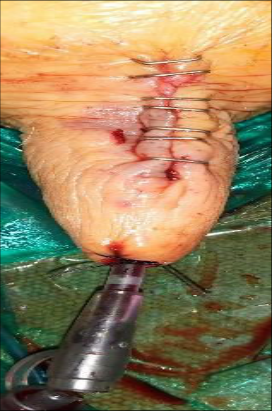 Figure 4
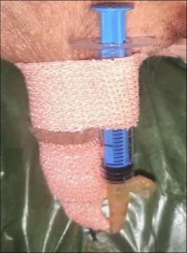 Figure 5
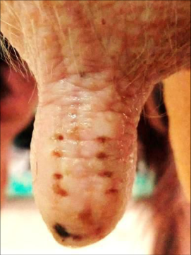 Figure 6
Introduction
Udder and teat health care is increasingly important for dairy farmers since disease condition involving them ultimately affects the productivity causing heavy economic loss. The cases of teat laceration with fistula are considered as emergency because of any delay in repair of such teat can lead to mastitis or even necrosis of the teat (Singh et al., 2003) [7] . Teat fistula may be acquired secondary to full thickness teat injuries that enter the teat cistern or may be even congenital (Thomas et al., 1995) [8] . The present case discusses the successful management of teat fistula in a crossbred jersey cow.
Case History and observation
All the animals were jersey crossbred pleuriparous cows (six) weighing around 290 kg to 360 kg body weight calved one to 3 months back were presented to the Department of Veterinary Surgery and Radiology, Veterinary clinical complex, RIVER with a history of teat injury on left hind teat (3), right fore teat (2) and left for teat (1) with milk leaking from the injured site for past one day. On Physical examination of the teat, showed 50 percent vertical cut and the lacerated wound (length X width ranging around 2.5cm x 1 cm) extended into the teat canal, resulting in leakage of milk from the middle of the teat (Fig. 1). The Teat was soiled and swollen. All other physiological parameters were found to be within normal range. Preoperative ultrasound scanning revealed a hypoechoic line of discontinuity in the skin and mucosal layer of the teat with linear probe of 7.5 MHz under water bath method.
Treatment
Following preoperative evaluation, food and water was withheld for 12-18 hours prior to surgery. The animals were sedated with Inj. Xylazine administered at the dose rate of 0.1mg/kg bodyweight intravenously and animals were placed in lateral recumbency with affected teat on the top. Local analgesia of the teat was achieved by ring block using lignocaine hydrochloride (2%). The teat was washed with normal saline followed by 0.5 percent povidone iodine solution. Under aseptic condition, the wound margins were debrided using No.10 scalpel blade and rinsed with normal saline solution. A sterile stainless-steel teat canula was inserted into the teat canal and the fistulation was located from the base to mid of the teat (Fig. 2). A 3-layer closure was performed. The muscular layer was sutured in double layer suture by simple continuous suture pattern using polyglactin 910 of size 3-0 (Fig. 3). Skin apposition (Fig. 4) was made using disposable stainless steel skin staples (Acos, Sunmedix) (Al-Mubarak et al., 2013) [1] in all the cows. A sterile modified polyvinyl tube (Infant feeding tube No. 10) was placed in the teat canal to maintain patency and fixed with stay suture. A sterile disposable 2ml syringe was connected to the modified polyvinyl tube and closed. The suture site was covered by povidone iodine gauze and adhesive tape (Dynafix) without any contamination (Fig. 5). Postoperatively Inj. Meloxicam @ 0.5mg/kg body weight I/M for 2 days and Inj. Streptopenicillin 2.5g was administered intramuscularly for 7 days. The owner was advised to drain out milk by opening the cap of the infant feeding tube at every two-hour interval with aseptic precaution, followed by intramammary infusion of Inj. Gentamicin 5ml mixed in 15 ml of Inj.Metronidazole. Dressing of the staples site was done on the 3 rd and 6 th postoperative days. On the 14 th post-operative day, the staples were removed using a staple remover (Fig. 6). Complete healing of the site was noticed with minimal scar, normal milking and milk yield. Postoperative healing was confirmed by ultrasound scanning, which revealed a hyperechoic area of mucosa to the teat wall.
Result and Discussion
The udder and teats are vulnerable to external trauma or injury because of their anatomical location, an increase in size of the udder and teats during lactation (Weaver et al., 2005) [10] . Teat injury caused by the barbed wire is a common aetiology for teat fistula (Nicholas et al., 2008) [5] . A fistula on the teat is exceedingly difficult to close during lactation, owing to the large amount of milk constantly passing through it. Infective organisms gain entry through this wound, leading to mastitis (Kumar, 2000) [4] . Surgical intervention on the teat is best performed during the first 12 hours following injury. Later, swelling of the teat can be too severe to permit adequate reconstitution of the tissue. In the present case study, all the six cows had an acute teat laceration with fistulation, and the surgery was attempted immediately after presentation (within 24 hours). Fistula was closed by suturing the muscle layer in a double-layer suture pattern using a simple continuous suture pattern using polyglactin 910 of size 2-0 (Velavan et al., 2014) [9] . A sterile modified polyvinyl tube (Infant feeding tube No. 10) was placed in the teat canal to maintain patency and fixed with stay suture (Aruljothi et al., 2012) [2] , which was effective in draining the milk. A sterile disposable 2ml syringe was connected to the modified polyvinyl tube and closed (Aruljothi et al., 2012) [2] to prevent mastitis. The suture site was covered by povidone iodine gauze and adhesive tape (Dynafix) (Gurunathan et al., 2019) [3] . The skin was apposed using disposable stainless steel skin staples which had better cosmetic results compared with conventional sutures (Premsairam et al., 2018) [6] . Postoperative pain and infection were very minimal, and the use of staples for skin closure allowed early healing (Premsairam et al., 2018) [6] .
Conclusion
From the present study, teat fistula in cows needs immediate surgical treatment to avoid delayed wound healing and the occurrence of complications such as wound dehiscence and infections. Use of disposable skin staples was effective in producing less scar formation and good apposition of the wound edges.
Acknowledgement
AcknowledgementThe authors are thankful to Dean, RIVER, for providing the necessary facilities to conduct the study.
References
10 in the original paper- Al-Mubarak L, Al-Haddab M. Cutaneous and wound closure materials: an overview and update. J Cutan Aesthet Surg. 2013;6(4):178-188.
- Aruljothi N, Balagopalan TP, Rameshkumar B, Alphonse RMD. Teat fistula and its surgical management in bovines. Intas Polivet. 2012;13(1):40-41.
- Gurunathan N, Aruljothi N, Udayakumari B, Balagopalan TP. Surgical management of fibropapiloma of teat in a cow: a case report. Int J Curr Microbiol App Sci. 2019;8(4):2131-2134.
- Kumar A. Surgical management of teat laceration or fistula. In: Veterinary surgical techniques. New Delhi: Vikas Publishing House Pvt. Ltd.; 2000. p. 223-226.
- Nichols S. Teat laceration repair in cattle. Vet Clin North Am Food Anim Pract. 2008;24(2):295-305.
- Premsairam C, Aruljothi N, Balagopalan TP, Alphonse RMD, Abiramy P. Application of skin staples for external wounds on teat. J Entomol Zool Stud. 2018;6(4):1378-82.
- Singh P, Singh J, Sharma PD. Surgical conditions of udder and teats in buffaloes. Intas Polivet. 2003;4:362365.
- Thomas JD, Simon FP, William CR. Diseases of the body system. In: Rebhun's diseases of dairy cattle. 2nd ed. St. Louis: Elsevier Health Sciences; 1995. p. 348.
- Velavan AP, Selvaraj, Rajasundaram RC. Surgical repair of recurrent teat fistula using sutures and tissue adhesive in a cow: a case report. Int J Food Agri Vet Sci. 2014;4(1):152-154.
- Weaver DA, St. Jean G, Steiner A. Teat surgery. In: Bovine surgery and lameness. 2nd ed. Oxford: Blackwell Publishing; 2005. p. 158-166.