
Research article · 2026 · Full text
International Journal of Biology SciencesMixed Gastrointestinal Parasitism in a free-ranging Nomadic Sheep Flock: A field study from Cauvery Delta
Volume 8(3) · Pages 53–57 · DOI: 10.33545/26649926.2026.v8.i3a.594 ↗
Research areasParasite Biology and Host InteractionsPaleopathology and ancient diseasesParasitic infections in humans and animals
Abstract
Author-writtenA pattanam sheep from a flock of twenty maintained in a free-range grazing system presented with a history of anorexia, dullness, blanched mucous membrane and diarrhea for past few days. During treatment, animal collapsed suddenly and was subjected to postmortem examination. On postmortem examination, numerous parasites were recovered from the gastrointestinal tract and identified as Haemonchus spp., Trichuris spp. and amphistomes. This implies that the animal was affected with mixed gastrointestinal infections mainly due to poor pasture management and absence of strategic anthelmintic intervention. The combined effects of the parasites induced severe anaemia, malabsorption of nutrients and fluids and led to hypovolemic shock. This case highlights the need for early diagnosis and importance of strategic deworming to prevent the economic loss and to increase the productivity of the farmers.
Summary
Source-checkedPlain-language reading of the full paper, prepared with AI and checked against the text. Each point links to the authors' own words.
Why it matters
- Gastrointestinal parasitism is a widespread problem for sheep globally, especially those kept in extensive or free-range grazing systems. View evidence ↗
- Helminth parasites lead to economic losses by impairing growth, productivity, and fertility, and can cause death in severe instances. View evidence ↗
- Sheep are more susceptible to parasitic infections than goats due to their specific grazing behaviors. View evidence ↗
The question
- The study aims to investigate a case of sudden mortality in a sheep from a flock affected by mixed helminthic infections. View evidence ↗
What they did
- Researchers performed a detailed post-mortem examination of the gastrointestinal tract, including the rumen, abomasum, and intestines, immediately after the animal's death. View evidence ↗
- Parasites were identified by morphological characteristics, and random fecal samples from the rest of the flock were analyzed using sedimentation and floatation techniques. View evidence ↗
What they found
- The abomasum showed a heavy infection of Haemonchus contortus, identified by its characteristic 'barber's pole' appearance and tongue-shaped vulval flap. View evidence ↗
- Trichuris sp. (whip-like worms) were identified in the large intestine, characterized by a posterior section thicker than the anterior section. View evidence ↗
- Gastrothylax crumenifer (amphistomes) were found in the rumen, identified by the crossing over of the uterus at the middle level. View evidence ↗
- The animal's carcass exhibited marked pallor of the intestinal mucous membrane, which indicated severe anemia. View evidence ↗
- Fecal tests on other sheep in the flock confirmed a mixed parasitic infection, as eggs of strongyles, amphistomes, and Trichuris sp. were detected. View evidence ↗
- Treatment of the flock with a combination of oxyclozanide and levamisole resulted in fecal samples being negative for parasitic eggs 7 days later. View evidence ↗
- The combination of haemonchosis, amphistomosis, and trichuriosis caused blood loss and digestive impairment, leading to hypovolemic shock and the animal's death. View evidence ↗
What it means
- The authors conclude that strategic deworming based on fecal exams, combined with rotational grazing, hygiene, and nutritional support, can prevent mixed infections and improve productivity. View evidence ↗
- The study emphasizes that early diagnosis and strategic deworming are essential to prevent economic losses for farmers. View evidence ↗
Visual summary
1 visualsIllustrations explain the paper; they do not show study data.
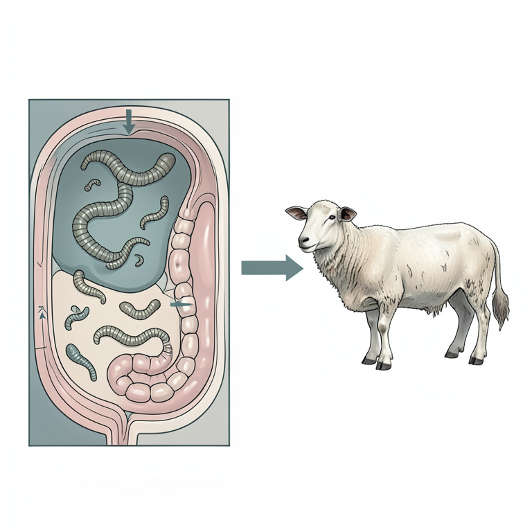
Pathological Synergy of Mixed Parasitism
AI-generated conceptual illustration (sheep digestive system). It explains the idea and does not show study data.
View source evidence ↗Figures & tables
From the original paper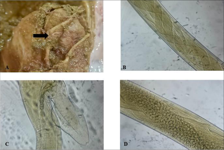 Figure 1
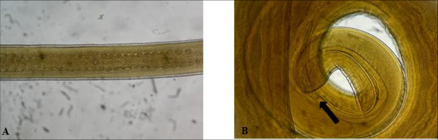 Figure 2
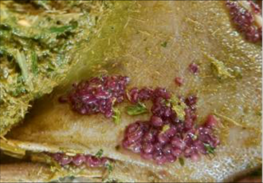 Figure 3
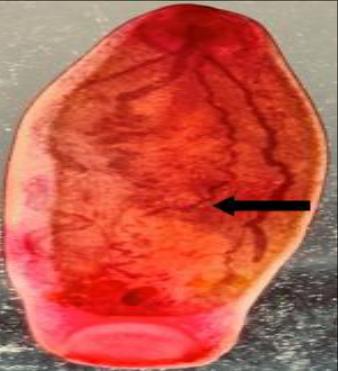 Figure 3
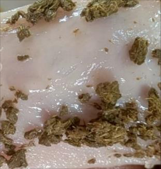 Figure 4
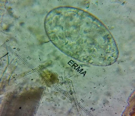 Figure 5A
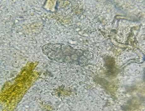 Figure 5B
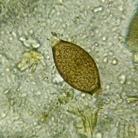 Figure 5C
Introduction
Gastrointestinal parasitism is one of the most prevalent issues affecting the sheep population worldwide, particularly under free-range and extensive grazing systems. Helminth parasites cause economic losses by reducing feed intake, growth rate, productivity and fertility of the animals and death in severe cases (Win et al., 2020) [15] . Due to grazing behavior of sheep, they are more susceptible to parasitic infection than goats, and climatic conditions also influence the occurrence and severity of the gastrointestinal parasitism which results in morbidity and mortality of the animals (Singh et al., 2017) [9] . Sheep grazing on contaminated pastures result in mixed parasitic infection due to long-term exposure to different stages of infective eggs and larvae (Soulsby, 1982) [10] . Haemonchus contortus is the most pathogenic parasite causing anaemia and sudden death which is highly prevalent and most important parasite inhabiting the abomasum of sheep (Khalafalla et al., 2011 and Besier et al., 2016) [6,2] . Trichuris sp. parasitizes the large intestine, mainly the caecum and colon, causing enteritis which results in wasting (Wang et al., 2022) [14] . Amphistomes or rumen flukes, mostly seen in herbivores during the rainy season, cause digestive disturbances and significantly aggravate the disease when co-existing with other roundworm infections (Huson et al., 2017 and Nyagura et al., 2024) [4,8] . This study highlights the sudden mortality of sheep from a flock with mixed helminthic infection.
Materials and Methods
A pattanam sheep from a flock of twenty animals maintained on a free-range grazing system in Trichy, Tamil Nadu was presented to the Veterinary Clinical Complex, VCRI, Orathanadu, TANUVAS with a history of dullness, reduced appetite, lethargy, blanched mucous membranes, diarrhoea and no history of anthelmintic intervention in the flock. The animal appeared severely debilitated and was subjected to the emergency treatment but collapsed during treatment. A detailed post-mortem examination was carried out immediately after death. During thorough examination of the gastrointestinal tract, parasites were observed in the rumen, abomasum and intestines. The parasites were collected in 70% ethanol and submitted for identification in Veterinary Parasitology Laboratory.
The worms were processed using standard procedures for identification based on morphological characteristics (Soulsby, 1982) [10] . In addition, faecal samples were collected from the remaining sheep in the flock randomly and examined for detection of endoparasites using sedimentation and floatation techniques.
Results and Discussion
Post-mortem findings revealed mixed gastrointestinal parasitism in the sheep. Heavy infection of reddish worms from the abomasum was identified as Haemonchus contortus based on the barber's pole appearance of the intestine and uterus, and the presence of a tongue-shaped vulval flap (Fig 1). Whip like worms were present in the large intestine identified as Trichuris sp., which is characterized by having a posterior part thicker than the anterior part of the body, and in the case of male worms, the posterior end is curved with a single spicule in a sheath ( indicating the presence of mixed parasitic infection in the flock. The flock was dewormed with a combination of oxyclozanide and levamisole for eliminating the parasites (Soulsby, 1982) [10] . Fecal samples were negative for parasitic eggs 7 days after the treatment, and increased activity of the animals indicated successful recovery in the flock. Mixed helminth infection is most commonly seen in sheep maintained under extensive and free-range systems, which causes a major problem particularly in tropical and subtropical regions and results in severe economic loss by decreasing productivity and causing morbidity and mortality (Waller, 2006) [12] . In the present study, the detection of Haemonchus contortus, Trichuris sp. and Gastrothylax crumenifer indicated the multiple parasitism of the gastrointestinal tract of the sheep. Among these, haemonchosis is one of the most ubiquitous parasitic diseases of sheep due to blood-sucking activity (0.05 ml/day/worm) and high burden lead to severe anaemia, haemorrhagic abomasitis, hypoproteinaemia, submandibular oedema and often cause sudden death (Gadahi et al., 2009 and Arsenopoulos et al., 2021) [3,1] . The marked pallor of the mucous membrane observed during the present study strongly indicates acute haemonchosis as the primary cause of mortality in the sheep. Amphistomes are digenetic trematodes which inhabit the rumen and reticulum and impair the normal digestive function of the animals. They cause significant damage to the ruminal mucosa, loss of ruminal papillae in severe cases which impacts both ruminal motility and fermentation, leading to anorexia, malabsorption of nutrients, and contributing to gradual wasting (Tandon et al., 2014 and Wani et al., 2024) [11,13] . Immature amphistomes are pathogenic, causing haemorrhagic enteritis and diarrhoea (Mondal et al., 2002) [7] . In chronic conditions, animals exhibit signs such as anorexia, weight loss and severe debilitation, thereby resulting in reducing the animal's ability to withstand concurrent infection. Trichuris ovis, known as the ovine whipworm, is a caecal parasite mainly causing the thickening of the caecal mucous membrane, haemorrhages, congestion, nodule formation and ulceration of the large intestine. Heavy burdens impair the absorption of fluids by the intestine and cause diarrhoea resulting in dehydration (Iliev et al., 2017 and Bulbul et al., 2020) [5,13] . The concurrent infection of haemonchosis, amphistomosis and trichuriosis causes the combined clinical consequences such as blood loss, digestive disturbances and impairment in fluid absorption, which led to hypovolemic shock and death in this case. The current findings of the study highlight the lifethreatening effects of mixed parasitic infection in sheep maintained in free-range conditions. The continuous exposure of animals to a contaminated pasture especially in extensive and free-range grazing systems, absence of deworming and climatic conditions play a major role in the development of mixed helminthic infection. The combination of anthelmintics after proper diagnosis using faecal examination will always help the farmers to avoid concurrent infections in animals. which predispose to the mixed infectionslack of deworming, contaminated pasture, climatic conditions, and poor nutritionsignificantly increase the susceptibility of animals to the heavy parasitic burdens. This led to the death of animals and caused economic loss to the farmers. The strategic deworming based on faecal examination, pasture management, rotational grazing, maintaining proper hygiene and providing nutritional support not only prevents the mixed parasitic infection but also improves the productivity of the farmers.
Acknowledgement
AcknowledgementThe authors are thankful to the Dean, VCRI, Orathanadu and the Professor and Head, Veterinary Clinical Complex and the Professor and Head, Dept. of Veterinary Medicine for providing necessary facilities and support for the study.
Figures and tables from the paper
References
15 in the original paper- Arsenopoulos KV, Fthenakis GC, Katsarou EI, Papadopoulos E. Haemonchosis: A challenging parasitic infection of sheep and goats. Animals (Basel). 2021 Feb 1;11(2):363. DOI: 10.3390/ani11020363. PMID: 33535656; PMCID: PMC7912824.
- Besier RB, Kahn LP, Sargison ND, Van Wyk JA. Diagnosis, treatment and management of Haemonchus contortus in small ruminants. Adv Parasitol. 2016;93:181-238. DOI: 10.1016/bs.apar.2016.02.024. Epub 2016 May 10. PMID: 27238006.
- Gadahi JA, Arshed MJ, Ali Q, Javaid SB, Shah SI. Prevalence of gastrointestinal helminths of sheep and goat in and around Rawalpindi and Islamabad, Pakistan. Vet World. 2009;2:51-53.
- Huson KM, Oliver NAM, Robinson MW. Paramphistomosis of ruminants: An emerging parasitic disease in Europe. Trends Parasitol. 2017 Nov;33(11):836-844. DOI: 10.1016/j.pt.2017.07.002. Epub 2017 Jul 25. PMID: 28754416.
- Iliev PT, Ivanov A, Kirkova Z, Hristov K, Dinkova K, Ananiev J. Some parasitological, pathological and immunohistochemical examinations in sheep naturally infected by Trichuris ovis. The Journal of Supercomputing. 2017;15:174-178.
- Khalafalla RE, Elseify MA, Elbahy NM. Seasonal prevalence of gastrointestinal nematode parasites of sheep in the northern region of Nile Delta, Egypt. Parastol Res. 2011;337-340.
- Mondal MMH, Alim Md, Shahiduzzaman Md, Farjana Thahsin, Islam Md Khoyrul, Das PM. Pathology of amphistomiasis in ruminants in Bangladesh. Bangladesh Veterinary Journal. 2002;36:103-108.
- Nyagura I, Malatji MP, Ngcamphalala PI, Mukaratirwa S. A systematic review and meta-analysis on the prevalence of amphistome infection in domestic ruminants in the sub-Saharan Africa region. Journal of Helminthology. 2024;98:e80. DOI: 10.1017/S0022149X24000725.
- Singh E, Kaur P, Singla LD, Bal MS. Prevalence of gastrointestinal parasitism in small ruminants in western zone of Punjab, India. Vet World. 2017 Jan;10(1):61-66. DOI: 10.14202/vetworld.2017.61-66. Epub 2017 Jan 17. PMID: 28246448; PMCID: PMC5301180.
- Soulsby EJ. Helminths, Arthropods and Protozoa of Domesticated Animals. 7 th ed. Baillière Tindall; London: 1982. p. 1-772.
- Tandon V, Roy B, Shylla JA, Ghatani S. Amphistomes. In: Toledo R, Fried B, editors. Digenetic Trematodes. Advances in Experimental Medicine and Biology, vol 766. Springer; New York, NY: 2014. p. 113-123. DOI: 10.1007/978-1-4939-0915-5_11.
- Waller PJ. From discovery to development: current industry perspectives for the development of novel methods of helminth control in livestock. Vet Parasitol. 2006 Jun 30;139(1-3):1-14. DOI: 10.1016/j.vetpar.2006.02.036. Epub 2006 May 3. PMID: 16675128.
- Wani Z, Shah S, Rather SA, Allaie I, Shafi M, et al. Pathomorphological changes associated with Paramphistome infection in the rumen of buffalo. Indian Journal of Animal Health. 2024;10:36062/ijah.2023.02823.
- Wang L, Zhang G, Fu Y, Ning C, Li Z, Wang H, et al. Prevalences and characteristics of Trichuris spp. infection in sheep in pastoral areas of the Tianshan, Xinjiang, China. J Vet Res. 2022 Dec 3;66(4):591-597. DOI: 10.2478/jvetres-2022-0056. PMID: 36846028; PMCID: PMC9944990.
- Win SY, Win M, Thwin EP, Htun LL, Hmoon MM, Chel HM. Occurrence of gastrointestinal parasites in small ruminants in the central part of Myanmar. J Parasitol Res. 2020 Nov 25;2020:8826327. DOI: 10.1155/2020/8826327. PMID: 33294216; PMCID: PMC7714589.