
Research article · 2026 · Full text
International Journal of Biology SciencesSurgical affections of eye in cattle - A clinical study (24 cows)
Volume 8(3) · Pages 125–130 · DOI: 10.33545/26649926.2026.v8.i3b.602 ↗
Research areasOcular Surface and Contact LensOcular Infections and TreatmentsVeterinary Pharmacology and Anesthesia
Abstract
Author-writtenIn this study, 24 cattle which were presented with growth, worms and foreign bodies in the eye, corneal opacity and ulcers were taken. The animals aged between 3 and 12 years and breeds such as cross-bred jersey, Hallikar, and kangayam were reported. Complete examination of the eyes and ultrasound scanning were carried out to study the ocular lesions. Surgical procedure for removal of growth, foreign body and worms in the affected eye was carried out by restraining animals in standing sedation using Inj. xylazine. Local anaesthesia was achieved using 2% lignocaine hcl given as auriculopalpebral nerve block and Peterson nerve block. The growths were resected using electrocautery (80 °C), foreign bodies and worms by surgical karatotomy. In some cases, enucleation of the eye was carried out as the lesions involved the complete eyeball. Histopathological examination of growths revealed squamous cell carcinoma (n=8), fibrosarcoma (n=2), adenocarcinoma and basal cell carcinoma (n=1) each. Corneal opacity and ulcer cases were treated with autologous PRP subconjunctival injections and topical eye drops. Post operatively the parental antibiotic therapy and Topical application of chloromycetin eye ointments were advised. On follow-up, recurrence of the tumours was reported in squamous cell carcinoma cases and in an adenocarcinoma case. Healing of corneal opacity and ulcers was observed in all cases. Early surgical treatment is necessary for better success of treatment and to prevent culling of animals.
Summary
Source-checkedPlain-language reading of the full paper, prepared with AI and checked against the text. Each point links to the authors' own words.
Why it matters
- Eye diseases in cattle are a major concern because they negatively affect both the welfare of the animal and the economic productivity of the livestock. View evidence ↗
- Bovine ocular affections range from common disorders like conjunctivitis and corneal ulcers to less frequent but critical conditions such as tumors and traumatic injuries. View evidence ↗
The question
- The study aimed to evaluate the clinical presentation and surgical management of 24 cattle suffering from ocular growths, worms, foreign bodies, corneal opacity, and ulcers. View evidence ↗
What they did
- The study included cattle aged 3 to 12 years, including Hallikar, Kangayam, and cross-bred Jersey breeds. View evidence ↗
- Animals were managed with standing sedation using xylazine (0.05 mg/kg IM) and local anesthesia via 2% lignocaine HCl using Peterson and auriculopalpebral nerve blocks. View evidence ↗
- Autologous platelet-rich plasma (PRP) was prepared by a two-step centrifugation process, retaining the lower one-third of the plasma containing the platelet pellet. View evidence ↗
- Cattle with corneal ulcers (n=6) and opacity (n=4) received subconjunctival injections of autologous PRP on days 0, 7, and 14. View evidence ↗
What they found
- Squamous cell carcinoma was the most common ocular lesion, occurring in 8 cases, treated primarily by enucleation (n=7) and one radical resection. View evidence ↗
- Enucleation of tumor masses was successful with no complications or recurrences reported within the first two months post-treatment. View evidence ↗
- Tumor recurrence occurred in two cases where eyelid masses (squamous cell carcinoma and adenocarcinoma) were resected rather than enucleated. View evidence ↗
- Surgical extraction of Thelazia rhodensi (eye worm) led to restored corneal transparency and improved vision over two weeks without complications. View evidence ↗
- Subconjunctival PRP injections in cases of corneal ulcers and opacity resulted in improved transparency and epithelial healing over two weeks. View evidence ↗
- A wooden foreign body was successfully extracted via a limbus opening, with the resulting superficial corneal erosion healing spontaneously within one week. View evidence ↗
What it means
- The authors conclude that early surgical intervention is essential to improve treatment success and prevent the need to cull affected animals. View evidence ↗
- The study emphasizes that combining precise surgical techniques with regenerative therapies, such as PRP, is an effective approach for managing bovine eye diseases. View evidence ↗
Visual summary
1 visualsIllustrations explain the paper; charts are replotted from the paper's own table values.
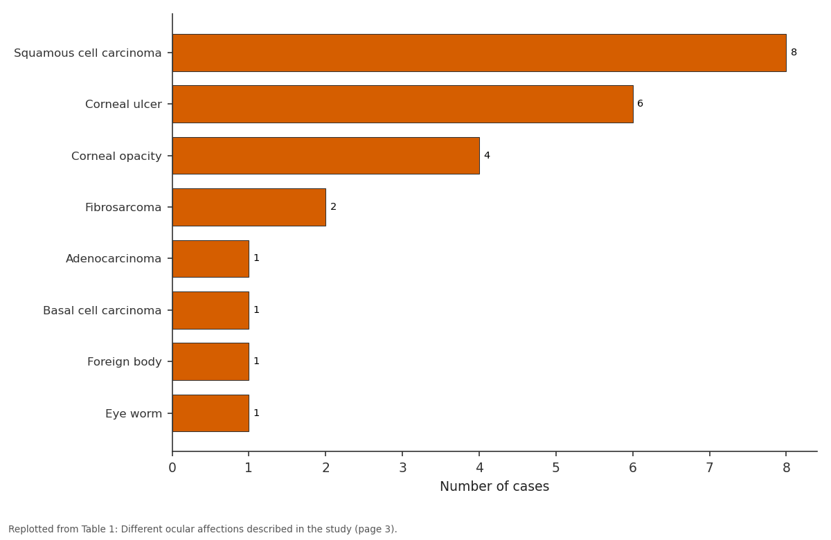
Number of cases: Squamous cell carcinoma vs Corneal ulcer vs Corneal opacity vs Fibrosarcoma vs Adenocarcinoma
Replotted from Table 1 (page 3) of the original paper. Every bar is the paper's own table value.
View source evidence ↗Figures & tables
From the original paper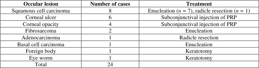 Table 1
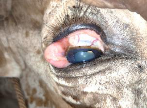 Figure 1
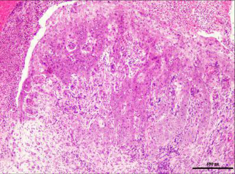 Figure 2
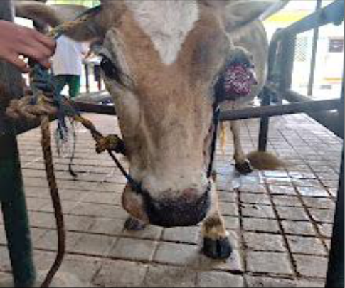 Figure 3
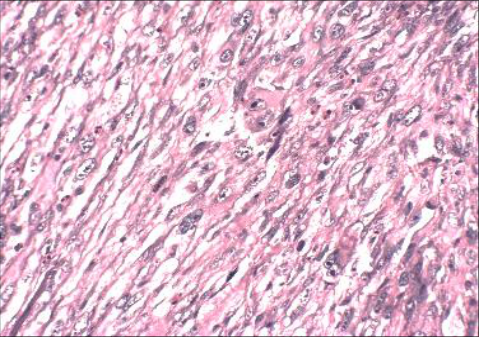 Figure 4
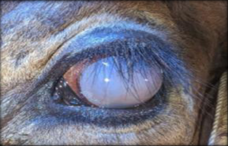 Figure 5
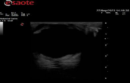 Figure 6
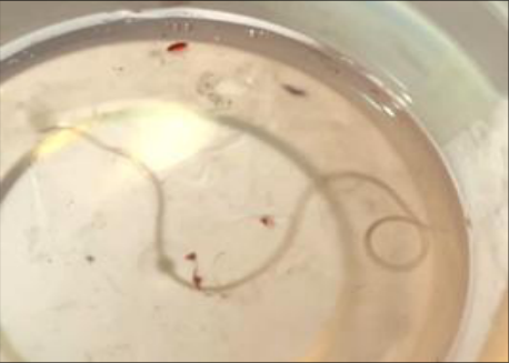 Figure 7
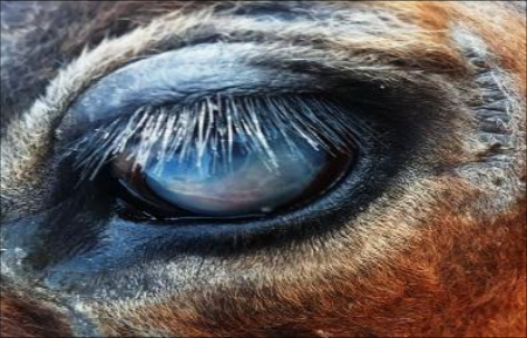 Figure 8
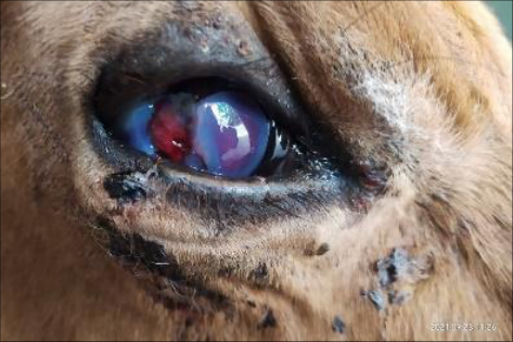 Figure 9
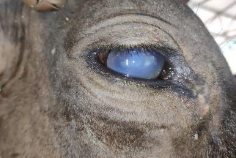 Figure 10
Introduction
The eye is an important special sense organ in vertebrate animals, which depends on it to survive and interact with the environment. Ocular diseases in bovines represent a significant concern for veterinarians and cattle owners alike, impacting animal welfare and economic productivity. comprehensive overview of the diverse ocular affections observed in bovines, encompassing a wide range of conditions affecting the eye and adnexa. Common ocular disorders such as infectious keratitis, conjunctivitis, corneal ulcers, and uveitis are explored, alongside less frequent but clinically important conditions, including ocular neoplasms and traumatic injuries.
Materials and Methods
Animal details 24 cattle were reported in this study, aged between 3 and 12 years and breeds such as crossbred jersey, Hallikar and Kangayam.
Clinical symptoms
The clinical signs varied depending on the condition, but several signs were commonly observed in most cases. These included intense pain when the eye was exposed to direct sunlight. The ocular lesions were extremely painful, interfering with grazing behaviour and leading to reduced performance and even weight loss. Additional complications seen in cases with delayed treatment included tear staining on the face, accumulation of pus matting the eyelashes and facial hair, severe conjunctivitis, both superficial and deep corneal vascularisation, and corneal ulceration.
Ocular examination
Ocular affections can be diagnosed by external and internal examination. External examination includes naked eye examination, smear, culture, Schirmer tear test, fluorescent dye test, slit lamp biomicroscopy, gonioscopy, and tonometer. An internal examination was achieved through ultrasonography.
Anaesthesia
Xylazine was administered as a premedication at a dose of 0.05 mg/kg intramuscularly to achieve standing sedation and facilitate restraint of the animals for procedures such as the removal of growths, foreign bodies, or worms from the eye. For proper ocular examination, eyelid movement was reduced by performing an auriculopalpebral nerve block using a subcutaneous injection of 5 mL of 2% lidocaine hydrochloride over the dorsal surface of the zygomatic arch. Also, Peterson's technique was used to block the five nerves that supply the eye. Surgical procedures on the eye were carried out in conscious animals employing a four-point retrobulbar nerve block. This was achieved by injecting 2% lidocaine with an 18-gauge, 9 cm needle at the dorsal, lateral, ventral, and medial aspects of the globe (12, 3, 6, and 9 o'clock positions, respectively), as described by Ivany.
Platelet-rich plasma preparation
Approximately 8-10 mL of whole blood was collected aseptically from the jugular vein into a sterile syringe containing 1 mL of 3.8% sodium citrate solution as an anticoagulant (maintaining a 9:1 ratio of blood to anticoagulant) or an ACD vial. The collected blood was transferred into sterile centrifuge tubes and subjected to the first centrifugation at 2600 rpm for 10 minutes (soft spin) to separate the blood components. Following centrifugation, three layers were obtained: the lower red blood cell layer, the middle buffy coat containing platelets and leukocytes, and the upper plasma layer. The plasma and buffy coat layers were carefully aspirated using a sterile pipette and transferred to another sterile tube, avoiding disturbance of the red blood cell layer. The separated plasma was then centrifuged again at 3600 rpm for 15 minutes (hard spin) to concentrate the platelets. After the second centrifugation, the upper twothirds of the plasma, identified as platelet-poor plasma (PPP), was discarded, while the lower one-third portion containing the platelet pellet was retained and gently resuspended to obtain platelet-rich plasma (PRP). The prepared PRP was either used immediately for therapeutic application or stored at 4 °C for up to 6 hours before use.
Pre-operative management Anti-inflammatory/ analgesic therapy
The extent of anti-inflammatory treatment required was generally based on the nature of the disease and its level of invasiveness. Flunixin meglumine was administered intravenously at a dose of 1.1 mg/kg immediately before performing the eye enucleation procedure
Antibiotic therapy
Broad-spectrum systemic antibiotic (Procaine penicillin G at a dose rate of 20.000 I.U/kg B.W., and streptomycin at a dose rate of 10 mg/kg B.W.) therapy was administered in all cases, especially on those that underwent eye enucleation.
Surgical site preparation
The hair surrounding the eye was clipped, and the skin was disinfected using 5% povidone iodine. The ear was draped, and the lateral part of the halter was covered to minimise contamination of the surgical area. A saline rinse was used instead of antiseptic scrubs to avoid corneal irritation or injury. In procedures other than enucleation, an ophthalmic ointment was applied to the cornea before aseptic preparation to provide additional protection.
Surgical technique
Enucleation was performed in ten cases (squamous cell carcinoma (n = 7), fibrosarcoma (n = 2) and basal cell carcinoma (n = 1) as per the standard technique for transpalpebral approach. The eyelids were sutured together with a simple continuous pattern by non-absorbable suture material (silk size 2). A circumferential incision was made about one centimetre away from the eyelid margins. Dissection was carried out using both blunt and sharp techniques through the orbicularis oculi muscle, fascia, and subcutaneous tissues surrounding the eye, using the inner surface of the bony orbit as a guide. The medial and lateral canthal ligaments were sharply cut to allow access to the deeper, caudal portion of the orbit. The retrobulbar muscles and the optic nerve sheath were transected as far back as possible. The eyeball was removed and bleeding was carefully arrested, either with a hemostat if it was minor or via ligation using cat gut 3-0 suture material. The surgical wound was closed in a horizontal mattress suture pattern using braided silk size 1, leaving a small opening near the medial canthus to allow drainage of exudates and for daily placement of sterile gauze soaked in antiseptic. In two cases (adenocarcinoma (n = 1), Squamous cell carcinoma (n = 1) (Fig. 1) growths were on eyelid and the growths were resected using electrocautery (80 °C) in cutting mode and bleeding was arrested in coagulation mode. In one animal, which was suffering from a corneal foreign body, the foreign body (wooden piece) was extracted through a stab incision at the level of the limbus and it was removed using a mosquito artery forceps. The incision was closed using PGA 5-0 by simple interrupted suture pattern. In one animal with corneal edema and opacity and loss of vision, ultrasonographic examination of the eye revealed eye worm. A stab incision was made on the limbus at the 6 o'clock position and the worm was removed using mosquito artery forceps. The incision was closed using PGA 5-0 by simple interrupted suture pattern. Sub conjunctival injection of autologous platelet rich plasma was done on the bulbar conjunctiva for corneal ulcer (n = 6) and corneal opacity (n = 4) cases on 0 th , 7 th and 14 th day of presentation using a 24G needle.
Post-operative care
Animals were kept in a confined area to prevent selfmutilation. Broad-spectrum antibiotic (Procaine penicillin G at a dose rate of 20.000 IU/kg B.W., and streptomycin at a dose rate of 10 mg/kg B.W.) was injected IM for 5 consecutive days, and topical application of chloromycetin eye ointments was advised. Daily monitoring of the surgical site and assessment of general well-being were performed until suture removal. Sutures were removed in 10 to 14 days to allow for complete healing of the skin.
Histopathological examination
The whole tumour mass was removed, cut into pieces and fixed in (10%) neutral buffer formalin. The tissue specimens were processed in a tissue processor, and paraffin blocks were made (5-6 µm) with a microtome and stained with Hematoxylin and Eosin dyes. The stained slides were examined under a light microscope. Table (1) shows different cases admitted to the clinic with ocular lesions with various ocular affections. Enucleation was a successful treatment for ocular tumour masses. There were no intra-operative and post-operative complications and also no recurrence of tumour recorded in any of the cases during the first two months after treatment. Histopathological examination revealed squamous cell carcinoma (n = 7) (Fig 1, 2), fibrosarcoma (n = 1) (Fig 3,4), and basal cell carcinoma (n = 1). In two animals with masses on the eyelids, the mass was resected, resulting in recurrence.
Results
On histopathological examination, it was found to be squamous cell carcinoma and adenocarcinoma.
In one case with a corneal foreign body (wooden piece), the foreign body was successfully extracted through the limbus opening, and the site of the foreign body left a superficial erosion on the cornea, which disappeared spontaneously after one week without any complications.
In one animal with corneal edema and opacity and loss of vision (Fig. 5), ultrasonographic examination of the eye revealed eye worm (Fig. 6); the eye worm (Thelazia rhodensi) was successfully extracted through the limbus opening (Fig. 7) and showed gradual healing of the corneal defect with reduction in opacity and inflammation, restoration of corneal transparency and improvement of vision over a period of two weeks (Fig. 8) without any complication.
In case of animals with corneal ulcer (Fig. 9) and corneal opacity (Fig. 10), subconjunctival injection of autologous platelet-rich plasma in bulbar conjunctiva resulted in enhanced epithelial healing, reduction of corneal inflammation and vascularisation, and gradual improvement in corneal transparency over a period of two weeks without any complication.
Discussion
Ocular Squamous Cell Carcinoma (OSCC), also known as "cancer eye," is a primary epithelial tumor that can develop in various ocular and periocular tissues, including the palpebral skin, corneal and conjunctival epithelium, the third eyelid, and the limbus (Fornazari et al., 2017) [4] . It is one of the most common neoplasms in cattle worldwide and represents the leading cause of enucleation among all ocular diseases (Shruthi et al., 2018) [13] . The development of OSCC is multifactorial, influenced by genetic, phenotypic, and environmental factors such as prolonged exposure to ultraviolet radiation, as well as infections with Bovine Papillomavirus (BPV) and Bovine Herpesvirus (BHV) (Fornazari et al., 2017; Priyanka et al., 2021) [4,11] . The condition is often accompanied by inflammatory changes and complications such as glaucoma, ulceration, and anterior synechia (Lalitha et al., 2024) [7] .
In the present study, enucleation was performed using either Peterson's or retrobulbar nerve blocks, consistent with earlier reports that described surgical excision as the treatment of choice for OSCC in cattle (Ali et al., 2015; Gautam et al., 2016; Scheck, 2005; Edwards, 2010) [1,17,18,19] . Foreign body penetration into the anterior chamber generally carries a guarded prognosis due to the high likelihood of secondary endophthalmitis, particularly when caused by plant material (Irby, 2004) [6] . In corneal conditions associated with pain, the use of a mydriatic cycloplegic agent is recommended (Williams et al., 2002) [16] . In this study, topical atropine and parenteral administration of a COX-2 inhibitor (meloxicam) were used, improving the animal's comfort. The foreign body was surgically removed under general and regional anaesthesia (Fazili et al., 2010) [3] . Eye worm (ocular thelaziasis) is another common ocular surgical condition in cattle. Diagnosis is usually straightforward based on clinical signs such as lacrimation, photophobia, blepharospasm, corneal opacity, and the visible presence of worms in the anterior chamber (Vigneswari et al., 2019) [15] . While medical therapy is generally ineffective due to slow absorption of dead parasites and associated immune reactions (Lavach, 1990; Moore et al., 1983) [8,10] , surgical removal under general or regional anesthesia remains the most effective treatment (Das et al., 2021; Tuntivanich et al., 2011) [2,14] . In the present study, surgical extraction was successfully performed under both general anesthesia and regional nerve blocks. For chronic ocular conditions, antibiotic and antiinflammatory eye drops often provide limited benefit. However, blood-derived products such as platelet-rich plasma (PRP) have proven to be effective and safer alternatives for chronic conjunctivitis and corneal erosions (Ranjan et al., 2020) [12] . Serum-derived eye drops have shown efficacy in treating ocular surface disorders including severe dry eye, persistent epithelial defects, and neurotrophic keratitis (Lee et al., 2016) [9] . Alio et al. (2007) [20] found that autologous PRP promoted healing of chronic corneal ulcers while alleviating pain and inflammation. PRP contains significantly higher concentrations of platelets, Epidermal
Growth Factor (EGF), and transforming growth factor-beta (TGF-β) than whole blood, enhancing tissue regeneration (Lee et al., 2016) [9] . Similarly, Hartwig et al. (2004) [5] highlighted the potential of platelet-derived growth factors in ocular surface repair. In our study, this regenerative property of PRP was utilised through subconjunctival injection of autologous PRP into the bulbar conjunctiva of cows suffering from corneal ulceration and opacity.
Conclusion
Ocular disorders in cattle, including ocular squamous cell carcinoma (OSCC), foreign body injuries, and parasitic infestations such as ocular thelaziasis, represent significant challenges in veterinary ophthalmology due to their impact on animal welfare, productivity, and vision. Among these, OSCC remains the most prevalent neoplasm of the bovine eye, often necessitating surgical intervention such as enucleation for effective management. Early diagnosis and appropriate surgical procedures, performed under regional or general anesthesia, have proven to yield favorable outcomes and minimize postoperative complications. Supportive medical management plays an equally important role in improving recovery and comfort, as demonstrated through the use of cycloplegic agents, non-steroidal antiinflammatory drugs (NSAIDs), and antibiotic therapies. However, in chronic and refractory ocular conditions, the introduction of regenerative therapies such as autologous platelet-rich plasma (PRP) offers a promising alternative. PRP, rich in growth factors and cytokines, has shown encouraging results in promoting corneal healing, reducing inflammation, and restoring ocular integrity.
Overall, this study underscores the importance of integrating surgical precision with emerging regenerative treatments in the management of bovine ocular diseases. Continuous clinical observation, early detection, and the adoption of advanced therapeutic approaches can significantly enhance the prognosis and quality of life of affected animals, while contributing to improved productivity in the livestock sector.
Figures and tables from the paper
| Occular lesion | Number of cases | Treatment |
|---|---|---|
| Squamous cell carcinoma | 8 | Enucleation (n = 7), radicle resection (n = 1) |
| Corneal ulcer | 6 | Subconjunctival injection of PRP |
| Corneal opacity | 4 | Subconjunctival injection of PRP |
| Fibrosarcoma | 2 | Enucleation |
| Adenocarcinoma | 1 | Radicle resection |
| Basal cell carcinoma | 1 | Enucleation |
| Foreign body | 1 | Keratotomy |
| Eye worm | 1 | Keratotomy |
| Total | 24 |
Table text extracted from the original PDF; superscript letters are shown inline. The original table image is below.
View original table image
References
20 in the original paper- Ali MM, Sadan MA, Ibrahim A. Ocular field surgery in ruminants. International Journal of Veterinary Medicine: Research & Reports. 2015;2015:709104. DOI:10.5171/2015.709104.
- Das J, Kuldeep SK, Singh R. Surgical removal of eyeworm in indigenous cow. Journal of Entomology and Zoology Studies. 2021;9(1):1487-1489.
- Fazili MUR, Buchoo BA, Bhattacharyya HK. Successful management of a delayed case of a corneal foreign body in a cow. Turkish Journal of Veterinary and Animal Sciences. 2010;34(3):295-298. DOI:10.3906/vet-0804-12.
- Fornazari GA, Kravetz J, Kiupel M, Sledge D, Filho IRDB, Montiani-Ferreira F. Ocular squamous cell carcinoma in Holstein cows from the South of Brazil. Veterinary World. 2017;10(12):1413-1420.
- Hartwig D, et al. Effect of platelet-derived growth factors on ocular surface defects. Journal of Ophthalmic Research. 2004;41(4):367-375.
- Irby NL. Surgical diseases of the eye in farm animals. In: Fubini SL, Ducharme NG, editors. Farm Animal Surgery. Philadelphia: Saunders; 2004. p. 429-468.
- Lalitha CH, Ramaprasad MBV, Durgaprasad M, Manasa BB, Kumari LR, Kumar RA. Bovine ocular squamous cell carcinoma in cattle: a case report. International Journal of Veterinary Sciences and Animal Husbandry. 2024;9(4):348-350. DOI:10.22271/veterinary.2024.v9.i4f.1570.
- Lavach JD. Large animal ophthalmology. St. Louis: Mosby; 1990.
- Lee JH, Kim MJ, Ha SW, Kim HK. Autologous plateletrich plasma eye drops in the treatment of recurrent corneal erosions. Korean Journal of Ophthalmology. 2016;30(2):101-107.
- Moore CP, Sarazan RD, Whitley RD, Jackson WF. Equine ocular parasites: a review. Equine Veterinary Journal. 1983;2(Suppl):76-85. doi:10.1111/j.20423306.1983.tb04565.x.
- Priyanka N, Nagaraja BN, Nagaraju N, Manjunath DR, Balapannavar BR. Study on occurrence of ocular tumors in cattle. The Pharma Innovation Journal. 2021;10(7):329-333.
- Ranjan R, Singh GD, Kumar A, Gattani A. Therapeutic management of chronic bulbar conjunctivitis in cattle using autologous platelet-rich plasma. Haryana Veterinarian. 2020;59(2):274-275.
- Shruthi PJ, Sujatha K, Srilatha CH, Rayulu VC. Incidence of different tumors in bovines. Open Access Journal of Science. 2018;2(4):220-222.
- Tuntivanich N, Tiawsirisup S, Tuntivanich P. Success of anterior chamber paracentesis as a treatment for ocular setariasis in equine eye: case report. Journal of Equine Veterinary Science. 2011;31(1):8-12. DOI:10.1016/j.jevs.2010.11.017.
- Vigneswari M, Sankar P, Kathirvel S. Surgical management of ocular thelaziasis in cattle. Indian Journal of Animal Sciences. 2019;21(1):139-140.
- Williams DL, Barrie K, Evans TF. Veterinary ocular emergency. 1st ed. Oxford: Butterworth-Heinemann; 2002.
- Gautam PK, Gautam RK, Banerjee S, Chattopadhyaya MC, Pandey JD. Heavy metals in the environment: fate, transport, toxicity and remediation technologies. Nova Sci Publishers. 2016;60:101-30.
- Scheck P, Nelson TO. Lack of pervasiveness of the underconfidence-with-practice effect: boundary conditions and an explanation via anchoring. Journal of Experimental Psychology: General. 2005 Feb;134(1):124.
- Edwards A. Being an expert professional practitioner: The relational turn in expertise. Springer science & business media; 2010 Aug 5.
- Alio JL, Colecha JR, Pastor S, Rodriguez A, Artola A. Symptomatic dry eye treatment with autologous plateletrich plasma. Ophthalmic research. 2007 Mar 19;39(3):124-129.