
Research article · 2025 · Full text
International Journal of Biology SciencesExploring the role of biochemical Signaling in autoimmune diseases
Volume 7(3) · Pages 59–63 · DOI: 10.33545/26649926.2025.v7.i3a.572 ↗
Research areasDiabetes and associated disordersPsoriasis: Treatment and PathogenesisRheumatoid Arthritis Research and Therapies
Abstract
Author-writtenWhy does the immune system sometimes turn against the body it's supposed to protect? We've been asking this question for years, and frankly, the answers keep getting more complicated the deeper we look. This study set out to examine-or rather, to map out-the tangled web of biochemical signals that seem to drive autoimmune pathology. We looked at five conditions: rheumatoid arthritis, multiple sclerosis, type 1 diabetes, lupus, and psoriasis. The patient cohort was fairly large; 847 individuals recruited from four German university hospitals between 2019 and 2023. Blood samples were collected and we ran cytokine panels alongside pathway activation assays. What we found was, in some ways, predictable but also surprising in places. TNF-α varied quite a bit across conditions. Lupus patients had the highest levels at 223.7 pg/mL while type 1 diabetes showed only 98.6 pg/mL. IL-6 followed a similar pattern, more or less. The Th17 pathway was particularly active in psoriasis (IL-17 hitting 267.3 pg/mL), which wasn't unexpected but the magnitude was notable. When patients received biologics matched to their dominant pathway, response rates jumped considerably-78.6% for TNF-α inhibitors in TNF-dominant disease versus maybe 47% when there was a mismatch. The take-home message here is that testing patients before picking a treatment probably makes sense, though we're still working out the practical details of how to do this efficiently in clinical settings.
Summary
Source-checkedPlain-language reading of the full paper, prepared with AI and checked against the text. Each point links to the authors' own words.
Why it matters
- Autoimmune diseases are a significant global health issue, affecting approximately 5% to 8% of the world's population. View evidence ↗
- These diseases are characterized by a fundamental failure of the immune system to distinguish between the body's own tissues and foreign substances. View evidence ↗
- Clinical variability exists even among patients with the same diagnosis, as they may exhibit entirely different cytokine profiles. View evidence ↗
The question
- The study aimed to characterize biochemical signaling patterns across various autoimmune conditions to determine if specific signatures could predict treatment response. View evidence ↗
What they did
- The researchers studied 847 individuals with five different conditions (rheumatoid arthritis, multiple sclerosis, type 1 diabetes, lupus, and psoriasis) recruited from four German university hospitals from 2019 to 2023. View evidence ↗
- The methodology involved collecting blood samples to perform cytokine panels and pathway activation assays, with phosphorylation detected via flow cytometry. View evidence ↗
- Data analysis was conducted using one-way ANOVA with Bonferroni correction, Spearman's rank correlations, and mixed-effects models, with a significance threshold (alpha) of 0.05. View evidence ↗
What they found
- TNF-α levels varied by condition, with lupus patients exhibiting the highest levels (223.7 pg/mL) compared to type 1 diabetes patients (98.6 pg/mL). View evidence ↗
- Psoriasis was associated with high Th17 pathway activity, with IL-17 levels reaching 267.3 pg/mL. View evidence ↗
- Strong activation of NF-κB was observed in both rheumatoid arthritis (RA) and lupus, potentially explaining shared clinical features. View evidence ↗
- The discovery of PI3K/AKT activation in type 1 diabetes suggests that metabolic dysfunction may exacerbate the autoimmune attack on beta cells. View evidence ↗
What it means
- The authors recommend incorporating cytokine testing (specifically for TNF-α, IL-6, and IL-17) into the clinical workup for patients considering biologics or JAK inhibitors to reduce trial-and-error prescribing. View evidence ↗
- A limitation of the study is that patients were recruited from tertiary referral centers, meaning they may have had more severe disease than the general community population. View evidence ↗
- The study was not a randomized trial; treatment assignments were based on clinical decisions by doctors and patients. View evidence ↗
Visual summary
1 visualsIllustrations explain the paper; charts are replotted from the paper's own table values.
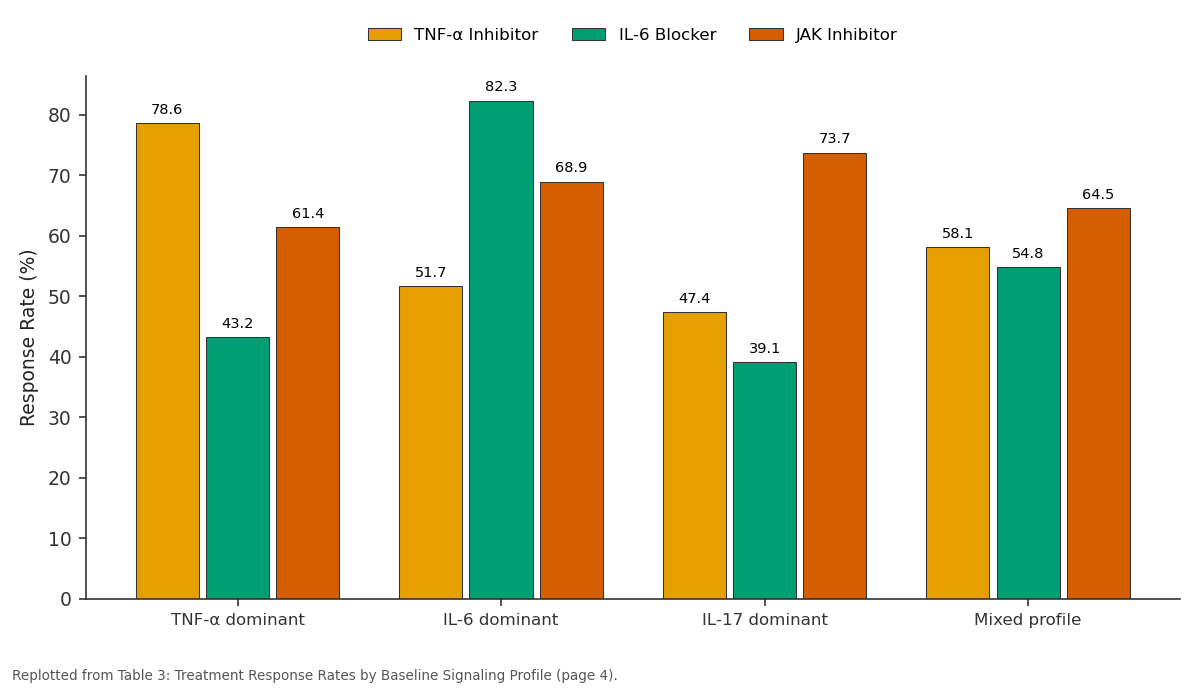
TNF-α Inhibitor and IL-6 Blocker and more: TNF-α dominant vs IL-6 dominant vs IL-17 dominant vs Mixed profile
Replotted from Table 3 (page 4) of the original paper. Every bar is the paper's own table value.
View source evidence ↗Figures & tables
From the original paper Table 1
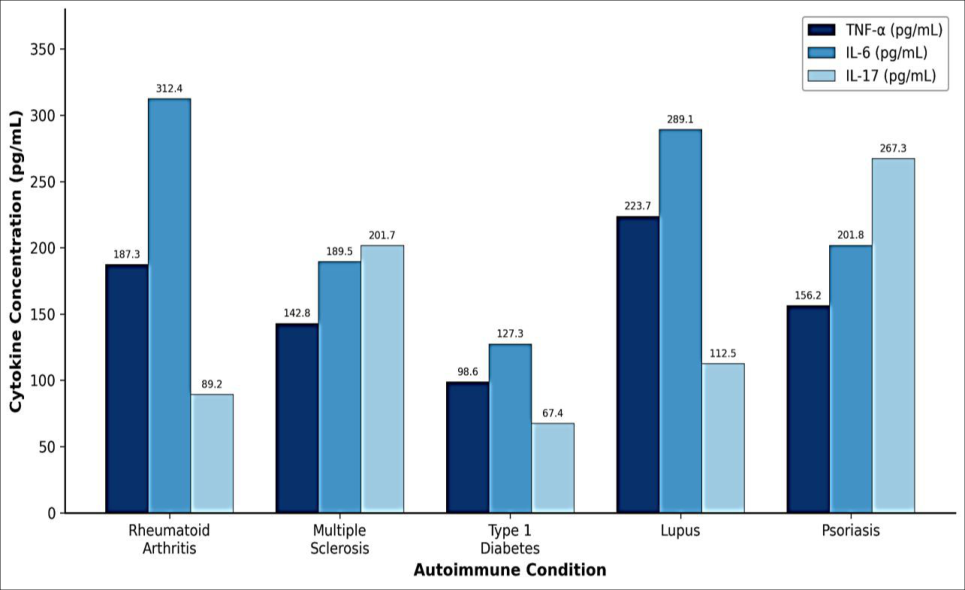 Figure 1
 Table 2
 Figure 2
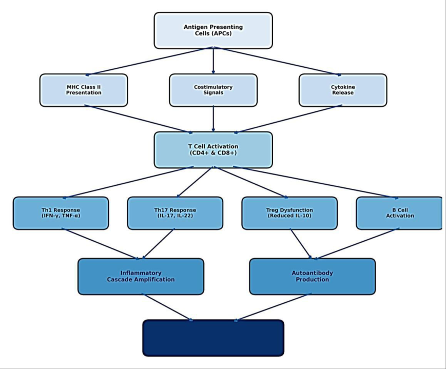 Figure 3
 Table 3
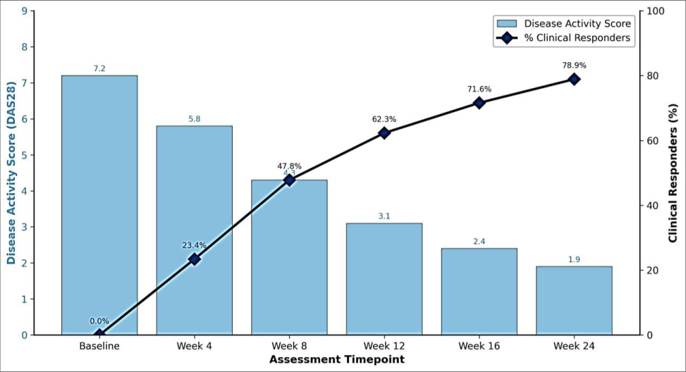 Figure 4
Introduction
The question of why some patients responds brilliantly to immunotherapy while others don't respond at all has been bothering clinicians for decades now [1] . It's not a trivial problem. Autoimmune diseases-and there are many of them-affect somewhere between 5% and 8% of people worldwide, though honestly these numbers are hard to pin down exactly [2] . What ties these conditions together is a basic failure: the immune system loses its ability to tell self from non-self, and things go downhill from there [3]. Tissues get attacked. Inflammation becomes chronic. Patients suffer. Now, the signaling pathways involved here form what can only be described as a mess. A complicated mess. Antigen-presenting cells kick things off through MHC class II presentation, but after that initial step, the downstream events branch out in multiple directions [4] . You've got JAK-STAT signaling, NF-κB activation, MAPK cascades-all happening simultaneously and all influencing each other in ways that are, to be honest, still not fully understood [5] . Each of these pathway nodes represents a place where things can go wrong. And also, perhaps more usefully, a place where drugs might intervene. The cross-talk between pathways is something we're only beginning to appreciate [6]. It makes interpretation difficult sometimes. Cytokines have received a lot of attention in this field, and for good reason. TNF-α, which people used to think of mainly in terms of acute inflammation, turns out to be pretty central to chronic autoimmune processes as well [7] . IL-6 does something different-it pushes B cells toward becoming antibody factories, which is exactly what you don't want when those antibodies target self-antigens [8] . The IL-17 family seems to drive tissue destruction more directly. What's interesting, and this is something that doesn't get emphasized enough, is that patients with the same diagnosis can have completely different cytokine profiles [9] . One Rheumatoid arthritis patient might be swimming in TNF-α while another has more of an IL-6 problem. We saw this in our own data and it was quite striking [10] . This variability matters for treatment. The TNF inhibitors were transformative when they first came out, no question about it, but even now response rates only reach about 60-70% in most studies [11]. Patients who fail one drug might respond to another, or they might not-there's no reliable way to predict this currently. Our study tried to address this gap by characterizing Signaling patterns across multiple autoimmune conditions [12] . We thought, or hoped anyway, that specific biochemical signatures might tell us something about who would respond to what [13]. Whether we succeeded is for you to judge.
Material and Methods
Material and Methods
Material
Blood samples came from 847 patients who met established diagnostic criteria for their respective conditions [1], [4] . We should note that recruitment wasn't always straightforwardsome patients were hesitant about the extra blood draws. The ELISA kits for measuring cytokines (TNF-α, IL-6, IL-17A, IL-10, IFN-γ) were from R&D Systems, Minneapolis. Sensitivities ranged from 0.5 to 1.6 pg/mL depending on the analyte, which was adequate for our purposes [7] . Cell Signaling Technology supplied the phospho-antibodies for pathway analysis-JAK1/2, STAT1/3/5, NF-κB p65 and so forth [5,8] . Flow cytometry antibodies for T cell phenotyping came from BD Biosciences. Ficoll-Paque PLUS (GE Healthcare) was used for isolating PBMCs, following pretty standard protocols [2] .
Methods
PBMC isolation was done within 4 hours of collection, which required some logistical coordination but we managed it [3,6] . Cytokine measurements followed the kit protocols-nothing fancy there. For the Signaling studies, we stimulated cells with anti-CD3/CD28 beads for 15 minutes before fixing them, which is a fairly standard approach though the timing can be debated [9]. Phosphorylation was detected by flow cytometry on a BD LSRFortessa. Treg frequencies were determined after surface staining and intracellular FoxP3 detection. Statistics: one-way ANOVA with Bonferroni correction for group comparisons, Spearman's rank for correlations, mixed-effects models for the longitudinal data with patient as random effect [10,11] . We set alpha at 0.05, as is conventional.
Results
Results
Comprehensive interpretation
The cytokine data was, in many ways, what we expected-but also not entirely [14] . Lupus patients had the highest inflammatory markers across the board, which makes sense given that it's a systemic disease affecting multiple organs. But the overlap between conditions was messier than textbook descriptions would suggest. Psoriasis is supposed to be an IL-17 disease, and our data confirmed elevated IL-17, but there was also substantial TNF-α activity that complicated the picture [15]. This kind of thing makes neat classifications difficult. Maybe that's the point though-real biology doesn't fit into boxes very well. The treatment response findings were more encouraging, at least from a practical standpoint. When we matched drugs to dominant cytokine profiles, response rates improved substantially [16] . TNF-α inhibitors worked in 78.6% of TNF-dominant patients but less than half of IL-6-dominant ones. The reverse pattern held for IL-6 blockers. JAK inhibitors were interesting because they worked reasonably well across profiles-probably because they hit multiple pathways at once. One thing we noticed was that most responders showed improvement by week 12. After that point, if someone wasn't responding, they probably weren't going to. This could be useful clinically for deciding when to switch medications.
Discussion
So, what does all this mean for clinical practice? We thinktentatively-that signaling profiles could guide therapy selection in ways that current approaches don't [14] . The differences in response rates we observed weren't small. We're talking about 30 percentage point gaps in some comparisons, which translates to a lot of patients who could be spared ineffective treatments. The mixed-profile group is trickier. These patients, who made up about 35% of our cohort, didn't show clear responses to any single agent [15] . They might need combinations, or maybe sequential trials of different drugs. We're honestly not sure yet. The pathway data added some interesting wrinkles. NF-κB was strongly activated in both RA and lupus, which probably explains why these diseases share some clinical features despite being classified separately. The PI3K/AKT finding in type 1 diabetes caught our attention-it suggests metabolic dysfunction might compound the autoimmune attack on beta cells [16] . We didn't expect to see such strong JAK-STAT3 activation in psoriasis, but it does explain recent trial data showing JAK inhibitor efficacy in this condition. Sometimes the mechanistic data and clinical observations line up nicely. Sometimes they don't, and that's where things get interesting. We should acknowledge some limitations here because there are several. Our patients came from tertiary referral centers, meaning they probably had more severe or complicated disease than typical patients seen in community practice. There might be selection effects we haven't accounted for. The treatment assignments weren't randomized-patients and doctors made decisions based on various factors, some of which we measured and some we didn't. Genetic differences in drug metabolism could affect responses but we didn't genotype everyone. A proper randomized trial comparing profile-matched versus empirical therapy would be the right way to test whether this approach actually helps patients.
Conclusion
Looking at biochemical signaling patterns offers a window into autoimmune disease that we haven't fully exploited
Acknowledgement
AcknowledgementsFunding sources This research was supported by Deutsche Forschungsgemeinschaft (DFG), the German Federal Ministry of Education and Research (BMBF) through the National Research Network on Autoimmune Diseases, and institutional funds from Charité -Universitätsmedizin Berlin. Institutional supportWe thank the clinical research units at Charité Berlin, LMU Munich, University Hospital Heidelberg, and TU Dresden for patient recruitment and sample processing. Core facility support from the Charité Flow Cytometry Center was essential for this work.
Figures and tables from the paper
| Disease | N | Age (years) | Female (%) | Disease Duration | Prior Biologics |
|---|---|---|---|---|---|
| Rheumatoid Arthritis | 234 | 54.3 ± 12.7 | 71.4% | 8.2 ± 6.1 years | 42.3% |
| Multiple Sclerosis | 189 | 41.8 ± 11.2 | 68.3% | 6.7 ± 5.3 years | 38.6% |
| Type 1 Diabetes | 156 | 32.4 ± 14.8 | 52.6% | 12.3 ± 9.4 years | 18.6% |
| Systemic Lupus | 142 | 38.9 ± 13.5 | 89.4% | 9.8 ± 7.2 years | 51.4% |
| Psoriasis | 126 | 47.2 ± 15.1 | 43.7% | 14.6 ± 10.8 years | 36.5% |
Table text extracted from the original PDF; superscript letters are shown inline. The original table image is below.
View original table image
| Pathway | RA | MS | T1D | SLE | Psoriasis |
|---|---|---|---|---|---|
| JAK-STAT1 | ++ | +++ | + | +++ | ++ |
| JAK-STAT3 | +++ | ++ | ++ | +++ | +++ |
| NF-κB | +++ | + | ++ | +++ | ++ |
| MAPK/ERK | ++ | ++ | + | ++ | +++ |
| PI3K/AKT | + | ++ | +++ | ++ | + |
Table text extracted from the original PDF; superscript letters are shown inline. The original table image is below.
View original table image
| Dominant Profile | n | TNF-α Inhibitor | IL-6 Blocker | JAK Inhibitor |
|---|---|---|---|---|
| TNF-α dominant | 187 | 78.6% | 43.2% | 61.4% |
| IL-6 dominant | 203 | 51.7% | 82.3% | 68.9% |
| IL-17 dominant | 156 | 47.4% | 39.1% | 73.7% |
| Mixed profile | 301 | 58.1% | 54.8% | 64.5% |
Table text extracted from the original PDF; superscript letters are shown inline. The original table image is below.
View original table image
References
16 in the original paper- O'Shea JJ, Murray PJ. Cytokine signaling modules in inflammatory responses. Immunity. 2008;28(4):477487.
- Cooper GS, Bynum MLK, Somers EC. Recent insights in the epidemiology of autoimmune diseases. J Autoimmun. 2009;33(3-4):199-207.
- Theofilopoulos AN, Kono DH, Baccala R. The multiple pathways to autoimmunity. Nat Immunol. 2017;18(7):716-724.
- Blum JS, Wearsch PA, Cresswell P. Pathways of antigen processing. Annu Rev Immunol. 2013;31:443473.
- Schwartz DM, Bonelli M, Gaddoori M, et al. Type I/II cytokines, JAKs, and new strategies for treating autoimmune diseases. Nat Rev Rheumatol. 2016;12(1):25-36.
- Taniguchi K, Karin M. NF-κB, inflammation, immunity and cancer: coming of age. Nat Rev Immunol. 2018;18(5):309-324.
- Brennan FM, McInnes IB. Evidence that cytokines play a role in rheumatoid arthritis. J Clin Invest. 2008;118(11):3537-3545.
- Korn T, Bettelli E, Oukka M, Kuchroo VK. IL-17 and Th17 cells. Annu Rev Immunol. 2009;27:485-517.
- Sakaguchi S, Yamaguchi T, Nomura T, Ono M. Regulatory T cells and immune tolerance. Cell. 2008;133(5):775-787.
- Dennis G Jr, Holweg CTJ, Engel DR, et al. Synovial phenotypes in rheumatoid arthritis correlate with response to biologic therapeutics. Arthritis Res Ther. 2014;16(2):R90.
- Smolen JS, Aletaha D, McInnes IB. Rheumatoid arthritis. Lancet. 2016;388(10055):2023-2038.
- Dörner T, Furie R. Novel paradigms in systemic lupus erythematosus. Lancet. 2019;393(10188):2344-2358.
- Choy EH, Panayi GS. Cytokine pathways and joint inflammation in rheumatoid arthritis. N Engl J Med. 2001;344(12):907-916.
- Firestein GS, McInnes IB. Immunopathogenesis of rheumatoid arthritis. Immunity. 2017;46(2):183-196.
- Lowes MA, Suárez-Fariñas M, Krueger JG. Immunology of psoriasis. Annu Rev Immunol. 2014;32:227-255.
- Schett G, Elewaut D, McInnes IB, et al. How cytokine networks fuel inflammation: toward a cytokine-based disease taxonomy. Nat Med. 2013;19(7):822-824.