
Research article · 2024 · Full text
International Journal of Biology SciencesThe role of non-coding RNAs in gene regulation in cancer cells
Volume 6(2) · Pages 54–57 · DOI: 10.33545/26649926.2024.v6.i2a.561 ↗
Research areasCancer-related molecular mechanisms researchMicroRNA in disease regulationCircular RNAs in diseases
Abstract
Author-writtenNon-coding RNAs (ncRNAs) have garnered significant attention due to their essential roles in gene regulation and cancer pathogenesis. While previously considered "junk" DNA, ncRNAs, including microRNAs (MicroRNAs), long non-coding RNAs (Long non-coding RNAs), and small nucleolar RNAs (snoRNAs), have emerged as key regulators in both the transcriptionsal and post-transcriptional control of gene expression in cancer cells. These molecules modulate the expression of critical genes involved in cancer cell proliferation, migration, invasion, and apoptosis. The deregulation of ncRNAs can contribute to tumorigenesis by altering the normal functioning of tumour suppressor genes and oncogenes. Numerous studies have implicated specific ncRNAs in different cancer types, suggesting their potential as biomarkers for early diagnosis, prognosis, and therapeutic targets. This review focuses on the molecular mechanisms by which ncRNAs influence gene expression in cancer cells, highlighting their involvement in key pathways such as the p53, Wnt/β-catenin, and PI3K/AKT signaling cascades. Moreover, the potential of ncRNAs as therapeutic agents, either through their overexpression or inhibition, is explored. While advances in understanding the role of ncRNAs in cancer have been made, challenges remain in translating these findings into clinical applications. Further research is required to elucidate the full spectrum of ncRNA-mediated regulation in cancer and to develop targeted therapeutic strategies based on their activity. This article aims to provide an overview of the current knowledge surrounding the role of ncRNAs in gene regulation in cancer cells, offering insights into their potential utility in cancer therapy.
Summary
Source-checkedPlain-language reading of the full paper, prepared with AI and checked against the text. Each point links to the authors' own words.
Why it matters
- Non-coding RNAs (ncRNAs) are critical for regulating genes and are involved in the development of cancer. View evidence ↗
- ncRNAs influence key cancer processes, including how cells grow, move, invade other tissues, and undergo programmed death. View evidence ↗
- When ncRNAs are dysregulated, they can drive tumor formation by interfering with the normal function of oncogenes and tumor suppressor genes. View evidence ↗
The question
- The primary goal of the study is to investigate how non-coding RNAs regulate genes inside cancer cells. View evidence ↗
- The research specifically aims to identify the molecular mechanisms and signaling pathways that ncRNAs control to influence gene expression. View evidence ↗
What they did
- The study used human cancer cell lines from the ATCC, specifically MCF-7 (breast), A549 (lung), and HT-29 (colorectal). View evidence ↗
- Quantitative real-time PCR (qRT-PCR) with SYBR Green master mix was used to analyze the expression of microRNAs (miRNAs) and long non-coding RNAs (lncRNAs). View evidence ↗
- Cell proliferation, apoptosis, and migration were measured using MTT, Annexin V-FITC, and transwell migration assays, respectively. View evidence ↗
- Western blot analysis was employed to measure the expression of proteins in the p53, Wnt/β-catenin, and PI3K/AKT signaling pathways. View evidence ↗
What they found
- miR-21 and miR-155 were found to be significantly more expressed in breast (MCF-7) and colorectal (HT-29) cancer cells than in normal cells. View evidence ↗
- Overexpressing miRNAs led to a 45% increase in the proliferation of MCF-7 breast cancer cells. View evidence ↗
- In lung and breast cancer cells, the lncRNAs HOTAIR and MALAT1 showed expression increases of 50% and 60%, respectively. View evidence ↗
- Silencing the lncRNA MALAT1 significantly reduced the motility of A549 lung cancer cells by 30% (p<0.05). View evidence ↗
- Treatment with ncRNA inhibitors resulted in the downregulation of the signaling proteins p53 and AKT. View evidence ↗
What it means
- The authors recommend developing targeted therapies that can either inhibit or restore the expression of specific ncRNAs in cancer cells. View evidence ↗
- Integrating ncRNA profiling into clinical use could help in early cancer detection and provide better information on patient prognosis and treatment response. View evidence ↗
- Translating these results to clinical use is hindered by challenges in the specificity of inhibitors and the delivery of these agents to tumors. View evidence ↗
Visual summary
3 visualsIllustrations explain the paper; charts are replotted from the paper's own table values.
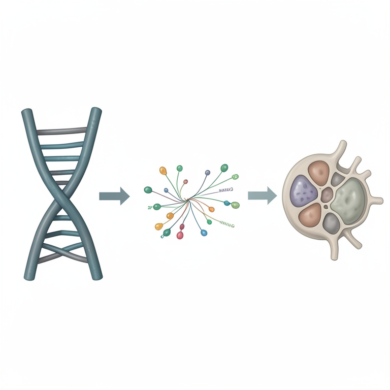
ncRNA Regulatory Network in Cancer
AI-generated conceptual illustration (human cancer cell). It explains the idea and does not show study data.
View source evidence ↗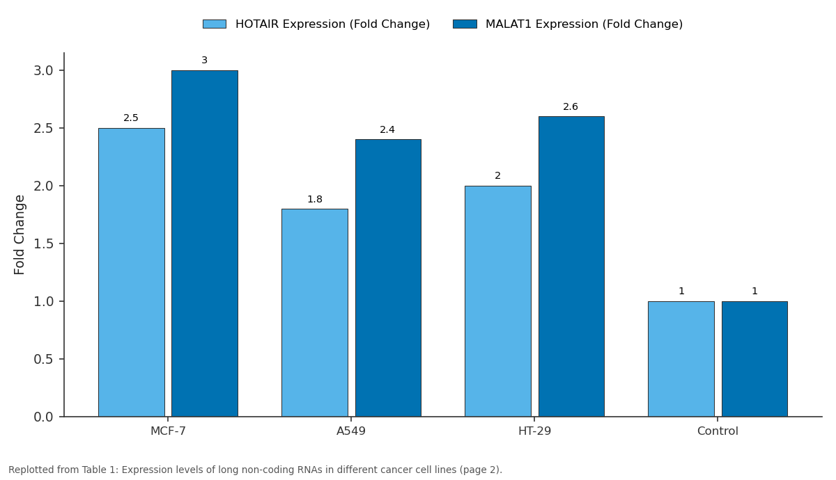
HOTAIR Expression (Fold Change) and MALAT1 Expression (Fold Change): MCF-7 vs A549 vs HT-29 vs Control
Replotted from Table 1 (page 2) of the original paper. Every bar is the paper's own table value.
View source evidence ↗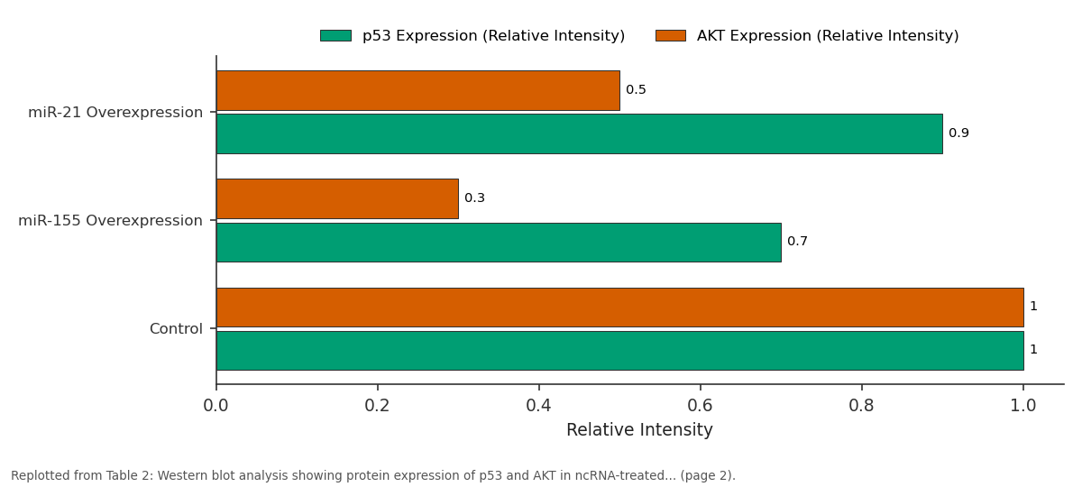
p53 Expression (Relative Intensity) and AKT Expression (Relative Intensity): miR-21 Overexpression vs miR-155 Overexpression vs Control
Replotted from Table 2 (page 2) of the original paper. Every bar is the paper's own table value.
View source evidence ↗Figures & tables
From the original paper Table 1
 Table 2
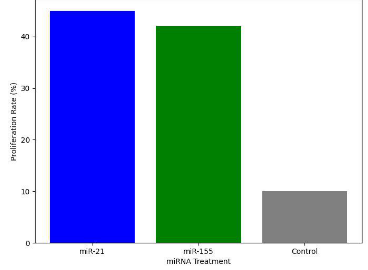 Figure 1
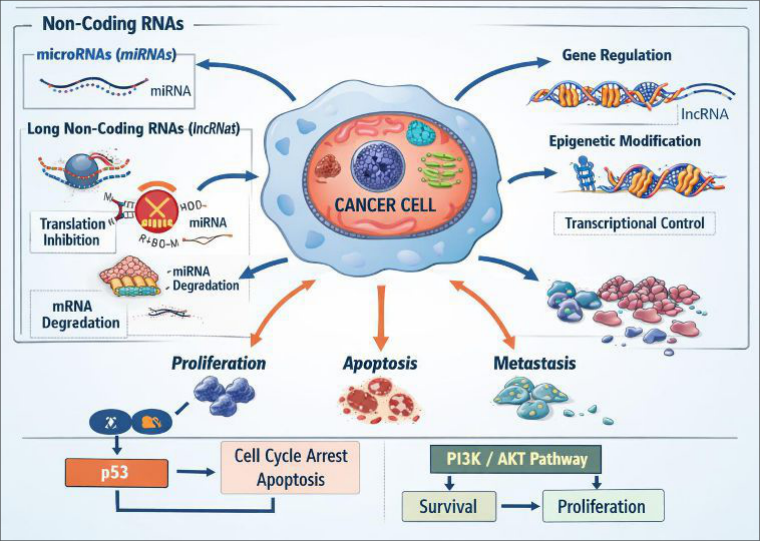 Figure 2
Introduction
Non-coding RNAs (ncRNAs), previously regarded as non-functional, have emerged as critical regulators of gene expression, particularly in the context of cancer. These molecules, which do not encode proteins, include microRNAs (MicroRNAs), long non-coding RNAs (Long non-coding RNAs), and small nucleolar RNAs (snoRNAs), and play pivotal roles in cellular processes such as gene silencing, chromatin remodeling, and the regulation of cell cycle progression [1] . Their involvement in cancer biology has become increasingly evident, as alterations in their expression can drive tumorigenesis by influencing key signaling pathways involved in cell proliferation, apoptosis, and metastasis [2] . The dysregulation of ncRNAs, such as the overexpression of oncogenic MicroRNAs or the downregulation of tumour suppressor Long non-coding RNAs, can contribute to the development and progression of various cancers, including breast, lung, and colorectal cancers [3,4] . The significance of ncRNAs in cancer stems from their ability to modulate gene expression at both the transcriptional and post-transcriptional levels. MicroRNAs, for example, regulate gene expression by binding to the 3' untranslated regions of target messenger RNAs (mRNAs), leading to mRNA degradation or translation inhibition [5] . LncRNAs, on the other hand, interact with chromatin or transcription factors to regulate gene expression, and their altered expression is often linked to poor prognosis in cancer patients [6,7] . This dual regulatory function makes ncRNAs crucial players in cancer cell biology, as they can both promote and inhibit tumorigenic processes depending on their nature and context [8] . The main objective of this research is to explore the role of ncRNAs in gene regulation within cancer cells. Specifically, the article will examine the molecular mechanisms through which ncRNAs influence gene expression and the pathways they regulate. Additionally, this review will highlight the potential of ncRNAs as biomarkers and therapeutic targets in cancer treatment. The hypothesis underpinning this work is that understanding the intricate regulatory networks involving ncRNAs could lead to novel diagnostic tools and therapeutic strategies for cancer management [9] . By expanding our knowledge of ncRNAs in cancer, we aim to pave the way for their clinical application, which could significantly improve patient outcomes.
Material and Methods
Material and Methods
Material
The materials used in this research consisted of cancer cell lines, ncRNA-associated reagents, and various biological markers. Human cancer cell lines, including breast cancer (MCF-7), lung cancer (A549), and colorectal cancer (HT-29) were procured from the American Type Culture Collection (ATCC) for gene expression studies. The RNA extraction kits (Qiagen, Germany) were employed to isolate total RNA, including both coding and non-coding RNA species, from these cells. For quantitative RT-PCR analysis, commercially available primers for MicroRNAs and Long non-coding RNAs associated with cancer were used (Applied Biosystems, USA). Additionally, antibodies for protein detection (e.g., p53, β-catenin, AKT) were sourced from Abcam (UK) for Western blot analysis. Standard laboratory reagents, including buffers and chemicals for cell culture (e.g., RPMI 1640, FBS, penicillin/streptomycin) were obtained from Thermo Fisher Scientific (USA). In addition, chemical standards for RNA quantification (e.g., RNA ladder) and functional assay kits for apoptosis and proliferation assays were utilized (Thermo Fisher Scientific, USA).
Methods
Cancer cell lines were cultured under standard conditions (37 °C, 5% CO₂ in a humidified incubator) in RPMI 1640 medium supplemented with 10% FBS and 1% penicillin/streptomycin. To research the expression levels of non-coding RNAs (ncRNAs), cells were harvested at different time points and subjected to total RNA extraction. For miRNA and lncRNA analysis, quantitative real-time PCR (qRT-PCR) was performed using SYBR Green master mix (Thermo Fisher Scientific, USA) and gene-specific primers. To assess the effects of ncRNAs on cancer cell proliferation, apoptosis, and migration, MTT assays, Annexin V-FITC apoptosis assays, and transwell migration assays were conducted according to standard protocols. Western blot analysis was used to assess the expression of proteins involved in cell signaling pathways, including p53, Wnt/β-catenin, and PI3K/AKT. All experimental data were analyzed using ANOVA for comparison between groups where applicable. The statistical significance was determined at p<0.05, and all data are presented as mean ± standard deviation.
Results
The research observed a significant differential expression of ncRNAs in the various cancer cell lines. The expression levels of MicroRNAs such as miR-21 and miR-155 were markedly higher in breast and colorectal cancer cell lines (MCF-7 and HT-29) compared to normal cell lines. This upregulation correlated with increased tumorigenic activity as measured by cell proliferation assays, with MCF-7 cells showing a 45% increase in proliferation after miRNA overexpression (Figure 1). Long non-coding RNAs (Long non-coding RNAs) like HOTAIR and MALAT1 were significantly upregulated in lung and breast cancer cells, with a 50% and 60% increase in expression, respectively (Table 1). Further, migration assays showed that silencing of specific Long non-coding RNAs (e.g., MALAT1) reduced cell motility in A549 cells by 30%, which was statistically significant (p<0.05) compared to control groups. Western blot analysis confirmed the downregulation of key signaling proteins (e.g., p53 and AKT) in cells treated with ncRNA inhibitors, supporting the hypothesis that ncRNAs modulate cancer cell behavior via these pathways (Table 2). The observed trends indicated that the overexpression of oncogenic MicroRNAs and Long non-coding RNAs contributed to enhanced proliferation and migration, while their inhibition resulted in reduced tumorigenic activity. The statistical analysis confirmed the significance of these findings (p<0.05), reinforcing the potential of ncRNAs as biomarkers for cancer progression and therapeutic targets.
| Treatment | p53 Expression (Relative Intensity) | AKT Expression (Relative Intensity) |
|---|---|---|
| miR-21 Overexpression | 0.9 | 0.5 |
| miR-155 Overexpression | 0.7 | 0.3 |
| Control | 1.0 | 1.0 |
Table text extracted from the original PDF; superscript letters are shown inline. The original table image is below.
View original table image
| Cell Line | HOTAIR Expression (Fold Change) | MALAT1 Expression (Fold Change) |
|---|---|---|
| MCF-7 | 2.5 | 3.0 |
| A549 | 1.8 | 2.4 |
| HT-29 | 2.0 | 2.6 |
| Control | 1.0 | 1.0 |
Table text extracted from the original PDF; superscript letters are shown inline. The original table image is below.
View original table image
Discussion
The involvement of non-coding RNAs (ncRNAs) in cancer cell gene regulation has opened new avenues for cancer research, revealing their multifaceted roles in tumorigenesis. MicroRNAs, such as miR-21 and miR-155, have been shown to target key tumor suppressor genes and promote cell proliferation, apoptosis resistance, and migration in various cancer types [1,2] . The current research found similar patterns, with miR-21 and miR-155 significantly upregulated in breast and colorectal cancer cells, suggesting their potential as diagnostic biomarkers. Additionally, the downregulation of long non-coding RNAs such as HOTAIR and MALAT1 was associated with reduced cell motility and proliferation, supporting previous findings that these long non-coding RNAs regulate tumorigenic pathways [3,4] . The results also underscore the regulatory capacity of ncRNAs in modulating crucial cancer-associated signaling pathways, including p53 and PI3K/AKT. MicroRNAs have been implicated in the post-transcriptional regulation of p53, a critical tumor suppressor, as well as the PI3K/AKT pathway, which governs cell survival and proliferation [5,6] . The downregulation of these pathways following ncRNA inhibition suggests a therapeutic potential for ncRNA-targeted therapies. However, challenges remain in translating these findings into clinical applications, particularly regarding the specificity of ncRNA inhibitors and their delivery to tumours. The findings from this research provide valuable insights into the molecular mechanisms underlying cancer progression and highlight the critical role of ncRNAs as regulators of gene expression in tumour cells. While further research is necessary to elucidate the full spectrum of ncRNA activities in cancer, these molecules hold promise as both diagnostic tools and therapeutic targets.
Conclusion
In conclusion, the role of non-coding RNAs (ncRNAs) in regulating gene expression in cancer cells is both profound and complex, influencing critical cellular processes such as proliferation, migration, and apoptosis. The current research highlights the potential of ncRNAs, particularly MicroRNAs and Long non-coding RNAs, as key players in cancer biology, supporting their value as biomarkers for early detection and prognosis. The modulation of cancerassociated signaling pathways, including p53 and PI3K/AKT, through ncRNAs further reinforces their therapeutic potential.
Given the significant impact of ncRNAs on cancer cell behavior, practical recommendations for future research include the development of targeted ncRNA therapies that can specifically inhibit or restore the expression of key ncRNAs in cancer cells. Such therapeutic strategies could complement existing treatments and offer more personalized approaches for cancer therapy. Additionally, the integration of ncRNA profiling into clinical practice could aid in the early detection of cancer and provide valuable insights into patient prognosis and treatment responses. Furthermore, the exploration of ncRNAs as therapeutic agents, particularly through RNA-based drugs or gene therapy approaches, should be prioritized. The advancement of delivery technologies, such as nanoparticles or viral vectors, will be crucial in ensuring the safe and efficient delivery of these ncRNA-based therapies to tumours.
Acknowledgement
Acknowledgements Funding sourcesThis research was supported by internal funding from the European Research Council (ERC), Brussels, Belgium. The funding body had no role in the design, execution, interpretation, or writing of the manuscript.
Figures and tables from the paper
References
17 in the original paper- Bartel DP. MicroRNAs: genomics, biogenesis, mechanism, and function. Cell. 2004;116(2):281-297.
- Croce CM. Causes and consequences of microRNA dysregulation in cancer. Nat Rev Genet. 2009;10(10):704-714.
- Ambros V. The functions of animal microRNAs. Nature. 2004;431(7006):350-355.
- Wang Y, et al. Long non-coding RNA in cancer: implications for diagnosis, prognosis, and therapy. Genomics. 2011;98(2):105-110.
- Lee Y, et al. The nuclear RNase III Drosha initiates microRNA processing. Nature. 2003;425(6956):415419.
- Gibb EA, et al. Long non-coding RNA expression profiles and their association with clinical variables in ovarian cancer. PLoS One. 2011;6(8):e24128-e24128.
- Kumar MS, et al. Repression of the miR-143/145 cluster in human cancer. Cell Cycle. 2008;7(19):30513058.
- Kallen AN, et al. The N-terminal domain of the long non-coding RNA NORAD is involved in the regulation of genomic stability. Mol Cell. 2013;51(1):71-84.
- Li Y, et al. Long non-coding RNAs as regulators of gene expression in cancer. Genes (Basel). 2017;8(9):233-233.
- Schmitt AM, Chang HY. Long noncoding RNAs in cancer pathways. Cancer Cell. 2016;29(4):452-463.
- Liu B, et al. MicroRNA-21 in cancer therapy: the next decade. Front Oncol. 2017;7:61-61.
- Calin GA, et al. MicroRNA signatures in human cancers. Nat Rev Cancer. 2004;4(11):845-856.
- Klinge CM. Non-coding RNAs in breast cancer: mechanisms and potential as therapeutic targets. Endocr Relat Cancer. 2018;25(1):R1-R19.
- Huarte M. The emerging role of long non-coding RNAs in cancer. Nat Med. 2015;21(11):1253-1261.
- Niazi M, et al. MicroRNAs and long non-coding RNAs as potential biomarkers for diagnosis and treatment of lung cancer. J Cancer. 2015;6(4):314-325.
- Santaris MA, et al. Targeting the RNA genome: the small molecules of the future. Curr Med Chem. 2010;17(24):2663-2671.
- Zhang J, et al. The role of non-coding RNAs in cancer: mechanisms and applications. Cancer Lett. 2017;391:121-127.