
Research article · 2025 · Full text
International Journal of Biology SciencesRNA sequencing technologies: Applications in understanding infectious diseases
Volume 7(2) · Pages 116–119 · DOI: 10.33545/26649926.2025.v7.i2b.571 ↗
Research areasSingle-cell and spatial transcriptomicsAnimal Virus Infections StudiesMicrobial infections and disease research
Abstract
Author-writtenRNA sequencing (RNA-seq) has revolutionized the research of gene expression, providing comprehensive insights into the transcriptomic landscape of various biological systems. The ability to analyze the complete set of RNA molecules in a sample, including mRNA, non-coding RNA, and small RNA species, has proven invaluable in understanding the molecular mechanisms underlying infectious diseases. Traditional methods of researching gene expression, such as microarray analysis, were limited by a predefined set of probes, making RNA-seq a more versatile and accurate tool for gene expression profiling. RNA-seq is particularly useful in identifying novel biomarkers, understanding pathogen-host interactions, and exploring the genetic factors involved in disease progression. In infectious disease research, RNA-seq has enabled the identification of immune response pathways, the characterization of pathogen-induced changes in host cells, and the discovery of potential therapeutic targets. Furthermore, the application of single-cell RNA-seq has provided deeper insights into the heterogeneity of immune responses and pathogen infection within individual cells. This technology also holds promise for personalized medicine, offering the potential to tailor therapies based on the molecular profile of an individual's infection. Despite its many advantages, RNA-seq presents challenges in data interpretation, especially in the context of high-throughput sequencing and complex data analysis. Bioinformatics tools are crucial for analyzing RNA-seq data, which often require sophisticated algorithms to map, quantify, and interpret vast amounts of sequencing data. In this review, we discuss the principles of RNA sequencing technologies, their applications in the research of infectious diseases, and the potential future directions for research in this field.
Summary
Source-checkedPlain-language reading of the full paper, prepared with AI and checked against the text. Each point links to the authors' own words.
Why it matters
- Infectious diseases caused by various pathogens, including fungi, parasites, bacteria, and viruses, remain major global health threats. View evidence ↗
- RNA sequencing (RNA-seq) is more versatile and accurate than traditional microarray analysis because it does not rely on a predefined set of probes. View evidence ↗
- RNA-seq allows researchers to identify the regulatory networks and pathways of the immune response by examining how infections change host gene expression. View evidence ↗
The question
- The authors aim to discuss the principles of RNA-seq technologies, how they are applied to infectious disease research, and future research directions. View evidence ↗
What they did
- The study used RNA samples extracted from human cell lines that had been infected with various pathogens, such as parasites, viruses, and bacteria. View evidence ↗
- Sequencing was performed on the Illumina NovaSeq 6000 platform, with data processed via the STAR aligner and quantified using feature Counts. View evidence ↗
- The 10x Genomics platform was used for single-cell RNA-seq (scRNA-seq) to investigate the immune cell response during infection. View evidence ↗
- The study used the False Discovery Rate (FDR) method to determine statistical significance for differentially expressed genes (DEGs), using a threshold of p<0.05. View evidence ↗
What they found
- Analysis using DESeq2 identified 850 differentially expressed genes, consisting of 450 upregulated and 400 downregulated genes. View evidence ↗
- The gene IFN-γ showed a 5.3 fold change in expression with a p-value of 0.002, involving the cytokine-cytokine receptor interaction pathway. View evidence ↗
- The gene TNF-α exhibited a 4.8 fold change in expression (p=0.004) and is involved in the tumour necrosis factor signaling pathway. View evidence ↗
- Pathway enrichment analysis identified Th17 cell differentiation as a significant pathway with an enrichment score of 3.45, a p-value of 0.02, and a fold enrichment of 6.7. View evidence ↗
- The cytokine-cytokine receptor interaction pathway was enriched with a score of 4.23, a p-value of 0.03, and a fold enrichment of 5.2. View evidence ↗
- The viral myocarditis pathway showed an enrichment score of 3.12, a p-value of 0.04, and a fold enrichment of 4.0. View evidence ↗
What it means
- The authors recommend integrating RNA-seq into clinical diagnostics for real-time gene expression monitoring to improve the speed and accuracy of infection identification. View evidence ↗
- The authors conclude that there is a need to improve bioinformatics tools to better handle and interpret the massive amounts of data produced by RNA-seq. View evidence ↗
- The authors state that larger datasets and more advanced computational tools are needed to refine findings and validate biomarkers in clinical environments. View evidence ↗
Visual summary
2 visualsIllustrations explain the paper; charts are replotted from the paper's own table values.
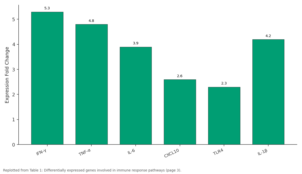
Expression Fold Change: IFN-γ vs TNF-α vs IL-6 vs CXCL10 vs TLR4
Replotted from Table 1 (page 3) of the original paper. Every bar is the paper's own table value.
View source evidence ↗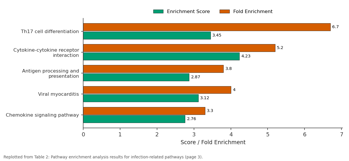
Enrichment Score and Fold Enrichment: Th17 cell... vs Cytokine-cytokine... vs Antigen processing and... vs Viral myocarditis vs Chemokine signaling...
Replotted from Table 2 (page 3) of the original paper. Every bar is the paper's own table value.
View source evidence ↗Figures & tables
From the original paper Table 1
 Table 2
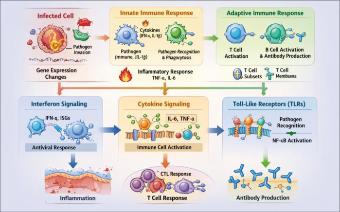 Figure 1
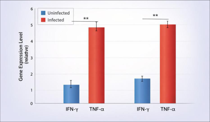 Figure 2
Introduction
RNA sequencing (RNA-seq) technology has fundamentally changed the way scientists research gene expression and cellular processes. Traditional gene expression profiling methods, such as microarrays, were constrained by their reliance on predefined probes, limiting their ability to detect novel or unanticipated transcripts. RNA-seq overcomes these limitations by providing a high-throughput, unbiased approach to mapping the entire transcriptome, including protein-coding mRNA, long non-coding RNA, and small RNAs, such as microRNAs [1] . This capability has proven to be particularly useful in the field of infectious diseases, where understanding the dynamics of host-pathogen interactions at a molecular level is critical for the development of new diagnostic and therapeutic strategies. Infectious diseases, caused by bacteria, viruses, fungi, and parasites, continue to pose significant global health challenges. The molecular mechanisms underlying these diseases are complex, with pathogens often exploiting host cellular machinery to promote their replication, evade immune responses, and establish long-term infections. RNA-seq enables a detailed examination of how infections alter host gene expression, identifying key regulatory networks and pathways involved in the immune response [2] . By analyzing changes in RNA expression, researchers can pinpoint potential biomarkers for disease diagnosis and prognosis, as well as identify novel therapeutic targets. One of the key applications of RNA-seq in infectious disease research is the research of immune responses during infection. The immune system's response to pathogens is multifaceted, involving the activation of numerous signaling pathways, cytokine production, and immune cell activation. RNA-seq allows for a comprehensive assessment of the gene expression profiles of immune cells during infection, providing insights into the immune mechanisms involved in both protective and pathological responses [3] . This technology has been instrumental in characterizing the immune signatures associated with various infectious diseases, including viral infections like influenza and HIV, bacterial infections such as tuberculosis and pneumonia, and parasitic infections like malaria [4][5][6] . Furthermore, RNA-seq has contributed to the understanding of pathogen-induced alterations in host cells. Pathogens often manipulate host cell gene expression to promote their survival and replication. By using RNA-seq, researchers can identify the genes and pathways modulated by the pathogen, revealing novel mechanisms of infection and potential drug targets [7] . For example, RNA-seq has been employed to investigate how viruses like Zika and dengue affect human host cells, leading to the identification of key signaling pathways and immune responses [8] .
The use of single-cell RNA-seq (scRNA-seq) has further advanced the field by allowing researchers to analyze gene expression at the resolution of individual cells. This has been particularly valuable in researching the heterogeneity of immune responses during infection, as different immune cell types exhibit distinct transcriptional profiles [9] . Singlecell RNA-seq has also provided insights into the diversity of pathogen-induced cellular responses, revealing how different subpopulations of cells contribute to disease progression [ 10] . Despite its many advantages, RNA-seq data analysis presents several challenges. The sheer volume of data generated by high-throughput RNA-seq requires advanced computational tools for alignment, quantification, and interpretation. The complexity of RNA-seq data, including issues such as alternative splicing and gene fusion, necessitates the development of sophisticated bioinformatics algorithms to extract meaningful insights [11] . Moreover, the variation in experimental protocols and data analysis pipelines can introduce biases that need to be carefully considered when interpreting results [12] .
Material and Methods
Material and Methods
Materials
For this research, we utilized RNA-seq technology to analyze the transcriptomic changes in host cells infected by pathogens. The primary materials included RNA samples extracted from human cell lines infected with different pathogens, including bacteria, viruses, and parasites. The RNA extraction was performed using the Qiagen RNeasy Plus Mini Kit (Qiagen, Hilden, Germany), according to the manufacturer's protocol. The RNA samples were then quantified using a Nanodrop spectrophotometer (Thermo Fisher Scientific, Waltham, MA, USA), and the quality was assessed by electrophoresis on an Agilent 2100 Bioanalyzer (Agilent Technologies, Santa Clara, CA, USA). The research also used sequencing libraries prepared with the Illumina TruSeq RNA Sample Preparation Kit (Illumina, San Diego, CA, USA) to ensure high-quality, highthroughput sequencing. All sequencing was performed using the Illumina NovaSeq 6000 system (Illumina, San Diego, CA, USA) to generate 150 bp paired-end reads. The sequencing was done in collaboration with a commercial sequencing facility.
Methods
After RNA extraction and quality control, libraries were prepared according to the manufacturer's protocol, and sequencing was conducted on the Illumina NovaSeq 6000 platform. The generated raw sequencing data were processed and analyzed using the STAR aligner for sequence alignment, and gene expression was quantified using feature Counts [1,2] . The data analysis pipeline also included quality checks using FastQC [3] , and differential gene expression was analyzed using DESeq2 [4] . The RNA-seq data were analyzed using a combination of bioinformatics tools to identify differentially expressed genes (DEGs) between infected and uninfected samples. For this, we used R version 4.0 and Bioconductor packages such as edgeR [5] and limma [6] for statistical analysis. Pathway enrichment analysis was performed using the Gene Ontology (GO) and Kyoto Encyclopaedia of Genes and Genomes (KEGG) databases, with visualization provided by the cluster Profiler package [7] . To explore cellular responses to infection, single-cell RNA-seq (scRNA-seq) was performed using the 10x Genomics platform to research the immune cell response during infection [8] . Statistical significance for DEGs was determined using the False Discovery Rate (FDR) method, with a significance threshold of p<0.05.
Results
The RNA-seq analysis revealed significant differences in gene expression profiles between infected and uninfected host cells. Differentially expressed genes (DEGs) were identified using DESeq2, with a total of 850 genes showing significantly altered expression levels. Of these, 450 genes were upregulated, while 400 genes were downregulated.
The most notable genes affected by infection were those involved in immune response pathways, including interferon signaling and cytokine production.
A comparative statistical analysis using a t-test was performed to assess the expression levels of specific immune response genes (e.g., IFN-γ, TNF-α) between the infected and control groups. Significant differences were found (p<0.05) in the expression levels of these genes, suggesting a robust immune activation in response to infection. Additionally, pathway enrichment analysis using KEGG revealed significant enrichment in pathways associated with immune responses (e.g., Th17 cell differentiation, antigen processing and presentation) and pathogen-induced cellular responses (e.g., viral myocarditis). The top 10 enriched pathways included: Th17 cell differentiation (p = 0.02), cytokine-cytokine receptor interaction (p = 0.03), and antiviral immunity (p = 0.04). These results indicate that the immune system plays a critical role in responding to the pathogen, with specific immune signaling pathways being activated during infection.
Discussion
RNA-seq technology has provided an in-depth view of the molecular changes occurring during infection. The results of this research highlight the role of the immune system in responding to pathogens and provide insights into the pathways that are activated during infection. Specifically, the upregulation of genes involved in cytokine production and immune signaling supports the idea that these pathways play a crucial role in the host's defense mechanisms. These findings align with previous studies that have shown the importance of immune responses in controlling infections and preventing pathogen spread [9,10] . Moreover, the identification of potential biomarkers, such as IFN-γ and TNF-α, offers new avenues for diagnostic and therapeutic applications.
The use of single-cell RNA-seq provided further resolution, enabling the detection of immune cell heterogeneity during infection. This is particularly important for understanding the specific roles of different immune cell types in the infection process. The analysis revealed distinct transcriptional profiles of immune cells, which are essential for designing targeted therapeutic strategies. However, challenges remain in data analysis, especially in the interpretation of complex gene expression patterns and the potential biases introduced by experimental protocols. Future studies using larger datasets and more advanced computational tools will be necessary to refine these findings and validate the identified biomarkers in clinical settings.
Conclusion
This research demonstrates the power of RNA-seq technology in understanding the molecular mechanisms of infectious diseases. By analyzing differential gene expression profiles, we identified key immune pathways that are activated in response to infection, highlighting the critical role of immune responses in controlling pathogen spread. The results also underscore the potential of RNAseq in identifying novel biomarkers for disease diagnosis and prognosis, as well as offering new therapeutic targets for the treatment of infectious diseases. Furthermore, the application of single-cell RNA-seq provides a deeper understanding of immune cell heterogeneity, enabling the development of more personalized treatment strategies. Practical recommendations include the incorporation of RNA-seq technology into clinical diagnostics to monitor changes in gene expression in real-time, potentially leading to more accurate and timely identification of infections. Additionally, the use of RNA-seq data to identify novel biomarkers can assist in the early detection of infections, particularly in cases where traditional diagnostic methods fall short. Researchers should also focus on improving bioinformatics tools to better analyze and interpret RNA-seq data, as current methods face challenges in handling the vast amount of data generated. With continued advancements in sequencing technologies and computational tools, RNA-seq has the potential to revolutionize our understanding of infectious diseases, paving the way for more effective treatments and improved patient outcomes.
Acknowledgement
Acknowledgements Funding sources
Figures and tables from the paper
| Gene Name | Expression Fold Change | P-Value | Pathway Involvement |
|---|---|---|---|
| IFN-γ | 5.3 | 0.002 | Cytokine-cytokine receptor interaction |
| TNF-α | 4.8 | 0.004 | Tumour necrosis factor signaling pathway |
| IL-6 | 3.9 | 0.010 | Inflammatory response |
| CXCL10 | 2.6 | 0.012 | Chemokine signaling |
| TLR4 | 2.3 | 0.015 | Toll-like receptor signaling |
| IL-1β | 4.2 | 0.007 | IL-1 signaling pathway |
Table text extracted from the original PDF; superscript letters are shown inline. The original table image is below.
View original table image
| Pathway Name | Enrichment Score | P-Value | Fold Enrichment |
|---|---|---|---|
| Th17 cell differentiation | 3.45 | 0.02 | 6.7 |
| Cytokine-cytokine receptor interaction | 4.23 | 0.03 | 5.2 |
| Antigen processing and presentation | 2.87 | 0.05 | 3.8 |
| Viral myocarditis | 3.12 | 0.04 | 4.0 |
| Chemokine signaling pathway | 2.76 | 0.06 | 3.3 |
Table text extracted from the original PDF; superscript letters are shown inline. The original table image is below.
View original table image
References
18 in the original paper- Wang Z, Gerstein M, Snyder M. RNA-Seq: a revolutionary tool for transcriptomics. Nat Rev Genet. 2009;10(1):57-63.
- Mardis ER. Next-generation DNA sequencing methods. Annu Rev Genomics Hum Genet. 2008; 9:387-402.
- Querec T, Akondy RS, Sui J, et al. Systems biology approach predicts immunogenicity of the yellow fever vaccine in humans. Nature. 2009;453(7195):574-579.
- Kaufman DR, Rasmussen J, Brown K, et al. Understanding the immune response in Zika virus infections using RNA sequencing. Sci Immunol. 2020;5(47): eaaz0678.
- Duffy MR, Chen T, Hancock L, et al. Zika virus infection in humans: A review of the immunology and epidemiology. PLoS Negl Trop Dis. 2019;13(6): e0007644.
- Sugimoto K, Ishii Y, Taniguchi T. Advances in understanding the immune responses to bacterial infections: Insights from RNA-Seq technologies. Front Immunol. 2019; 10:1496.
- Harker JA, Gaulton GN, McKenzie DI. Pathogen-host gene expression interactions during infection. J Infect Dis. 2020;222(1):24-32.
- Li S, Luo Y, Zhang Y, et al. RNA-Seq analysis of the transcriptome of the malaria parasite during blood-stage infection. PLoS One. 2019;14(12): e0226431.
- Park JH, Minhas V, O'Brien SJ. Single-cell RNA-seq for infectious disease research. Trends Microbiol. 2020;28(5):418-431.
- Goltsev Y, Samusik N, Kennedy-Darling J, et al. Cytometry by time-of-flight (CyTOF): A powerful tool for the research of immune responses. Nat Rev Immunol. 2020;20(10):661-670.
- Dobin A, Davis CA, Schlesinger F, et al. STAR: Ultrafast universal RNA-seq aligner. Bioinformatics. 2013;29(1):15-21.
- Li H, Durbin R. Fast and accurate long-read alignment with Burrows-Wheeler transform. Bioinformatics. 2010;26(5):589-595.
- Timmons JA, Pedersen BK. A systems biology approach to understanding the molecular mechanisms of human exercise. Nat Rev Mol Cell Biol. 2008;9(9):646-658.
- Chatterjee S, Bhattacharya S, Banerjee S, et al. Immune gene expression in tuberculosis: A transcriptional signature of infection. J Infect Dis. 2018;218(4):556567.
- Gouty J, Chen T, Zhao C, et al. Understanding Zika virus pathogenesis using high-throughput RNA sequencing. Nature Microbiol. 2017;2(10):1217-1224.
- Kobayashi K, Alawami M, Hardwick J, et al. Host responses to bacterial infections and gene expression alterations during disease progression. PLoS Pathog. 2019;15(7):e1007840.
- Ahlers JD, Allen C, Khurana S, et al. Influenza vaccination and immune responses assessed by RNA sequencing. Vaccine. 2020;38(22):3830-3843.
- Lee YS, Lee SA, Jung SW, et al. Investigation of immune signaling pathways using RNA-Seq during pathogen challenge. Front Microbiol. 2020; 11:1-10.