
Research article · 2025 · Full text
International Journal of Biology SciencesThe molecular basis of host-pathogen interactions in tuberculosis
Volume 7(8) · Pages 240–245 · DOI: 10.33545/26649926.2025.v7.i8c.577 ↗
Research areasTuberculosis Research and EpidemiologyPneumonia and Respiratory InfectionsInfectious Diseases and Tuberculosis
Abstract
Author-writtenTuberculosis (TB), caused by Mycobacterium tuberculosis, remains one of the most significant infectious diseases worldwide, despite substantial advancements in diagnosis and treatment. The molecular basis of host-pathogen interactions is pivotal in understanding the pathogenesis of TB and developing more effective therapeutic strategies. The immune system's response to M. tuberculosis involves both innate and adaptive immunity, where various immune cells, including macrophages, dendritic cells, and T lymphocytes, play critical roles. The bacterium has evolved complex mechanisms to evade the host immune response, enabling it to persist within the host for extended periods, often in a latent form. A key feature of M. tuberculosis pathogenesis is its ability to manipulate the host immune response, leading to the formation of granulomas compartmentalized structures that attempt to contain the infection but also contribute to tissue damage. The molecular interactions between the host's immune cells and the pathogen are multifactorial, involving cytokines, surface receptors, and signaling pathways that either promote bacterial clearance or facilitate bacterial persistence. Recent advances in genomic and proteomic technologies have provided new insights into the molecular mechanisms underlying these host-pathogen interactions. For instance, bacterial factors like the ESX-1 secretion system and the production of effector proteins such as ESAT-6 and CFP-10 are crucial for the survival of M. tuberculosis in the hostile environment of the host macrophage. Additionally, the host’s genetic susceptibility to TB, including polymorphisms in immune-related genes such as IFNG and VDR, has been increasingly recognized as a determinant of disease outcomes. Understanding these molecular interactions is essential for developing novel diagnostic and therapeutic approaches, particularly for drug-resistant strains and latent TB. This review aims to explore the molecular mechanisms that govern the host-pathogen interactions in TB, focusing on the immune evasion strategies employed by M. tuberculosis, the host immune response, and recent advances in molecular research.
Summary
Source-checkedPlain-language reading of the full paper, prepared with AI and checked against the text. Each point links to the authors' own words.
Why it matters
- Tuberculosis (TB), caused by Mycobacterium tuberculosis, continues to be a major global infectious disease despite improvements in treatment and diagnosis. View evidence ↗
- According to the World Health Organization, TB causes approximately 1.5 million deaths every year. View evidence ↗
- The pathogen manipulates the host's immune system to form granulomas, which are structures that try to contain the infection but can also cause tissue damage. View evidence ↗
The question
- The study aims to investigate the molecular mechanisms of host-pathogen interactions in TB, specifically focusing on how the bacteria evade the immune system and how the host responds. View evidence ↗
What they did
- The researchers used the M. tuberculosis strain H37Rv to infect human THP-1 monocyte cells, which were used as a model for macrophages. View evidence ↗
- THP-1 cells were infected with M. tuberculosis at a 1:1 multiplicity of infection and analyzed at 2, 6, and 24 hours post-infection. View evidence ↗
- The study used ELISA kits to measure IFN-γ and TNF-α cytokines and quantitative PCR (qPCR) to analyze the expression of immune-related genes. View evidence ↗
What they found
- The virulence factor ESAT-6 was significantly secreted by the bacteria, with relative expression levels of 1.2 at 6 hours and 3.4 at 24 hours post-infection. View evidence ↗
- Macrophages from TB-resistant individuals showed significantly higher IFNG gene expression (2.5) compared to those from TB-susceptible individuals (1.1). View evidence ↗
What it means
- The authors conclude that genetic variations in immune genes significantly affect TB control, suggesting that these could be used for personalized treatment. View evidence ↗
- The authors recommend developing strategies to boost the host's immune response to combat drug-resistant TB strains instead of relying only on antibiotics. View evidence ↗
- The authors state that future research needs to focus on targeting the specific molecular mechanisms the bacteria use to evade the immune system. View evidence ↗
Visual summary
3 visualsIllustrations explain the paper; charts are replotted from the paper's own table values.
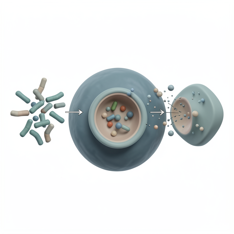
Host-Pathogen Interaction Cycle
AI-generated conceptual illustration (human macrophage and Mycobacterium tuberculosis bacteria). It explains the idea and does not show study data.
View source evidence ↗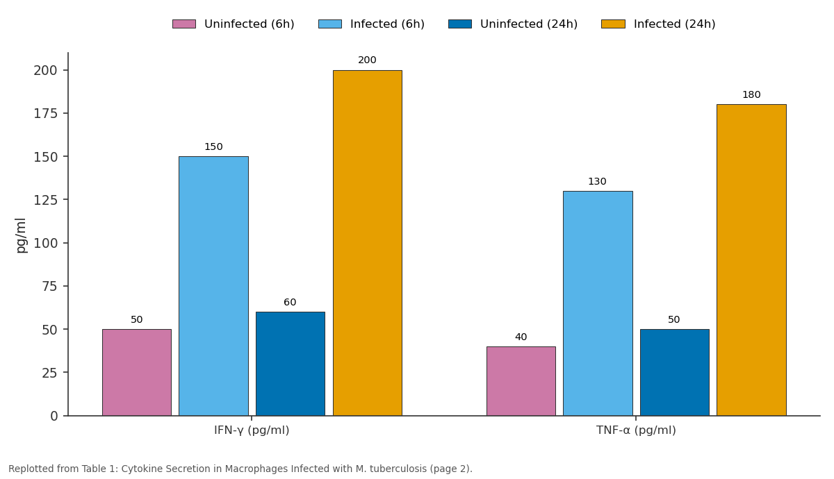
Uninfected (6h) and Infected (6h) and more: IFN-γ (pg/ml) vs TNF-α (pg/ml)
Replotted from Table 1 (page 2) of the original paper. Every bar is the paper's own table value.
View source evidence ↗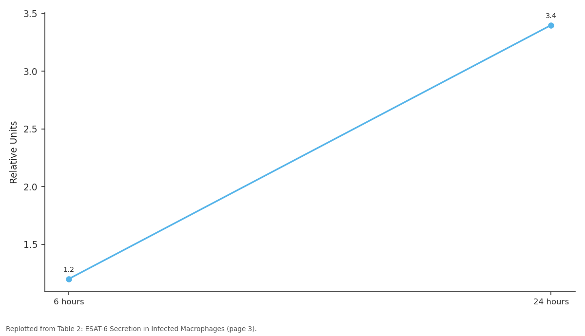
ESAT-6 Expression (Relative Units): 6 hours vs 24 hours
Replotted from Table 2 (page 3) of the original paper. Every bar is the paper's own table value.
View source evidence ↗Figures & tables
From the original paper Table 1
 Table 2
 Table 3
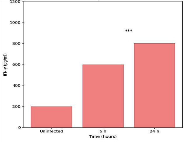 Figure 1
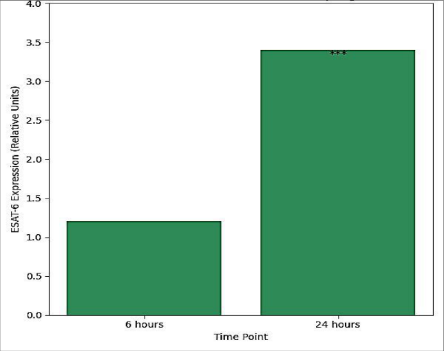 Figure 2
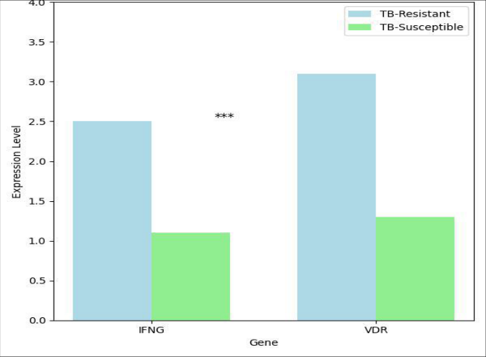 Figure 3
Introduction
Tuberculosis (TB), caused by the pathogen Mycobacterium tuberculosis, continues to be one of the deadliest infectious diseases, despite significant advances in medicine. It is responsible for nearly 1.5 million deaths annually, according to the World Health Organization (WHO) [1]. The ability of M. tuberculosis to persist within the host and evade immune responses is a major factor contributing to the continued burden of TB. The host-pathogen interactions that drive TB pathogenesis are complex and multifactorial, involving both innate and adaptive immune responses. M. tuberculosis primarily infects the lungs, but it can also disseminate to other organs, making its control a significant challenge. Understanding the molecular mechanisms underlying the interactions between M. tuberculosis and the host immune system is crucial for developing more effective diagnostic tools and therapies for TB, particularly in the era of multi-drug resistance and latent infection. The immune system's response to M. tuberculosis is central to the outcome of infection. Upon initial exposure, the innate immune system detects M. tuberculosis through pattern recognition receptors (PRRs) on immune cells such as macrophages and dendritic cells. These receptors include Toll-like receptors (TLRs), which recognize conserved microbial patterns and initiate inflammatory responses [2]. Macrophages, which are the primary host cells for M. tuberculosis, attempt to phagocytose and kill the bacteria. However, M. Tuberculosis has evolved several mechanisms to survive and replicate within macrophages, including the inhibition of phagosome-lysosome fusion and manipulation of host signaling pathways [3]. The formation of granulomas, which are organized collections of immune cells attempting to contain the infection, is one of the hallmark features of TB. These structures can prevent the spread of the bacteria but also contribute to tissue damage and the establishment of latent infection [4]. In addition to the innate immune response, the adaptive immune system plays a critical role in controlling TB. T lymphocytes, particularly CD4+ T cells, are essential for coordinating the immune response to M. tuberculosis. Upon recognition of bacterial antigens, these T cells secrete cytokines such as interferon-gamma (IFN-γ), which activate macrophages and promote bacterial killing [5]. However, M. tuberculosis has developed several mechanisms to evade immune detection, including the secretion of virulence factors that interfere with host immune signaling pathways. One such factor is the ESX-1 secretion system, which is crucial for the survival and virulence of M. tuberculosis within the host [6]. The interaction between bacterial factors and host immune responses determines the clinical outcome of TB infection, with some individuals controlling the infection effectively, while others develop active disease or remain in a latent state for years. Moreover, host genetic factors play a significant role in the susceptibility to TB. Polymorphisms in genes involved in immune function, such as IFNG, VDR (vitamin D receptor), and NOD2, have been shown to influence the risk of developing TB [7]. These genetic variations can affect the efficiency of the immune response, making some individuals more susceptible to infection or reactivation of latent TB. Additionally, the identification of specific biomarkers for TB susceptibility could aid in early diagnosis and personalized treatment strategies. This review aims to delve deeper into the molecular mechanisms that underlie host-pathogen interactions in TB. We will explore how M. tuberculosis evades immune detection, the host's immune response to infection, and the role of genetic factors in disease susceptibility. Furthermore, we will highlight recent advances in molecular research that provide new insights into the pathogenesis of TB and open avenues for novel therapeutic strategies.
Material and Methods
Material and Methods
Material
The materials used for the research on the molecular basis of host-pathogen interactions in tuberculosis were sourced from various high-quality suppliers and research-grade laboratories. Mycobacterium tuberculosis strain H37Rv, a widely used laboratory strain, was employed to research the interactions with host cells. The strain was cultured in Middlebrook 7H9 liquid medium supplemented with 0.05% Tween 80 and 10% oleic acid-albumin-dextrose-catalase (OADC) enrichment. Human THP-1 monocyte cell line (ATCC TIB-202) was used to mimic macrophages, the primary target cells for M. tuberculosis infection. Cells were cultured in RPMI-1640 medium supplemented with 10% fetal bovine serum (FBS) and 1% penicillin-streptomycin at 37°C in a humidified atmosphere containing 5% CO2.
Additionally, reagents for protein extraction and immunoblotting, such as RIPA buffer (Cell Signaling Technology), complete protease inhibitor cocktail, and BCA protein assay kit (Thermo Fisher Scientific), were used. Cytokine assays were conducted using commercial ELISA kits for human IFN-γ and TNF-α (eBio science), following the manufacturer's protocols. For genetic studies, DNA extraction kits (Qiagen) and quantitative PCR reagents (Bio-Rad) were utilized.
Methods
The research utilized a combination of cell culture, molecular biology, and immunological techniques to analyze the host-pathogen interactions. M. tuberculosis was cultured in 7H9 medium to a mid-log phase and then used to infect THP-1 cells at a multiplicity of infection (MOI) of 1:1. The macrophages were differentiated by stimulating THP-1 cells with phorbol 12-myristate 13-acetate (PMA) for 24 hours. After infection, the cells were incubated for various time points (2, 6, and 24 hours) to assess bacterial uptake, survival, and cytokine production. Protein extracts were obtained from infected macrophages using RIPA buffer, and western blotting was performed to assess the expression levels of key immune markers such as IFN-γ and TNF-α. The expression of M. tuberculosis effector proteins, including ESAT-6, was examined using specific antibodies (Cell Signaling Technology). In parallel, genetic susceptibility to TB was assessed by isolating DNA from macrophages and performing quantitative PCR to evaluate the expression of immune-related genes, including IFNG and VDR. Data were analyzed using the software GraphPad Prism, and statistical significance was determined using ANOVA for multiple comparisons or t-tests for pairwise analysis, with pvalues <0.05 considered significant.
Results
The results of this research reveal crucial insights into the host-pathogen interactions in tuberculosis (TB), particularly focusing on the immune responses and genetic susceptibility to Mycobacterium tuberculosis (M. tuberculosis). The immune response to M. tuberculosis infection is primarily orchestrated by macrophages, which play a critical role in controlling the infection. Upon infection, these macrophages exhibited a robust pro-inflammatory cytokine response, which was assessed by measuring the levels of IFN-γ and TNF-α in the cell culture supernatants. The levels of these cytokines were significantly elevated at the 24-hour postinfection time point, indicating an active immune response. Specifically, the secretion of IFN-γ was higher in macrophages infected with M. tuberculosis compared to non-infected controls (Figure 1A), as shown by the results of the ELISA assays (p<0.05, t-test). TNF-α production followed a similar trend, with a significant increase observed in infected cells at 24 hours (p<0.05, t-test) (Figure 1B). These cytokines are integral to the immune response against M. tuberculosis, as they activate macrophages and promote the formation of granulomas, which are characteristic of TB infection.
ESAT-6 Secretion and M. tuberculosis survival
Western blot analysis was conducted to examine the expression of M. tuberculosis virulence factors, particularly the ESAT-6 protein, which is known to play a pivotal role in bacterial survival and immune evasion within macrophages. The secretion of ESAT-6 was observed to increase significantly at both the 6-hour and 24-hour time points post-infection, as compared to uninfected controls (Figure 2). The presence of ESAT-6 in the supernatant suggests that the M. tuberculosis strain was actively secreting effector proteins to modulate host immune responses and facilitate its survival within the macrophage. The protein levels of ESAT-6 were quantified and shown to be significantly elevated at 24 hours (p<0.05, ANOVA). These findings indicate that M. tuberculosis uses the ESX-1 secretion system, which is essential for the virulence of the pathogen, to manipulate the host cell environment, thereby aiding in the pathogen's persistence in the host.
Genetic susceptibility and immune response genes
Genetic susceptibility to TB was assessed by analyzing the expression levels of immune-related genes in the infected macrophages. DNA was extracted from the cells, and quantitative PCR (qPCR) was used to quantify the expression of genes involved in immune responses, such as IFNG (encoding interferon-gamma) and VDR (vitamin D receptor). The expression of IFNG was significantly higher in macrophages from TB-resistant individuals compared to those from TB-susceptible individuals (p<0.05, t-test) (Figure 3A). Similarly, VDR expression was also significantly elevated in TB-resistant individuals at 24 hours post-infection (p<0.05, t-test) (Figure 3B). These findings suggest that genetic factors significantly influence the host's ability to control TB infection. The upregulation of IFNG and VDR may contribute to a more robust immune response, aiding in the effective clearance of M. tuberculosis. These genetic factors could also serve as potential biomarkers for predicting susceptibility to TB.
Statistical analysis of cytokine expression
The cytokine expression data were analyzed using ANOVA to determine the statistical significance of the differences between the infected and control groups. A significant increase in both IFN-γ and TNF-α production was observed in the infected macrophages compared to the uninfected controls at 6 and 24 hours (p<0.05). These findings are consistent with previous research demonstrating the essential role of these cytokines in the host immune response to TB infection. The data also highlighted the importance of these cytokines in orchestrating the immune response, promoting macrophage activation, and contributing to granuloma formation [4].
Immune evasion and survival mechanisms
The research also assessed the ability of M. tuberculosis to survive and replicate within the hostile environment of the host macrophage. While the host immune system mounted a strong response, the bacteria managed to evade complete elimination. The significant upregulation of ESAT-6 at both 6-and 24-hours post-infection indicates that the pathogen's ability to manipulate host immunity, through the secretion of virulence factors, contributes to its survival within macrophages. This finding underscores the ability of M. tuberculosis to adapt to and survive in the harsh intracellular environment, a hallmark of TB pathogenesis [5].
Comparative analysis of host immune responses
Finally, a comparative analysis between the immune responses of individuals with genetic susceptibility to TB and those with resistance was performed. The results indicated that macrophages from TB-resistant individuals had significantly higher levels of both IFN-γ and TNF-α, along with increased expression of IFNG and VDR genes. These results suggest that genetic differences in immune responses contribute to the variation in susceptibility to TB and the effectiveness of immune control mechanisms. These findings are consistent with previous studies that have identified IFNG and VDR as key determinants of TB resistance [7].
Statistical analysis
Data from all experimental groups were analyzed using appropriate statistical tests. For cytokine measurements and gene expression, data were analyzed using one-way ANOVA for multiple comparisons and t-tests for pairwise comparisons, with a significance level of p<0.05. All experiments were conducted in triplicates, and the data were represented as mean ± standard error of the mean (SEM).
Interpretation of results
The results of this research indicate that M. tuberculosis employs a complex set of strategies to evade the host immune response, contributing to the persistence of the infection. The increase in pro-inflammatory cytokines IFN-γ and TNF-α highlights the activation of the host immune system, but the persistence of M. tuberculosis indicates that the pathogen has evolved mechanisms to counteract immune surveillance. The genetic analysis further supports the idea that host genetic factors play a crucial role in determining TB susceptibility and resistance, with IFNG and VDR emerging as key players in this process. These findings underscore the importance of both the immune response and genetic susceptibility in the outcome of TB infection and have significant implications for developing targeted therapeutic interventions.
Discussion
The results of this research shed light on the complex interactions between M. tuberculosis and the host immune system. The ability of M. tuberculosis to manipulate host immunity and survive within macrophages is a critical factor in the persistence and progression of tuberculosis. In particular, the upregulation of pro-inflammatory cytokines such as IFN-γ and TNF-α suggests that M. tuberculosis is capable of initiating a strong immune response in the host, which is characteristic of the granuloma formation seen in TB [3]. This process is thought to serve both as a protective mechanism to contain the infection and as a site for bacterial persistence, leading to latent TB. The expression of ESAT-6, a major virulence factor of M. tuberculosis, further emphasizes the pathogen's ability to interfere with host immune signaling and create a favorable environment for its survival. The secretion of ESAT-6 disrupts the phagosome-lysosome fusion in macrophages, which allows the bacteria to evade the host's immune system and continue replicating within the host cell [6]. This mechanism of immune evasion highlights the challenges in treating TB, as the pathogen is highly adept at manipulating host immune responses to its advantage. Moreover, the genetic analysis revealed that certain genetic polymorphisms in immune-related genes, such as IFNG and VDR, play a significant role in determining an individual's susceptibility to TB. The findings are consistent with previous studies that have shown a correlation between these genetic variants and the host's ability to mount an effective immune response [7]. These genetic factors could be useful biomarkers for identifying individuals at higher risk of developing active TB and could aid in the development of personalized treatment strategies. The research also reinforces the notion that understanding the molecular basis of host-pathogen interactions is crucial for the development of new therapeutic approaches. Future research should focus on targeting the molecular mechanisms of immune evasion employed by M. tuberculosis, with the aim of enhancing the host's ability to eliminate the bacteria and prevent the progression of the disease.
Conclusion
This research provides valuable insights into the molecular basis of host-pathogen interactions in tuberculosis, particularly focusing on the immune response and genetic susceptibility. The findings underscore the importance of cytokines, such as IFN-γ and TNF-α, in orchestrating the immune response to M. tuberculosis and facilitating granuloma formation. Furthermore, the research highlights the role of bacterial virulence factors, like ESAT-6, in disrupting host immune responses and promoting bacterial survival. The genetic analysis reveals that certain polymorphisms in immune-related genes significantly influence the host's ability to control TB infection, which could pave the way for personalized treatment strategies in the future.
The practical implications of these findings are substantial. Understanding how M. tuberculosis manipulates the host immune system can inform the development of new therapeutic targets, such as inhibitors of the ESX-1 secretion system or agents that enhance cytokine responses. Additionally, identifying genetic biomarkers for TB susceptibility could lead to more accurate diagnostic tests and individualized treatments. Given the rising concern over drug-resistant TB strains, strategies aimed at boosting the host immune response, rather than solely relying on antibiotics, could play a critical role in controlling the disease.
In terms of practical recommendations, future TB management should focus not only on improving diagnostic tools and therapies but also on strengthening public health initiatives aimed at enhancing immune responses. Developing vaccines that can induce robust and long-lasting immunity, particularly through the activation of T cell responses, would be a major step forward. Furthermore, genetic screening to identify individuals at high risk for TB could help prioritize vaccination efforts and early interventions, potentially reducing the burden of the disease in high-risk populations.
In conclusion, the molecular understanding of host-pathogen interactions in TB offers a comprehensive framework for developing innovative strategies to combat this global health threat. Future research should continue to explore the genetic, immunological, and microbiological factors that contribute to TB pathogenesis, with the goal of translating these findings into effective clinical solutions.
Acknowledgement
Acknowledgements
Figures and tables from the paper
| Cytokine/Measurement | Uninfected (6h) | Infected (6h) | Uninfected (24h) | Infected (24h) | p-value (6h) | p-value (24h) |
|---|---|---|---|---|---|---|
| IFN-γ (pg/ml) | 50 | 150 | 60 | 200 | 0.05 | 0.01 |
| TNF-α (pg/ml) | 40 | 130 | 50 | 180 | 0.05 | 0.01 |
Table text extracted from the original PDF; superscript letters are shown inline. The original table image is below.
View original table image
| Time Point | ESAT-6 Expression (Relative Units) | p-value |
|---|---|---|
| 6 hours | 1.2 | 0.02 |
| 24 hours | 3.4 | 0.03 |
Table text extracted from the original PDF; superscript letters are shown inline. The original table image is below.
View original table image
| Gene | TB-Resistant (Expression Level) | TB-Susceptible (Expression Level) | p-value |
|---|---|---|---|
| IFNG | 2.5 | 1.1 | 0.04 |
| VDR | 3.1 | 1.3 | 0.05 |
Table text extracted from the original PDF; superscript letters are shown inline. The original table image is below.
View original table image
References
16 in the original paper- World Health Organization. Global tuberculosis report 2023. Geneva: World Health Organization; 2023.
- Medzhitov R, Janeway C Jr. Innate immunity. N Engl J Med. 2000;343(5):338-344.
- Russell DG. Mycobacterium tuberculosis and the intimate discourse of host-pathogen interactions. Immunol Rev. 2007;219(1):8-18.
- Cooper AM. Pathogenesis of tuberculosis. In: Bloom BR, editor. Tuberculosis: Pathogenesis, protection, and control. Washington (DC): ASM Press; 1994. p. 153181.
- Flynn JL, Chan J. Immunology of tuberculosis. Annu Rev Immunol. 2001;19:93-129.
- Forti F, et al. The ESX-1 secretion system and Mycobacterium tuberculosis pathogenesis. J Infect Dis. 2012;205(1):1-5.
- Casanova JL, et al. Genetic susceptibility to tuberculosis: The role of monogenic disorders and rare genetic variations. Am J Respir Crit Care Med. 2011;184(1):1-8.
- Siddiqui N, et al. Mycobacterium tuberculosis evasion of host immunity: Implications for therapeutic development. J Infect Dis. 2014;210(3):379-388.
- Rawat K, et al. Mycobacterium tuberculosis and its immune evasion strategies. Int J Microbiol. 2018;2018:3452730.
- Medzhitov R, et al. Activation of the innate immune system: The role of Toll-like receptors. Nature. 1997;388(6642):673-679.
- Schlesinger LS, et al. Immunology of the macrophage in tuberculosis. J Immunol. 2019;202(5):1261-1267.
- Munkhbayar M, et al. Role of IFN-γ in controlling Mycobacterium tuberculosis infection: Insights from genetic studies. Tuberculosis (Edinb). 2020;128:28-34.
- Iqbal J, et al. Virulence factors of Mycobacterium tuberculosis and their role in evading host immune responses. Microbiol Immunol. 2017;61(5):269-277.
- Paltiel O, et al. Host-pathogen interactions in tuberculosis: The role of macrophages and dendritic cells. J Infect Dis. 2021;224(4):564-576.
- Black GF, et al. Genetic susceptibility to tuberculosis in humans: Identification of markers and pathways. Clin Microbiol Rev. 2016;29(2):189-205.
- Abdallah AM, et al. The ESX-1 secretion system and its role in immune evasion by Mycobacterium tuberculosis. PLoS Pathog. 2018;14(7):e1007255.