
Research article · 2025 · Full text
International Journal of Biology SciencesIncidance and management of white muscardine disease in the silkworm (Bombyx mori L.)
Volume 7(8) · Pages 62–65 · DOI: 10.33545/26649926.2025.v7.i8a.449 ↗
Research areasSilkworms and Sericulture ResearchSilk-based biomaterials and applicationsBiological Stains and Phytochemicals
Abstract
Author-writtenWhite muscardine is a fungal infection frequently observed in silkworms, with higher incidence during the rainy and winter seasons. Diseases in silkworms occur throughout the year in all sericulture regions, affecting Bombyx mori L. Among the major diseases-grasserie, flacherie, muscardine, and microsporidiosis white muscardine is particularly severe. It is caused by the entomopathogenic fungus Beauveria bassiana, also referred to as mycosis. The disease mainly targets the larval and pupal stages, resulting in considerable losses to cocoon production. Favorable conditions for its development include low temperatures (below 25 °C) and high relative humidity (90-95%). Infected larvae generally die within four to five days, and within 24 hours their bodies become mummified and covered with white fungal spores. Occasionally, infected larvae manage to spin cocoons but die inside cocoon, preventing moth emergence. The pathogen spreads through spores released from infected cadavers, with environmental factors such as high humidity and low temperature facilitating its transmission within rearing trays. This study highlights the symptoms, epidemiology, and environmental factors contributing to the occurrence, spread of white muscardine disease and management.
Summary
Source-checkedPlain-language reading of the full paper, prepared with AI and checked against the text. Each point links to the authors' own words.
Why it matters
- The mulberry silkworm (Bombyx mori L.) is the main species used in sericulture to produce commercially valuable silk. View evidence ↗
- Diseases cause significant economic impact in India, with annual cocoon crop losses estimated at 30-40%, and white muscardine specifically contributing 10-40% of those losses. View evidence ↗
- White muscardine is a severe fungal disease caused by the entomopathogenic fungus Beauveria bassiana. View evidence ↗
The question
- The study aims to describe the symptoms, epidemiology, environmental drivers, and management strategies for white muscardine disease. View evidence ↗
What they did
- The researchers used newly ecdysed fourth instar silkworm larvae for their experiments. View evidence ↗
- Larvae were infected by spraying a conidial suspension of 1 × 10⁶ conidia/mL at a rate of 1 mL per 100 larvae. View evidence ↗
- The infected larvae were maintained in plastic trays at a temperature of 25 ± 1 °C and a relative humidity of 90–95%. View evidence ↗
- The fungal pathogen was cultured on Potato Dextrose Agar (PDA) using conidia collected from mummified larvae. View evidence ↗
What they found
- Symptoms of the white muscardine infection became visible four days after the larvae were inoculated. View evidence ↗
- Infected larvae typically exhibit vomiting and die within five days of infection. View evidence ↗
- Dead larvae mummify into chalky white structures within 48 hours of death. View evidence ↗
- Some infected larvae can successfully spin cocoons, but they fail to emerge as moths because of secondary infections during the pupal stage. View evidence ↗
- Microscopic analysis of haemolymph and mummified larvae confirmed the presence of mycelial hyphae and cylindrical blastospores. View evidence ↗
- Global crop losses attributed to white muscardine are reported to range from 5% to 50%. View evidence ↗
What it means
- The authors conclude that the disease can be effectively prevented through strict hygiene, the use of lime powder after each moult, and the application of bed disinfectants such as Vijetha and Ankush. View evidence ↗
- The authors recommend dusting beds with fungicides (such as Diethane M-45, captol, or bavistin) mixed with slaked lime powder at a rate of 5gm-ft² after each moult. View evidence ↗
Visual summary
1 visualsIllustrations explain the paper; they do not show study data.
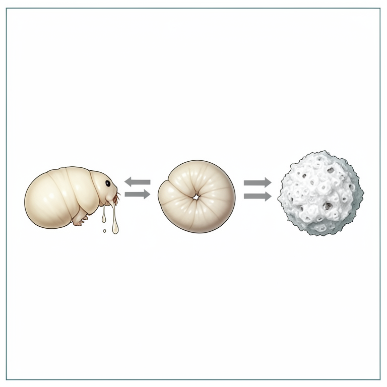
Timeline of White Muscardine Infection
AI-generated conceptual illustration (silkworm larvae (Bombyx mori)). It explains the idea and does not show study data.
View source evidence ↗Figures & tables
From the original paper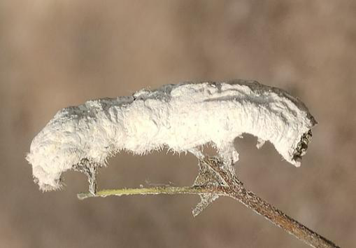 Figure 1
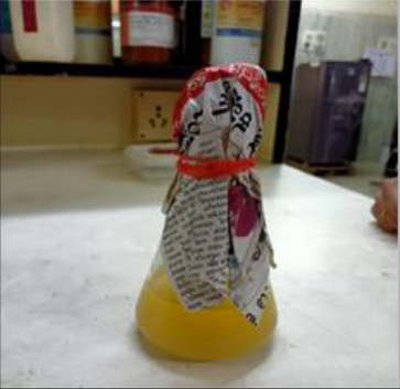 Figure 2
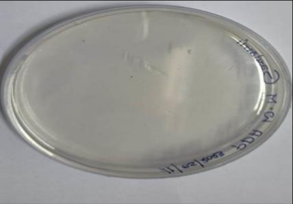 Figure 3
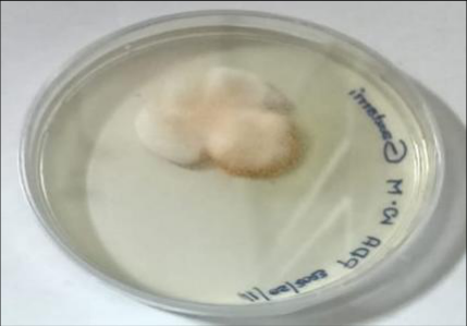 Figure 4
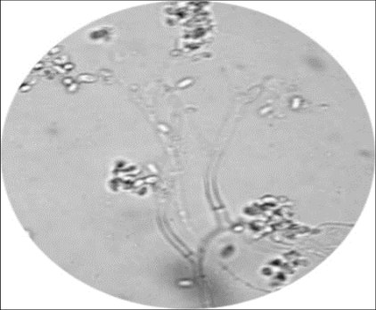 Figure 5
Introduction
Silk production represents the ultimate objective of sericulture, with the mulberry silkworm (Bombyx mori L.) being the primary species responsible for producing commercially valuable silk, known for its superior quality, elegance, and sheen (Nataraju et al., 2005) [14] . India holds the unique distinction of being the only country endowed with all four commercially exploited silkworm species-Mulberry, Eri, Muga, and Tasar. Among these, mulberry sericulture is predominantly practiced in states such as Karnataka, Andhra Pradesh, Tamil Nadu, Kerala, Assam, Bihar, Madhya Pradesh, Uttar Pradesh, Maharashtra, Punjab, Rajasthan, Gujarat, Odisha, Himachal Pradesh, and the northeastern states including Nagaland, Meghalaya, Mizoram, Arunachal Pradesh, and Tripura. Globally, approximately 95% of silk originates from mulberry, with China, India, and Japan ranking as the leading producers. However, diseases pose a major challenge to cocoon production, frequently leading to outbreaks and crop losses, particularly in tropical regions. The mulberry silkworm is susceptible to various pathogens. Dasgupta (1950) [2] identified the major silkworm diseases as Grasserie (caused by viruses), Flacherie (caused by bacteria), Muscardine (caused by fungi), and Pebrine (caused by protozoa/microsporidia). These diseases remain prevalent throughout the year across sericulture areas, causing significant larval mortality at different stages. In India, annual cocoon crop losses are estimated to range between 30-40% due to these diseases, with white muscardine alone accounting for 10-40% of the losses (Janakiraman, 1961) [4] . White muscardine is a fungal disease of silkworms primarily caused by the entomopathogenic fungus Beauveria bassiana. The term "muscardine" is derived from the Italian word "moscardino," meaning musk or grape confit, while "calcino" refers to the white powdery growth characteristic of this disease. The disease was first reported by the Italian entomologist Agostino Basi in 1763, who later, in 1835, formally described it. In Karnataka, it is locally known as Sunnakaddi or Sunnakattu roga (Janakiraman, 1961) [4] , and in West Bengal, it is referred to as Chuna-Kete.
There are various forms of muscardine, categorized based on the color of the conidia that develop on infected silkworm larvae, such as white, green, yellow, brown, and black. Over a thousand fungal species have been identified as muscardine pathogens (Yokohama, 1954) [18] . Among the four major microbial diseases of silkworms, muscardine (fungal mycosis) is considered the most lethal and destructive, as it is highly contagious and causes severe economic losses to cocoon production worldwide (Steinhaus, 1949) [17] . In India, the prevalence of white muscardine varies by region and season. It is particularly severe during the winter in Karnataka (Anonymous, 1975) [5] and during the rainy season in West Bengal (Mukherji, 1912) [11] . Seasonal and agro-climatic variations significantly influence disease outbreaks (Pringle, 1984) [15] . Low temperatures combined with high humidity create favorable conditions for disease development, especially during winter. Crop losses due to white muscardine can range from 5% to 50% across different countries (Jayaramaiah & Kuberappa, 1987) [10] . The fungus primarily infects third and fourth instar larvae, with symptoms becoming visible in the later stages of infection. However, it can affect all stages of the silkworm's life cycle. Farmers across various sericulture regions in India have frequently reported significant cocoon crop losses mainly attributed to this disease (Samson et al., 1990) [16] .
Materials and Methods
Materials and Methods
Experimental Materials
The study utilized Bombyx mori larvae, mulberry leaves as feed, conidia of the fungal pathogen Beauveria bassiana, analytical-grade chemicals, sterile glassware, and standard silkworm rearing apparatus.
Preparation of media, culture, stock dilution, and rearing techniques
Potato Dextrose Agar (PDA) medium was prepared by dissolving the required amount in double-distilled water. The medium was sterilized at 121 °C for 45 minutes in an autoclave and poured into sterile Petri dishes to solidify. Conidia were collected from mummified silkworm larvae using a sterile inoculating loop and cultured on the PDA plates. These plates were incubated at 25 °C to promote fungal growth. The culture was purified by the monohyphal tip method, and all procedures were carried out under aseptic conditions using a laminar airflow chamber. Fresh conidia of Beauveria bassiana were harvested from pure cultures and suspended in sterile distilled water to obtain the desired concentration. The conidial density was determined using a Neubauer haemocytometer following the method of Cantwell (1973). The experiment was conducted on newly ecdysed fourth instar larvae (first day after the third moult). A conidial suspension (1 × 10⁶ conidia/mL) was applied percutaneously by spraying 1 mL per 100 larvae. Treated larvae were reared in plastic trays lined with polyethylene sheets under controlled conditions (25 ± 1 °C temperature and 90-95% relative humidity), maintained using wet foam pads or moist paper folds, as described by Chandrashekaran and Nataraju (2008).
Disease Diagnosis:
For confirmation, a drop of the conidial suspension was placed on a glass slide, stained with lactophenol cotton blue, and examined under an electron microscope to observe germinating conidia. Fungal growth on PDA and microscopic conidia of B. bassiana were recorded.
Results and Discussion
A disease is an abnormal condition that arises due to physical or physiological disturbances, ultimately affecting the entire body system of the insect. Unfavorable environmental factors, such as high humidity and low temperature, create favorable conditions for pathogens to infect silkworms. These factors not only weaken the host but also promote the rapid multiplication of pathogens, thereby facilitating disease spread and progression. The primary source of pathogenic microorganisms is usually infected silkworms, and the intensity of the disease often increases due to secondary infections. During the experimental observations, progressive symptoms of infection were recorded daily following the treatment. Visible signs of disease appeared on the fourth day postinoculation. The fungal pathogen Beauveria bassiana infects the silkworm larvae through direct contact, with its conidia penetrating the integument of the host's body.
Visual Diagnosis
Infected silkworm larvae exhibit reduced appetite, sluggish behavior, and loss of movement. As the infection progresses, their bodies lose elasticity and develop moist, oily spots on the surface. The larvae may vomit and typically die within five days. Initially, the bodies of dead larvae remain soft, but they gradually stretch, become rubbery, harden, and eventually mummify as white fungal conidia cover the surface. Within 48 hours of death, the larvae turn into chalky white structures. The mummified stage is highly infectious, as the entire body, except for the chitinous head region, is enveloped in white mycelial growth that releases millions of conidia. Unlike larvae affected by grasserie, flacherie, or pebrine, mummified larvae remain hard and do not decay or emit odor. Some infected larvae that survive may spin cocoons but fail to emerge as moths due to secondary infections occurring during the pupal stage. Infected pupae respond sluggishly to external stimuli, die inside the cocoon, and display shriveled thoraxes, wrinkled abdomens, and bodies covered with aerial white hyphae. These pupae fail to emerge as adult moths.
Microscopic Diagnosis
Haemolymph and mummified larval samples were examined microscopically. Samples were stained with lactophenol cotton blue and observed under an electron microscope. The examination revealed the presence of mycelial hyphae and cylindrical blastospores branching from the conidia.
Causative Agent and Transmission
White muscardine is caused by the entomopathogenic fungus Beauveria bassiana (Bals.) Vuillemin, a pathogen known for its global distribution. The fungus infects silkworms through contamination of the body surface, followed by direct penetration of the germ tube. In young larvae, the disease progresses rapidly, whereas in mature larvae, it takes a chronic course. Transmission primarily occurs through spores produced on the dead larvae, which subsequently infect healthy worms. Infection can also spread through contaminated food, rearing appliances, or direct contact between infected and healthy worms. Low temperatures and high humidity significantly favours the spread of the disease. The pathogens may also be excreted by infected larvae, contributing to secondary infections in the rearing environment.
Epidemiology
Numerous studies conducted in India and other sericulture regions have confirmed that white muscardine is the most prevalent form of muscardine. The disease spreads more readily during periods of high humidity and low temperatures, conditions often present during rainy and winter seasons. Major sources of infection include mummified larvae, alternate insect hosts, contaminated mulberry leaves, and unclean rearing equipment.
Management and Control
Effective management of white muscardine requires strict hygiene practices, including thorough disinfection of rearing houses, appliances, and surrounding areas. Maintaining optimal temperature, humidity, and ventilation, along with proper bed spacing and regular cleaning, significantly reduces disease incidence. Supplying nutritious mulberry leaves and reducing humidity through heaters during damp seasons are also beneficial. The rapid spread of the disease is attributed to its wide host range, high sporulation rate, and poor management practices. To combat this, integrated disease control technologies have been developed, such as those introduced by CSR&TI, Mysore, which involve the use of chlorine dioxide, Anukush, and Vijetha. Regular application of bed disinfectants like Vijetha and Ankush, as well as the use of lime powder after each larval moult, coupled with maintenance of proper hygiene, effectively prevents the outbreak and spread of white muscardine.
Bed Disinfectants: Disinfectants play a key role of managing and controlling fungal diseases:
For Muscardine Control: Using sulfur, formalin chaff, and lime can help manage this disease. Fungicides: Products like Diethane M-45, captol, bavistin are mixed with slaked lime powder can be dusted on the bed (@ 5gm-ft²) in measured amounts after each moult. Bed Disinfectants: Products like RKO, Vetcare Vijetha Supplement, Ankush, Sanjeevani, Shakthi, Resham Jyothi, Labex, and Sericillin are effective in disease management. Preventing Fungal Entry: Ensuring that fungal pathogens do not enter the rearing room is critical. Controlling Alternate Hosts: Managing alternative hosts of fungal pathogens is vital in reducing the incidence of muscardine.
Acknowledgements
The author wishes to her gratitude to the Andhra Pradesh State Sericulture Research and Development Institute, Kirikera, Hindupur for providing lab facilities.
Figures and tables from the paper
References
18 in the original paper- Cantwell GE. Methods for determining the level of Nosema infection in honeybees. In: Cantwell GE, editor. Insect diseases. No. 2. New York: Marcel Dekker; 1973, p. 539-542.
- Dasgupta MR. Diseases of silkworm. Monograph on Cottage Industries, No 1. Calcutta: Government of India Press; 1950.
- Ishikawa Y, Miyajima S. Spread of the infectious flacherie in rearing trays of silkworm, Bombyx mori L. Appl Entomol Zool. 1964;8:86-88.
- Janakiraman AT. Diseases affecting the Indian silkworm races. J Silkworm. 1961;13:91-101.
- Anonymous. Annual report. Mysore: CSRTI; 1975, p. 89-92.
- Anonymous. Annual report 1991-1992. Mysore: CSRTI; 1992, p. 54.
- Bassi A. Del mal del sengno, calcinaccio o moscardino, malattia che affligge i bachi da seta e sul modo di liberarne le bigattaie anche le più infestate. Parte I, Teoria. Lodi: Orcesi; 1835, p. 1-67.
- Bulmer GS, Formtlgin RA. Pathogenic mechanism of mycotic agents. In: Howard D, editor. Fungi pathogenic for humans and animals. Part B. New York: Marcel Dekker; 1983, p. 259-266.
- Jayramaiah M, Kuberappa GC, Devaiah MC, Kalikal Y. White muscardine disease of silkworm and its management. Indian Silk. 1986;25(8):15-16.
- Jayaramaiah M, Kuberappa GC. Silkworm mycoses. AS Tech Ser. 1987;48:15-6. Bangalore: University of Agricultural Sciences; 1987, p. 85.
- Mukherji NG. Handbook of sericulture. Calcutta: Bengal Secretariat Book Depot; 1912, p. 74-86.
- Nataraju B, Datta RK. Application of textile dye-based dipstick immune diagnostic kit for management of infectious flacherie in silkworm rearing. In: Proceedings of XVII International Sericultural Commission Congress, Cairo, Egypt; 1999 Nov 23-27, p. 283-8.
- Nataraju B, Balavenkatasubbaiah M, Sharma SD, Selvakumar T, Thiagrajan V, Datta RK. A practical technology for diagnosis and management of diseases in silkworm rearing. Int J Indust Entomol. 2002;4(2):169-173.
- Nataraju B, Sathyaprasad K, Manjunatha D, Aswani Kumar C. A textbook on silkworm crop protection. Bangalore: Central Silk Board; 2005.
- Pringle Jameson A. Report on the diseases of silkworm in India. 1984, p. 1-78.
- Samson MV, Baig M, Sharma SD, Balavenkatasubbaiah M, Sasidharan TO, Jolly MS. Survey on the relative incidence of silkworm diseases in Karnataka, India. J Seric. 1990;29(2):248-254.
- Steinhaus EA. Principles of insect pathology. New York: McGraw-Hill; 1949.
- Yokohama T. Synthesised science of sericulture. Translated by Central Silk Board, India. Mumbai: Central Silk Board; 1962, p. 398.