
Research article · 2026 · Full text
International Journal of Biology SciencesMolecular detection of Buxtonella sulcata in Buffalo calves (Bubalus bubalis) from Orathanadu, Tamil Nadu
Volume 8(1) · Pages 107–110 · DOI: 10.33545/26649926.2026.v8.i1b.552 ↗
Research areasHelminth infection and controlProtist diversity and phylogenyMicrobial infections and disease research
Abstract
Author-writtenBuxtonella sulcata are intestinal ciliates commonly found in ruminants, yet their diagnosis is often complicated by morphological similarities with other ciliate genera. In the present study, Buxtonella cysts were detected in 44% (n=11) of fecal samples collected from buffalo calves in Orathanadu. Microscopically, the cysts observed were spherical, thick walled with kidney-shaped macronucleus and rounded micronucleus and measured 68–78 μm in length. The infection intensities ranged from 600 to 1000 cysts per gram of faeces. Due to the close morphological resemblance between Buxtonella and Balantidium cysts. Molecular identification and confirmation were carried out by extracting DNA from morphologically identified Buxtonella spp. positive faecal samples, followed by specific PCR targeting the ITS1–5.8S rRNA–ITS2 region of Buxtonella spp. A 530 bp amplicon was obtained from all 11 positive samples. One representative amplicon was randomly subjected for Sanger sequencing, and contigs were assembled using BioEdit v7.0.9 software. Nucleotide sequence analyses revealed that the Orathanadu isolate shared 94.1–100 % sequence similarity with previously published Buxtonella isolates from buffaloes and cattle. Phylogenetic analysis revealed that the Orathanadu isolate clustered closely with previously reported B. sulcata from buffaloes and cattle. This study provides molecular evidence for the presence of B. sulcata infection in buffalo calves and highlights the importance of molecular diagnostics for accurate species-level identification.
Summary
Source-checkedPlain-language reading of the full paper, prepared with AI and checked against the text. Each point links to the authors' own words.
Why it matters
- Buxtonella sulcata is a ciliate commonly found in the large intestines of domestic ruminants like cattle and buffaloes. View evidence ↗
- While usually commensal, these organisms can become opportunistic pathogens causing weight loss, diarrhea, and clinical enteritis during stress or poor hygiene. View evidence ↗
- Diagnosing Buxtonella is difficult because it looks morphologically similar to other ciliate genera. View evidence ↗
The question
- The study aimed to detect and molecularly identify Buxtonella cysts in buffalo calves from Orathanadu. View evidence ↗
What they did
- Researchers collected 25 fecal samples in June 2025 from diarrheic buffalo calves aged 2 months to 1 year in Orathanadu, Tamil Nadu. View evidence ↗
- Samples were analyzed using microscopy (direct wet mount, sedimentation, and flotation) and infection intensity was measured via the McMaster egg counting method. View evidence ↗
- PCR was used on 11 morphologically positive samples to amplify the ITS1-5.8S rRNA-ITS2 region using specific B5D and B5R primers. View evidence ↗
- One random PCR-positive amplicon was sequenced using Sanger sequencing to confirm the species identity. View evidence ↗
What they found
- The identified cysts were spherical and thick-walled, measuring 68-78 μm in length with a rounded micronucleus and kidney-shaped macronucleus. View evidence ↗
- Infection intensity in the positive samples ranged between 600 and 1000 cysts per gram of feces. View evidence ↗
- All 11 morphologically positive samples yielded a PCR amplicon of approximately 530 bp for the ITS1-5.8S rRNA-ITS2 region. View evidence ↗
- The Orathanadu isolate showed 94.1-100% nucleotide identity with previously reported Buxtonella isolates from cattle and buffaloes. View evidence ↗
- Maximum Likelihood phylogenetic analysis confirmed the isolate as B. sulcata, with bootstrap support over 90%. View evidence ↗
What it means
- The authors conclude that molecular diagnostics should be integrated into routine parasitological exams to ensure accurate differential diagnosis of protozoan infections. View evidence ↗
- The authors recommend continued surveillance and molecular characterization to better understand the host specificity and pathogenic potential of Buxtonella species. View evidence ↗
Visual summary
1 visualsIllustrations explain the paper; they do not show study data.
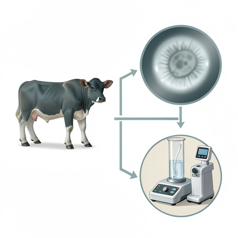
Diagnostic Workflow Comparison
AI-generated conceptual illustration (Buffalo calves (Bubalus bubalis) and Buxtonella sulcata ciliates). It explains the idea and does not show study data.
View source evidence ↗Figures & tables
From the original paper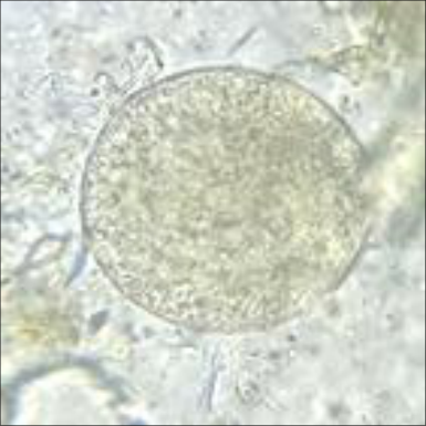 Figure 1
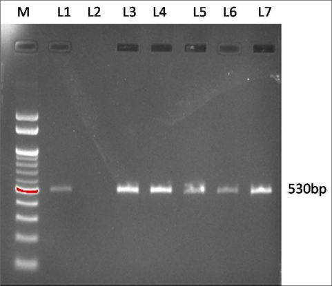 Figure 2
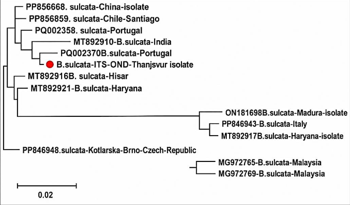 Figure 3
Introduction
Buxtonella species are intestinal ciliates belonging to the phylum Ciliophora, class Litostomatea, and family Balantidiidae, and are closely related to Balantidium coli, a parasite that infects humans and pigs [1] . These are large cyst-forming ciliates that infect ruminants and monkeys. Among domestic ruminants, Buxtonella sulcata is commonly reported in the large intestine of cattle and buffaloes [2] . These ciliates are generally regarded as commensal organisms inhabiting the caecum and colon; however, under conditions of stress, poor hygiene, or concurrent infections, they may become opportunistic pathogens leading to clinical enteritis, diarrhea, and weight loss [3,4] . The life cycle of B. sulcata comprises two stages: the trophozoite and the cyst. The trophozoite is the motile, feeding, and replicative stage found in the large intestine, while the cyst is the resistant, infective stage excreted with feces. Infection occurs through the fecal-oral route by ingestion of food or water contaminated with cysts [5] . Once ingested, excystation occurs in the host intestine, releasing trophozoites that colonize the large bowel. The prevalence of Buxtonella infection in buffaloes varies widely across regions and is influenced by climatic factors, husbandry practices, and the health status of animals. Studies from tropical and subtropical regions, including India, have reported prevalence rates ranging from 5% to 40% in cattle and buffaloes [2,6] . Although infections are often subclinical, Buxtonella has been associated with gastrointestinal disturbances and diarrhea, particularly in young or debilitated animals [7] . The severity of infection tends to increase in immunocompromised animals, resulting in chronic health complications, reduced production performance, and mortality in severe cases [8] .
Despite being reported for decades, Buxtonella infections in buffaloes remain under diagnosed due to their morphological resemblance to other intestinal protozoa, such as B. coli, and the lack of molecular diagnostic tools in routine veterinary parasitology [1]. So far, B. sulcata infection is not reported in humans. Given the economic importance of buffaloes as dairy and draught animals in many developing countries, understanding the epidemiology, pathogenic potential and control of Buxtonella infection is crucial for maintaining animal health and productivity.
Materials and Methods
Sample collection: A total of 25 faecal samples were collected from buffalo calves aged 2 months to 1 year during June 2025, all of which had a history of diarrhoea, at the Livestock Farm Complex, Veterinary College and Research Institute, Orathanadu, Tamil Nadu, southern India.
Faecal examination
The faecal samples were processed direct wet mount, sedimentation and flotation technique by standard protocols [3] . The samples were examined under microscopy and the parasite stages were identified based on their morphological characters [4] . The intensity of infection in positive fecal sample was measured using the McMaster egg counting method [9] .
Genomic DNA Isolation
Faecal samples positive for Buxtonella cysts were fixed in 70% ethanol. Total genomic DNA was extracted using the QIAamp DNA Stool Mini Kit (Qiagen, Germany) according to the manufacturer's instructions. Prior to extraction, cystpositive faecal samples were resuspended in 400 µL of lysis buffer (100 mM Tris-HCl [pH 8.0], 25 mM EDTA, 10% SDS), subjected to freeze-thaw cycles by freezing in liquid nitrogen for 3 min and thawing at 37 °C for 3 min. DNA concentration and purity were assessed by spectrophotometry (Nanodrop), and the extracted DNA was stored at -20 °C.
PCR amplification
PCR was performed on all 11 morphologically positive samples to amplify the internal transcribed spacer (ITS1-5.8S rRNA-ITS2) region of Buxtonella species using the published forward primer B5D (5′-GCT CCT ACC GAT ACC GGG T-3′) and reverse primer B5R (5′-ATA TGC TTA AGT TCA GCG GG-3′) as described by Ponce-Gordo et al. [10] . The reaction was carried out in a total volume of 20 µL, comprising 6 µL of nuclease-free water, 10 µL of Ampliqon Master Mix, 1 µL each of forward and reverse primers (20 pmol/µL), and 2 µL of template DNA. PCR amplification was performed under the following thermal cycling conditions: initial denaturation at 94 °C for 10 min; 30 cycles of denaturation at 94 °C for 1 min, annealing at 60 °C for 1 min, and extension at 72 °C for 1 min; followed by a final extension at 72 °C for 5 min. Amplified products were confirmed by electrophoresis on a 1.5% agarose gel using a submarine horizontal electrophoresis unit (Bio-Rad, USA).
Sequence analysis
One PCR-positive amplicon was randomly selected and subjected to Sanger sequencing at Eurofins Genomics Pvt. Ltd. (Bengaluru, India) to confirm the identity of Buxtonella species. The obtained nucleotide sequences were analyzed using BLAST and Gene Tool software. Sequences generated in the present study were aligned with reference Buxtonella sequences retrieved from the GenBank database using ClustalX v2.1. Phylogenetic analysis was performed using the Maximum Likelihood method with 1,000 bootstrap replicates in MEGA v7.0.7 to confirm species identity.
Results
In the present study, faecal examination revealed that 44% (n=11) of the faecal samples from buffalo calves were positive for Buxtonella cysts. Microscopically, the cysts observed were spherical, thick-walled with a kidney-shaped macronucleus and a rounded micronucleus, and measured 68-78 μm in length (Fig. 1). The cysts were morphologically similar to those of Balantidium species. The infection intensities in the positive faecal samples ranged from 600 to 1000 cysts per gram of faeces. The marked morphological similarity of cysts between the genera Buxtonella and Balantidium necessitated the use of molecular tools for accurate species identification. The ITS1-5.8S rRNA-ITS2 region of Buxtonella was successfully amplified in all 11 samples that were morphologically identified as Buxtonella species yielding an amplicon of approximately 530 bp, as visualized on a 1.5% agarose gel (Fig. 2). One randomly selected amplified product from the Orathanadu isolate was sequenced, and its phylogenetic relationship with previously published isolates was analyzed. BLAST analysis revealed that the ITS1-5.8S rRNA-ITS2 sequence of the Orathanadu isolate shared 94.1-100% nucleotide identity with isolates reported from buffaloes and cattle. Phylogenetic analysis using the Maximum Likelihood (ML) method demonstrated that the Orathanadu isolate clustered with published B. sulcata sequences with bootstrap support exceeding 90% (Fig. 3), thereby confirming molecular positivity.
Discussion
The present study confirmed the occurrence of Buxtonella sulcata infection in buffalo calves based on both morphological and molecular evidence. Microscopically, the cysts observed were spherical, thick-walled, and measured 68-78 μm, which closely corresponded with earlier descriptions of B. sulcata cysts reported in cattle and buffaloes [11]. However, reliance on cyst morphology alone had limited diagnostic value due to its strong resemblance to B. coli, frequently resulting in misidentification during routine fecal examinations [9,12] . Consequently, molecular techniques provided more reliable diagnostic tools, enabling accurate differentiation between these two ciliates and ensuring species-level confirmation [13] . In India, several studies had documented the morphological identification and prevalence of B. sulcata infection in cattle and buffaloes [14, 15] . Notably, Kalkal and Sangwan [15] reported an overall prevalence of 54.5% in buffaloes from the Hisar district of Haryana, while Maharana et al. [14] recorded prevalence rates of 4.57% and 15.79% in cattle and buffaloes, respectively, from Gujarat. Kumar et al. [8] reported an incidence of 35% from buffaloes of Gujarat. The variations in the percentages of infection could be due to many factors, such as environmental conditions, animal, farm management practices and stress factors.
In the present study, the infection intensity observed in faecal samples (600-1000 cysts/g) reflected a substantial cyst burden in calves. High cyst output had been associated with diarrheic conditions and poor nutritional status in young ruminants, although clinical manifestations varied depending on host immunity and the presence of co-existing enteric pathogens [16] . A previous study reported that animals exhibiting diarrheal symptoms had significantly higher infection rates than those with normal feces [12] . In animals with B. sulcata infection intensities exceeding 500 cysts per gram of feces, the frequency of diarrhea distinctly increased, and when intensities exceeded 2000 cysts per gram of feces, diarrhea frequency reached 100% [9] . B. sulcata has been recognized as an opportunistic pathogen in ruminants and its infections were reported to be highly prevalent among cattle and buffaloes worldwide [17] . Molecular characterization using the ITS1-5.8S rRNA-ITS2 region proved critical for species identification. The amplicon size (~530 bp) obtained in this study corresponded well with previous reports on Buxtonella ITS sequences from cattle and buffaloes [13,18] . ITS regions were known for their hypervariability, offering higher discriminatory power than conserved ribosomal genes and enabling clear differentiation among ciliate species [5] . The high sequence similarity (94.1-100%) between the Orathanadu isolates and published Buxtonella sequences supported the classification of these isolates within the Buxtonella genus. Phylogenetic analysis further demonstrated that the isolates clustered with Buxtonella sulcata from buffaloes and cattle, in agreement with earlier findings that ruminant Buxtonella species share a close evolutionary relationship despite host differences [1,18]. This close genetic relatedness suggests either crossspecies transmission or shared ancestral lineages within domestic ruminants. These findings contribute to the existing knowledge on protozoan infections in buffaloes and emphasize the importance of integrating molecular diagnostics into routine parasitological examinations and differential diagnosis. Accurate species-level molecular identification is particularly critical in regions where buffaloes are of significant economic importance and where mixed infections with other enteric pathogens are common. Continued surveillance and molecular characterization will help elucidate the epidemiology, host specificity and pathogenic potential of Buxtonella spp. in livestock populations. Such molecular data not only enhance diagnostic accuracy but also provide valuable insights into host specificity, geographical distribution, and the potential zoonotic importance of the parasite.
Acknowledgement
AcknowledgementWe are grateful to the Department of Biotechnology, Government of India, New Delhi for providing financial support for this research work.
References
16 in the original paper- Ponce-Gordo F, Martínez-Díaz RA. Taxonomy and phylogeny of Balantidium, Buxtonella and related ciliates. Protist. 2010;161(1):1-10.
- Radhakrishnan S, Nair SN, Mini M. Prevalence of Buxtonella sulcata infection in cattle and buffaloes. J Vet Parasitol. 2010;24(2):151-153.
- Ponce-Gordo F, Fonseca-Salamanca F, Martínez-Díaz RA. Genetic heterogeneity in internal transcribed spacer region of Balantidium coli and related species. Parasitol Res. 2011;108(4):979-987.
- Pradhan S, Singh A, Sahu S. Incidence of gastrointestinal protozoa in bovines of Odisha. Indian J Anim Res. 2019;53(5):665-668.
- Gupta P, Singla LD, Singh NK. Parasitic infections in dairy buffaloes: occurrence and management strategies. J Parasit Dis. 2018;42(3):446-452.
- Kumar B, Maharana BR, Prashad A, Joseph JP, Patel BR. Incidence of Buxtonella sulcata in Jaffrabadi buffaloes of south-western Gujarat, India. Buffalo Bull. 2017;36(4):623-628.
- Tomczuk K, Kurek L, Stec A, Studzinska M, Mochol J. Incidence and clinical aspects of colon ciliate Buxtonella sulcata infection in cattle. Bull Vet Inst Pulawy. 2005;49(1):29-33.
- Ponce-Gordo F, Jimenez-Ruiz E, Martinez-Diaz RA. Tentative identification of Balantidium species from ostriches (Struthio camelus) as Balantidium coli-like by polymorphic DNA analysis. Vet Parasitol. 2008;157:41-49.
- Timoney JF, Gillespie JH, Scott FW, Barlough JE. Hagan and Bruner's microbiology and infectious diseases of domestic animals. Ithaca (NY): Cornell University Press; 2017.
- Ganai A, Parveen S, Kaur D, Katoch R, Yadav A, Godara R, et al. Incidence of Buxtonella sulcata in bovines in R.S. Pura, Jammu. J Parasit Dis. 2015;39(3):446-447.
- Pomajbíková K, Oborník M, Horák A, Petrželková KJ, Grim JN, Modrý D. Buxtonella sulcata (Ciliophora, Trichostomatidae) from cattle and water buffalo: morphological and molecular comparison of isolates. Vet Parasitol. 2013;196(3-4):366-371.
- Maharana BR, Kumar B, Sudhakar NR, Behera SK, Patbandha TK. Prevalence of gastrointestinal parasites in bovines in and around Junagadh, Gujarat. J Parasit Dis. 2016;40(4):1174-1178.
- Kalkal H, Sangwan AK. Epidemiological investigations of Buxtonella sulcata in buffaloes of Hisar district, Haryana. J Entomol Zool Stud. 2019;7(6):1196-1199.
- Daugschies A, Najdrowski M. Eimeriosis in cattle: current understanding. J Vet Med. 2005;2(10):417-427.
- Huang CC, Wang LC, Pan CH, Yang CH, Lai CH. Investigation of gastrointestinal parasites of dairy cattle around Taiwan. J Microbiol Immunol Infect. 2014;47:70-74.
- Da Costa AC, de Almeida JC, de Carvalho AL. Molecular characterization of Buxtonella sulcata from cattle based on ITS rDNA sequences. Parasitol Res. 2019;118:155-162.