
Research article · 2026 · Full text
International Journal of Advanced Chemistry ResearchAntioxidant and Antibacterial Efficacy of Silver Nanoparticles Synthesized from Extract of Filamentous Cyanobacterium Scytonema sp
Volume 8(1) · Pages 23–27 · DOI: 10.33545/26646781.2026.v8.i1a.354 ↗
Research areasNanoparticles: synthesis and applicationsAlgal biology and biofuel productionAdvanced Nanomaterials in Catalysis
Abstract
Author-writtenContemporary algal based metallic nanoparticles with magnified biological potential have gained considerable attention. In the present study, silver nanoparticles (AgNPs) were synthesised by using cell-free extracts of a filamentous cyanobacterium Scytonema sp., isolated from the rooftop of Acharya Narendra Deo Kisan P.G. College, Babhnan, Gonda. The Scytonema sp. was harvested during the late exponential growth phase to advance the efficiency of nanoparticle synthesis. The distinct change in color and the emergence of a specific surface plasmon resonance (SPR) peak at 410 nm in the UV-visible spectrum was evidenced of AgNPs synthesis after 60 h of incubation. The antioxidant potential of AgNPs, including DPPH and ABTS radical scavenging methods, revealed quite similar pattern and significantly higher activity (about two folds higher than that of crude extract at 80 μm/mL concentration). Furthermore, the AgNPs exhibited pronounced, dose-dependent antibacterial activity against Escherichia coli (E. coli). The findings of present study point up the Scytonema sp. as an effective natural resource for the green synthesis of AgNPs with promising antioxidant and antibacterial utilization in biomedical and ecological spheres.
Summary
Source-checkedPlain-language reading of the full paper, prepared with AI and checked against the text. Each point links to the authors' own words.
Why it matters
- Conventional chemical and physical methods for synthesizing metallic nanoparticles can pose health hazards to humans and other living organisms. View evidence ↗
- There is a scientific push toward non-toxic, eco-friendly biosynthetic methods that use renewable resources and less energy. View evidence ↗
- Cyanobacteria are considered strong candidates for nanoparticle synthesis due to their high biomass productivity and growth rates. View evidence ↗
The question
- The study aimed to synthesize silver nanoparticles (AgNPs) using cell extracts from the cyanobacterium Scytonema sp. and evaluate their antibacterial and antioxidant activities. View evidence ↗
What they did
- Scytonema sp. was isolated from a rooftop in Gonda and grown in nitrogen-free BG-11 medium at 26 ± 2 °C with a 14:10 hour light/dark cycle. View evidence ↗
- Cells were harvested during the late exponential phase, centrifuged at 6000 rpm for 15 minutes, and sonicated at 30% amplitude for 15 minutes to prepare the extract. View evidence ↗
- The cell extract supernatant was mixed with 1 mM AgNO3 in 10 mL reaction mixtures and incubated at 37 °C for 60 hours. View evidence ↗
- Antioxidant activity was measured using DPPH and ABTS assays at concentrations of 20, 40, and 80 µg/mL. View evidence ↗
- Antibacterial efficacy was tested against E. coli using the disc diffusion method with AgNP concentrations of 5 µg/mL and 15 µg/mL. View evidence ↗
What they found
- The synthesis of stable AgNPs was confirmed by a visual color change to dark red and a surface plasmon resonance (SPR) peak at 410 nm. View evidence ↗
- The biosynthesized AgNPs exhibited a dose-dependent antibacterial effect against E. coli, with inhibition zones increasing as the concentration of AgNPs increased. View evidence ↗
What it means
- The authors conclude that Scytonema sp. is an effective, eco-friendly natural resource for synthesizing AgNPs with potential applications in pharmaceuticals. View evidence ↗
Visual summary
3 visualsIllustrations explain the paper; charts are replotted from the paper's own table values.
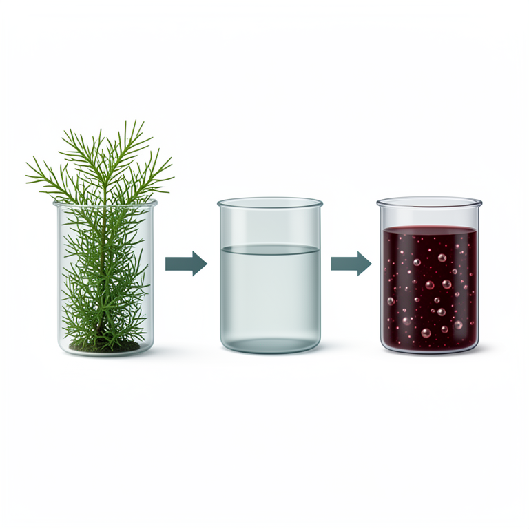
Green Synthesis Workflow
AI-generated conceptual illustration (Filamentous cyanobacterium Scytonema sp. and silver nanoparticles). It explains the idea and does not show study data.
View source evidence ↗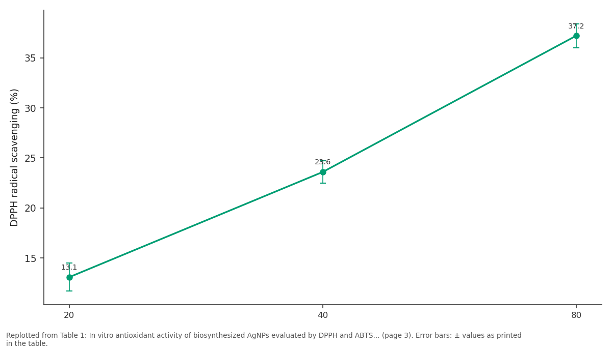
DPPH radical scavenging (%): 20 vs 40 vs 80
Replotted from Table 1 (page 3) of the original paper. Every bar is the paper's own table value.
View source evidence ↗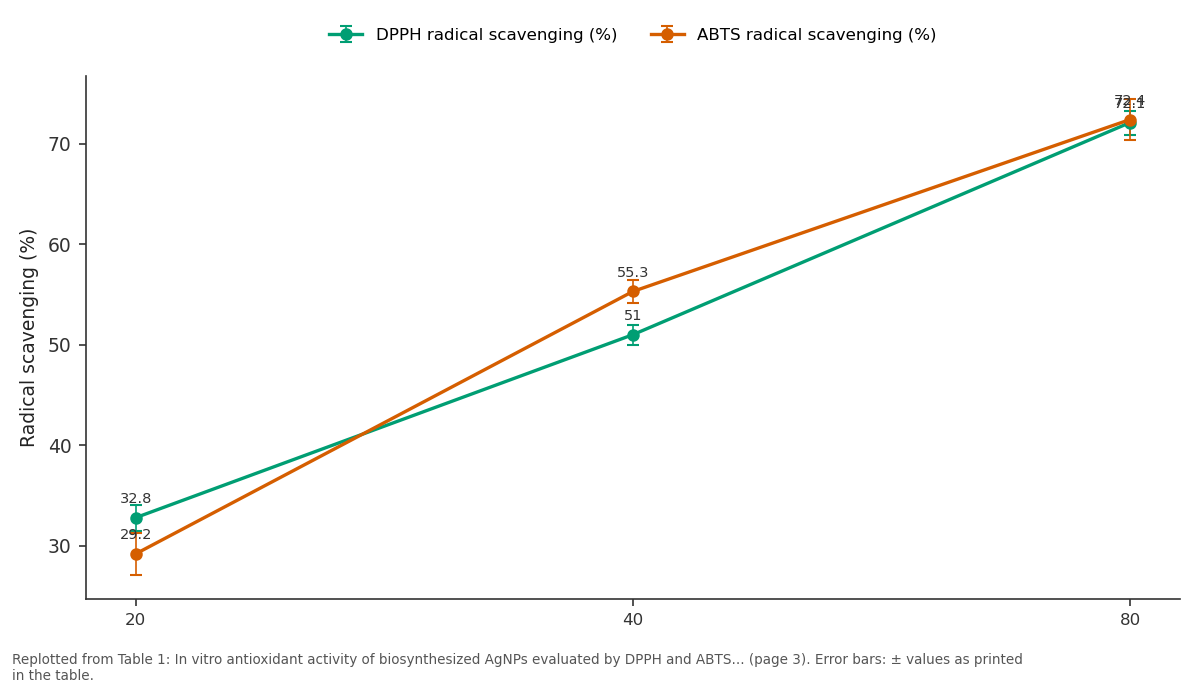
DPPH radical scavenging (%) and ABTS radical scavenging (%): 20 vs 40 vs 80
Replotted from Table 1 (page 3) of the original paper. Every bar is the paper's own table value.
View source evidence ↗Figures & tables
From the original paper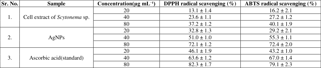 Table 1
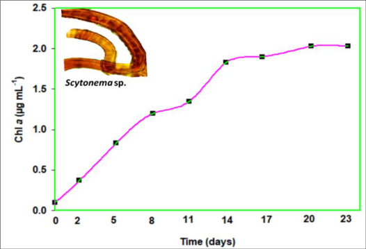 Figure 1
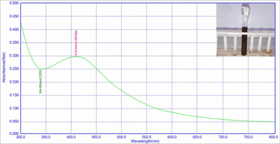 Figure 2
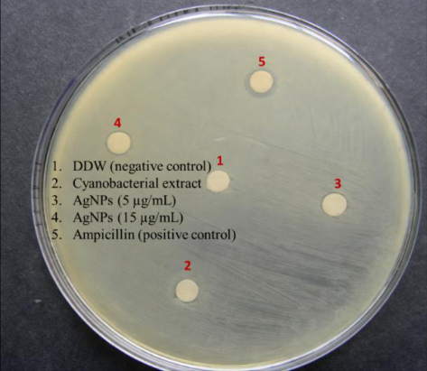 Figure 3
Introduction
Nanoparticles are materials that range from 1 to 100 nm in size. Recently, metallic nanoparticles have attracted considerable attention due to their wide range of applications in diverse fields (Pathak et al., 2019a, 2019b; Yiblet and Mintessnot; 2025) [15,16,26] . Despite novel physicochemical properties, metallic nanoparticles may pose potential health hazards to living organisms, including humans, especially when synthesized using conventional chemical and physical methods. Therefore, the growing concerns within the scientific community have led to the development of non-toxic, ecofriendly biosynthetic approaches that employ renewable resources and require lower energy input (Pathak et al., 2019a, 2019b; Bhardwaj et al., 2024) [15,16,4] . Among these, green-synthesized silver nanoparticles (AgNPs) have emerged as potent agents exhibiting antimicrobial, antifungal, antibacterial, anticancer, antioxidant, and anti-inflammatory activities (Bhardwaj and Naraian; 2021; Takci et al., 2023; Li et al., 2024) [3,24,9] . Cyanobacteria are a better biological candidate for nanoparticles synthesis because of having enhanced growth rate and biomass productivity (Pathak et al., 2019a, 2019b; Bhardwaj and Naraian; 2021) [15,16,3] . Use of cyanobacterial extracts for the nanoparticles biosynthesis is being widely practiced these days, as the extraction of nanoparticles formed inside the cell is a very costly and complex process. The cell extracts of the cyanobacterium Anabaena doliolum were utilized for the synthesis of AgNPs (Singh et al., 2014) [21] . Husain et al. investigated the biosynthesis of AgNPs from 30 different cyanobacterial strains (Husain et al., 2015) [7] . Sonker et al. (Sonker et al., 2017) [23] synthesized AgNPs utilizing the cell extract of the cyanobacterium Nostoc sp. strain HKAR-2 isolated from hot springs. Genus Scytonema has tremendous potential for nitrogen fixation and rich source of various biologically active compounds such as phycobilins, antibiotics, mycosporine-like amino acids (MAAs), scytonemin, toxins, etc. (Nowruzi et al., 2020) [11] . During the synthesis of AgNPs, these compounds act as reducing and stabilizing agents. In the present investigation, we used the cell extract of the rooftop-dwelling cyanobacterium Scytonema sp. for the synthesis of AgNPs. We also attempted to screen the in vitro antioxidant and antibacterial activity of the biosynthesized AgNPs.
Materials and methods
Materials and methods
Collection of Scytonema sp. and growth condition
The cyanobacterium Scytonema sp. was collected and purified from rooftop of Acharya Narendra Deo Kisan P.G College, Babhnan, Gonda and grown in BG-11 -medium (without nitrogen sources) (Rippka et al., 1979) [19] under axenic conditions, at temperature 26 ± 2 °C, light intensity 40-50 µmol photons m⁻² s⁻¹ and photoperiod 14:10 h (light/dark cycle).
Growth measurement
Growth of cyanobacteria was monitored by determining the changes in chlorophyll a (Chl a) content after desired time intervals. The Chl a was extracted in methanol. The recovered cyanobacterial sample was kept in 100% methanol overnight at 4 °C and its content was determined by method reported by Porra (Porra., 2002) [17] after observing absorption at 665.2 nm and 652 nm.
Chl a (μg/ml) = 16.29*A665.2 -8.54*A652 Where, A is the optical density (O.D.) at 665.2 and 652 nm, respectively.
Cell extract preparation and green synthesis of AgNPs
Cells of Scytonema sp. were harvested during the late exponential (log) phase. The collected cells were centrifuged (Remi mini centrifuge RM-02) at 6000 rpm for 15 minutes and the pellet was washed with double distilled water (DDW) and sonicated using ultrasonic vibration at 30% amplitude for 15 minutes. The homogenate was exposed to centrifugation (3 times), the supernatant was diluted through a series of dilutions with 1 mM AgNO3, and the reaction mixtures (10 mL) were incubated at 37 0 C for 60 h. Only the sample containing Scytonema cell extract served as the positive control. The formation of AgNPs was monitored visually at an interval of 12 h by observing the change in color of the reaction mixture.
Partial characterization of AgNPs by UV-VIS spectroscopy
The absorption spectrum was taken at different time interval (0, 12, 24, 36, 48, and 60 h) by using UV-Vis spectrophotometer (Shimadzu) with the resolution of 1 nm at the wavelength range of 200-800 nm, possessing a scanning speed of 200 nm/minutes.
2,2-Diphenyl-1-picrylhydrazyl (DPPH) radical scavenging assay
Free radical scavenging activity of desired concentrations (e.g., 20, 40 and 80 µg/mL) of Scytonema sp. extract and biosynthesized AgNPs was measured using a conventional DPPH radical scavenging assay (Shen et al., 2010) [20] .
2,2′azino-bis(3-ethylbenzothiazoline)-6-sulfonate (ABTS) radical scavenging assay
The antioxidant activity of Scytonema sp. extract and biosynthesized AgNPs of desired concentrations (20, 40 and 80 µg/mL) was evaluated using the ABTS radical scavenging assay following standard procedures provided by Re et al. (Re et al., 1999) [18] with minor modifications. The reduction of ABTS•⁺ was monitored spectrophotometrically at 734 nm, and scavenging activity was expressed as percentage inhibition.
Antibacterial activity
Antibacterial activity was assessed by the disc diffusion method against Gram negative bacterium E. coli following standard method with slight modification (Bauer et al., 1966) [9] . E. coli was grown in Muller Hinton Agar (MHA) medium at 37 °C. The desired concentration (05 μg/mL and 15 μg/mL) of AgNPs solution was soaked into separate discs on the Petri plates. Ampicillin served as positive control and double distilled water (DDW) was taken as negative control. After 24 h of incubation, the inhibition zones around the discs were measured.
Statistical analysis
The results express the mean values of three replicates of each experiment. One-way analysis of variance was performed for statistical analyses.
Results and Discussion
Growth profile of Scytonema sp. The growth pattern of Scytonema sp., monitored by evaluating Chl a content at regular intervals, showed a typical sigmoidal pattern with lag, exponential, and stationary phases (Fig. 1). The late exponential phase is often associated with enhanced production and release of extracellular biomolecules (Singh et al., 2014, San Keskin et al., 2016) [21,8] . These compounds are useful in biotechnological applications, including the biological synthesis of nanoparticles.
Partial characterization of AgNPs
The initiation of reduction of AgNO3 solution into AgNPs started after 12 h of the addition of 1 mM AgNO3 solution into the cell extract and completed after 60 h of incubation. The visual color change to dark red, and time-dependent increase in surface plasmon resonance (SPR) intensity at 410 nm confirm the successful extracellular biosynthesis of stable silver nanoparticles by Scytonema sp. extract (Harutyunyan et al., 2025) [5] (Fig. 2).
DPPH and ABTS assay
The results of antioxidant assay showed a clear concentration-dependent increase in radical scavenging activity of the AgNPs in both (DPPH and ABTS) assays (Table 1), indicating their effective antioxidant capacity. The free radical scavenging activity in DPPH and ABTS assay follow quite similar pattern and was about two folds higher than that of crude extract at 80 μm/mL concentration, though slightly lower than that of ascorbic acid at equivalent concentrations. This observation is consistent with previous reports (Bedlovičová et al., 2020) [2] . [22,14] [12,6,25,13,10] .
| Sr. No. | Sample | Concentration(µg mL⁻¹) | DPPH radical scavenging (%) | ABTS radical scavenging (%) |
|---|---|---|---|---|
| 1. | Cell extract of Scytonema sp. | 20 | 13.1 ± 1.4 | 16.2 ± 2.1 |
| 40 | 23.6 ± 1.1 | 27.2 ± 1.2 | ||
| 80 | 37.2 ± 1.2 | 40.1 ± 1.9 | ||
| 2. | AgNPs | 20 | 32.8 ± 1.3 | 29.2 ± 2.1 |
| 40 | 51.0 ± 1.0 | 55.3 ± 1.1 | ||
| 80 | 72.1 ± 1.2 | 72.4 ± 2.0 | ||
| 3. | Ascorbic acid(standard) | 20 | 46.1 ± 1.9 | 43.2 ± 1.0 |
| 40 | 63.6 ± 1.2 | 67.0 ± 1.4 | ||
| 80 | 82.3 ± 1.7 | 79.1 ± 2.3 |
Table text extracted from the original PDF; superscript letters are shown inline. The original table image is below.
View original table image
Conclusion
The present study explores a sustainable step towards the green synthesis of AgNPs using cell-free extracts of the rooftop dwelling cyanobacterium Scytonema sp. The harvesting of Scytonema sp. at late exponential phase enabled effective nanoparticle synthesis and was confirmed by a characteristic SPR peak at 410 nm. The results of antioxidant and antibacterial efficacy of AgNPs provide valuable insight and highlight its potential in the field of pharmaceuticals. These findings explore Scytonema sp. as an eco-friendly natural resource for the synthesis of versatile molecule AgNPs.
Figures and tables from the paper
References
26 in the original paper- Bauer AW, Kirby WM, Sherris JC, Turck M. Antibiotic susceptibility testing by a standardized single disc method. Am J Clin Pathol. 1966;45(4 Suppl):493-496.
- Bedlovičová Z, Strapáč I, Baláž M, Salayová A. A brief overview on antioxidant activity determination of silver nanoparticles. Molecules. 2020;25(14):3191.
- Bhardwaj AK, Naraian R. Cyanobacteria as biochemical energy source for the synthesis of inorganic nanoparticles: mechanism and potential applications. 3 Biotech. 2021;11(10):445.
- Bhardwaj H, Khute S, Sahu RK, Jangde RK. Emerging trends in hybrid nanoparticles: revolutionary advances and promising biomedical applications. Curr Drug Metab. 2024;25(4):248-265.
- Harutyunyan A, Hambardzumyan A, Gabrielyan L, Aghajanyan A, Kocharyan M, Avtandilyan N, et al. Silver nanoparticles fabricated using phycocyanin from Arthrospira platensis: green synthesis, physicochemical characterization, and biological activities. Microb Cell Fact. 2025;24(1):249.
- Huang X, Pang Y, Liu Y, Zhou Y, Wang Z, Hu Q. Green synthesis of silver nanoparticles with high antimicrobial activity and low cytotoxicity using catechol-conjugated chitosan. RSC Adv. 2016;6:6435764363.
- Husain S, Sardar M, Fatma T. Screening of cyanobacterial extracts for synthesis of silver nanoparticles. World J Microbiol Biotechnol. 2015;31(8):1279-1283.
- San Keskin NO, Kılıç NK, Dönmez G, Tekinay T. Green synthesis of silver nanoparticles using cyanobacteria and evaluation of their photocatalytic and antimicrobial activity. J Nano Res. 2016;40:120-127.
- Li H, Yang Z, Khan SA, Walsh LJ, Seneviratne CJ, Ziora ZM. Characteristics of metallic nanoparticles, especially silver nanoparticles, as anti-biofilm agents. Antibiotics. 2024;13(9):819.
- Michailidu J, Miškovská A, Jarošová I, Čejková A, Maťátková O. Antibacterial properties of silver and gold nanoparticles synthesized using Cannabis sativa waste extract against Pseudomonas aeruginosa. J Cannabis Res. 2025;7(1):20.
- Nowruzi B, Sarvari G, Blanco S. The cosmetic application of cyanobacterial secondary metabolites. Algal Res. 2020;49:101959.
- Park J, Lim DH, Lim HJ, Kwon T, Choi JS, Jeong S, et al. Size-dependent macrophage responses and toxicological effects of Ag nanoparticles. Chem Commun. 2011;47:4382-4384.
- Parmar S, Kaur H, Singh J, Matharu AS, Ramakrishna S, Bechelany M. Recent advances in green synthesis of Ag NPs for extenuating antimicrobial resistance. Nanomaterials. 2022;12(7):1115.
- Patel V, Berthold D, Puranik P, Gantar M. Screening of cyanobacteria for their ability to synthesize silver nanoparticles with antibacterial activity. Biotechnol Rep. 2014;5:112-119.
- Pathak J, Ahmed H, Singh DK, Pandey A, Singh SP, Sinha RP. Recent developments in green synthesis of metal nanoparticles utilizing cyanobacterial cell factories. In: Tripathi DK, Ahmad P, Sharma S, Chauhan DK, Dubey NK, editors. Nanomaterials in plants, algae, and microorganisms. Academic Press; 2019. p. 237-265.
- Pathak J, Sonker AS, Singh RV, Kumar D, Sinha RP. Synthesis of silver nanoparticles from extracts of Scytonema geitleri HKAR-12 and their in vitro antibacterial and antitumor potentials. Lett Appl Nanobio Sci. 2019;8(3):576-585.
- Porra RJ. The chequered history of the development and use of simultaneous equations for the accurate determination of chlorophylls a and b. Photosynth Res. 2002;73:149-156.
- Re R, Pellegrini N, Proteggente A, Pannala A, Yang M. Antioxidant activity applying an improved ABTS radical cation decolorization assay. Free Radic Biol Med. 1999;26(9-10):1231-1237.
- Rippka R, Deruelles J, Waterbury JB, Herdman M, Stanier RY. Generic assignments, strain histories and properties of pure cultures of cyanobacteria. J Gen Microbiol. 1979;111:1-61.
- Shen Q, Zhang B, Xu R, Wang Y, Ding X, Li P. Antioxidant activity in vitro of the selenium-contained protein from the Se-enriched Bifidobacterium animalis 01. Anaerobe. 2010;16:380-386.
- Singh G, Babele PK, Shahi SK, Sinha RP, Tyagi MB, Kumar A. Green synthesis of silver nanoparticles using cell extracts of Anabaena doliolum and screening of its antibacterial and antitumor activity. J Microbiol Biotechnol. 2014;24:1354-1367.
- Sondi I, Salopek-Sondi B. Silver nanoparticles as antimicrobial agent: a case study on Escherichia coli. J Colloid Interface Sci. 2004;275:177-182.
- Sonker AS, Pathak J, Kannaujiya VK, Sinha RP. Characterization and in vitro antitumor, antibacterial and antifungal activities of green synthesized silver nanoparticles using cell extract of Nostoc sp. strain HKAR-2. Can J Biotechnol. 2017;1:26-37.
- Takci DK, Ozdenefe MS, Genc S. Green synthesis of silver nanoparticles with antibacterial activity using Salvia officinalis aqueous extract. J Cryst Growth. 2023;614:127239.
- Tang S, Zheng J. Antibacterial activity of silver nanoparticles: structural effects. Adv Healthc Mater. 2018;7:1701503.
- Yiblet Y, Mintessnot M. Application of silver nanoparticles synthesized from species of green algae: a review. Disc Appl Sci. 2025;7(10):1215.