
Research article · 2026 · Full text
International Journal of Biology SciencesEthambutol-associated ocular toxicity: Pharmacological mechanisms and opportunities for prodrug design
Volume 8(1) · Pages 05–08 · DOI: 10.33545/26649926.2026.v8.i1a.535 ↗
Research areasDrug-Induced Ocular ToxicityCancer Treatment and PharmacologyRetinopathy of Prematurity Studies
Abstract
Author-writtenEthambutol remains an essential component of first-line antitubercular therapy despite being plagued by ocular toxicity, largely dependent on dosage and duration, most commonly ethambutol-induced optic neuropathy (AEON). For the past decade, an increasing number of clinical and experimental findings have associated damage to the retinal ganglion cells with host genetic susceptibility, oxidative stress, chelation of metal ions, and mitochondrial dysfunction. In parallel, prodrug strategies that preserve antimycobacterial potency while reducing off-target toxicity have taken on new interest following advances in the development of drugs for tuberculosis. This narrative review collates the literature related to the clinical range, pharmacological mechanisms, and translational prospects associated with ethambutol-associated ocular toxicity within the period from 2015 to 2025. Particular emphasis is placed on prodrug-based mitigation strategies.
Summary
Source-checkedPlain-language reading of the full paper, prepared with AI and checked against the text. Each point links to the authors' own words.
Why it matters
- Ethambutol is a critical drug for first-line tuberculosis treatment, but it is frequently associated with ocular toxicity, specifically ethambutol-induced optic neuropathy (AEON). View evidence ↗
- Even with recommended monitoring and dose limits, ocular toxicity can occur during long-term therapy or in susceptible patients at therapeutic concentrations. View evidence ↗
- Advances in mitochondrial biology, medicinal chemistry, and pharmacogenetics allow for the exploration of prodrugs to reduce eye exposure while maintaining antibacterial efficacy. View evidence ↗
The question
- The authors aim to review the clinical range, pharmacological mechanisms, and potential prodrug strategies to mitigate ethambutol-associated ocular toxicity based on literature from 2015 to 2025. View evidence ↗
What they did
- The study is a narrative review that synthesizes existing literature on the clinical and pharmacological aspects of ethambutol toxicity published between 2015 and 2025. View evidence ↗
What they found
- The risk of optic neuropathy is less than 1% for doses under 15 mg/kg/day, but increases to over 5% with higher cumulative doses or longer treatment durations. View evidence ↗
- Clinical presentation typically includes painless, bilateral, and symmetric visual loss, red-green color blindness (dyschromatopsia), and central or cecocentral scotomas. View evidence ↗
- While some patients recover after stopping the drug, incomplete recovery is linked to the loss of retinal ganglion cells, indicating irreversible injury in advanced cases. View evidence ↗
- Ethambutol's chemical structure allows it to strongly bind to divalent metal ions, such as zinc and copper. View evidence ↗
- The chelation of metal cofactors, particularly for cytochrome c oxidase, is proposed as a primary cause of mitochondrial dysfunction induced by ethambutol. View evidence ↗
- In neuronal and retinal cell models, ethambutol exposure reduces ATP synthesis, interferes with oxidative phosphorylation, and increases reactive oxygen species. View evidence ↗
- Optical coherence tomography (OCT) shows early thinning of the ganglion cell complex and retinal nerve fibre layer, confirming that the drug causes direct injury to retinal ganglion cells. View evidence ↗
- Experimental data indicates that esterifying the hydroxyl groups of ethambutol can control its antibacterial activity and allow for activation by microbial esterases. View evidence ↗
- Pathogen-activated prodrugs could theoretically reduce ocular exposure because retinal tissues lack the specific enzymes required to activate the drug, which would instead be released in infected macrophages. View evidence ↗
What it means
- The authors conclude that pathogen-activated ester prodrugs are a scientifically plausible way to reduce toxicity, though clinical benefits have not yet been proven. View evidence ↗
- The authors state that prodrug strategies require extensive preclinical ocular safety evaluations before they can be applied clinically. View evidence ↗
Visual summary
1 visualsIllustrations explain the paper; they do not show study data.
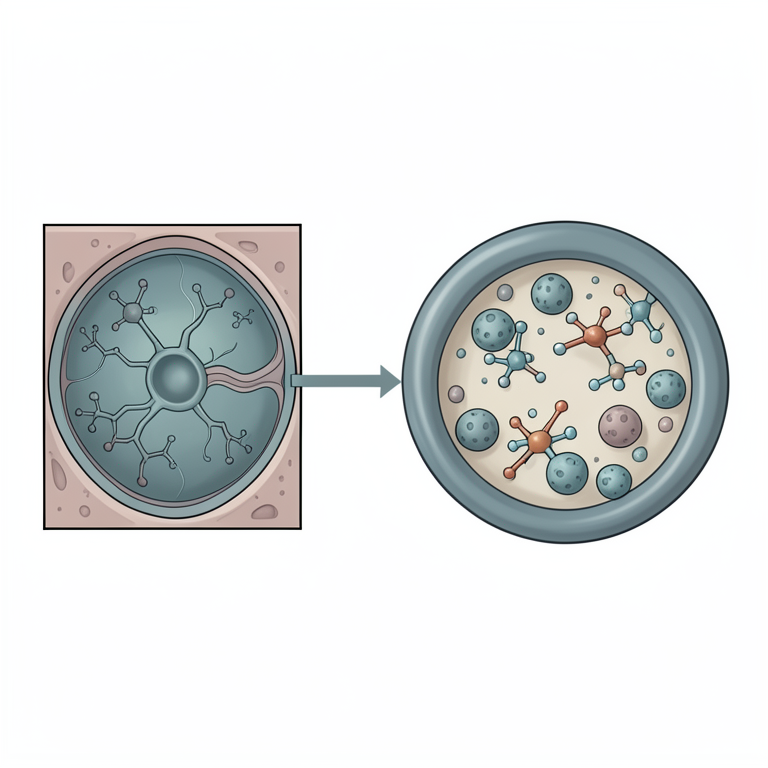
Prodrug Activation Concept
AI-generated conceptual illustration (retinal tissue and macrophages containing Mycobacterium tuberculosis). It explains the idea and does not show study data.
View source evidence ↗Introduction
EMB has been in use for more than 50 years as part of a multidrug regimen to treat drugsusceptible TB, yet it has been recognised for its toxicity to the optic nerve. The balance between its helpful effect and toxicity, specifically to the optic nerve, was recognised early in its history and confirmed in modern cohorts [1][2][3] . Ocular toxicity is apparent even at therapeutic concentrations during long-term treatment, and in more susceptible populations, despite dose limitations and regular ophthalmic monitoring currently recommended [5,9] . Emerging knowledge from pharmacogenetics, medicinal chemistry and mitochondrial biology offers the opportunity to revisit the mechanism of EMB toxicity and explore rational design strategies such as prodrugs to limit eye exposure without compromising the drug's antimycobacterial action [3,[12][13][14][15][16]23] .
Clinical Spectrum of Ethambutol-associated ocular toxicity 2.1 Epidemiology and Risk Factors
Ethambutol-induced optic neuropathy occurs in less than 1% of individuals receiving less than 15 mg/kg/day but greater than 5% of individuals receiving higher cumulative doses or for longer durations of therapy [2] . Cumulative dose, duration of therapy, older age, renal impairment, and pre-existing optic nerve, mitochondrial, or axonal function weakness have been associated with a statistically meaningful increased risk according to cohort and population-based studies.
Clinical Presentation
Ocular toxicity linked to ethambutol usually manifests as central or cecocentral scotomas, dyschromatopsia (typically red-green), and bilateral, symmetric, painless visual loss with decreased visual acuity [1,2,8] . Early in the course of the disease, fundoscopic findings may be normal, with optic disc pallor developing later [8] .
However, incomplete recovery is correlated with structural retinal ganglion cell loss detected by OCT, suggesting irreversible neuronal injury in advanced cases [8] .
Pharmacological and Pathophysiological Mechanisms
Ethambutol-associated ocular toxicity is best understood as a multifactorial process in which primary drug-specific chemical properties interact with host cellular vulnerability, particularly within Retinal Ganglion Cells (RGCs). Contemporary evidence supports a hierarchical model in which mitochondrial dysfunction and metal chelation represent primary initiating mechanisms, while oxidative stress, calcium dysregulation, and apoptotic signalling acting as downstream amplifiers of neuronal injury [3,6,9,10,25] .
Structural and Chemical Determinants of toxicity
Ethambutol has a bidentate structure with hydroxyl and amine functional groups that are close together. This gives it a strong ability to bind to divalent metal ions like copper and zinc [3] . This characteristic indirectly enhances its antimycobacterial efficacy by inhibiting arabinosyl transferases, yet simultaneously constitutes a pharmacological risk in mammalian tissues. Chelation of metal cofactors vital for mitochondrial enzymes, especially cytochrome c oxidase, has been suggested as a principal initiating factor in ethambutol-induced mitochondrial dysfunction [10,22] . Importantly, this chelating behaviour is intrinsic to the parent compound and is not shared to the same extent by other first-line antitubercular drugs, providing a mechanistic basis for ethambutol's unique association with optic neuropathy [10,22] .
Mitochondrial dysfunction as a central mechanism
Since 2015, ethambutol-induced optic neuropathy has been linked to mitochondrial impairment, which has been the subject of numerous experimental and clinical studies [6] . In models of neurones and retinal cells, ethambutol exposure has been demonstrated to interfere with oxidative phosphorylation, decrease ATP synthesis, and increase the production of reactive oxygen species [9] . Axonal transport and neuronal survival are compromised by these bioenergetic disruptions, which are especially important in long, unmyelinated optic nerve fibres. This mechanism is further supported by the similarities between ethambutol-induced optic neuropathy and inherited mitochondrial optic neuropathies, including Leber hereditary optic neuropathy [6,10,22] . Notably, even at standard doses, patients with nuclear gene variants or mitochondrial DNA mutations that impact mitochondrial dynamics are more vulnerable to ethambutol toxicity [6,7] .
Selective vulnerability of retinal ganglion cells
Retinal ganglion cells are uniquely vulnerable to mitochondrial insults due to their high energy demand, dense mitochondrial population, and long axonal projections extending from the retina to the lateral geniculate nucleus [10,22] . Even modest impairments in mitochondrial function can therefore precipitate RGC dysfunction and death. This selective vulnerability explains why ethambutol toxicity manifests predominantly as optic neuropathy rather than diffuse neurotoxicity.
Structural imaging studies using optical coherence tomography demonstrate early thinning of the retinal nerve fibre layer and ganglion cell complex, corroborating direct RGC injury rather than secondary demyelination [8] .
Metal Chelation and Enzyme Inhibition
In addition to causing general mitochondrial stress, ethambutol's ability to chelate copper and zinc might directly impede the function of metalloenzymes that are crucial for electron transport and antioxidant defence [3] . While there is no direct in vivo evidence from human optic nerve tissue, a combination of biochemical and clinical findings lends credence to this mechanism as a likely factor rather than a mere hypothesis [3] .
Downstream Pathways
Oxidative Stress and Calcium Dysregulation. Oxidative stress and the disruption of intracellular calcium levels seem to occur as a result of mitochondrial dysfunction. Studies using models of toxic optic neuropathy have shown that an increase in reactive oxygen species and calcium entry triggers apoptotic pathways in retinal ganglion cells [9] . These processes are not thought to be the main culprits on their own, but they probably speed up neuronal degeneration after mitochondrial damage starts.
Diagnostic and Monitoring Approaches
Functional assessments, including visual acuity, colour vision, and perimetry, continue to be fundamental in clinical monitoring [1,5] . Structural imaging using OCT has become a sensitive method for identifying early thinning of the retinal nerve fibre layer and ganglion cell complex, often before symptoms become apparent [8,41] . Electrophysiological methods, such as visual evoked potentials, can detect subclinical dysfunction, though they are not widely accessible [17] . Recent research into AIassisted screening represents emerging supplementary methods rather than established care standards [21] .
Rationale for prodrug design to mitigate ocular toxicity 5.1 Conceptual Framework
Prodrug strategies are commonly used to enhance the pharmacokinetics, tissue selectivity, and safety profiles of existing drugs [23] . For ethambutol, the goal of prodrug design is not to increase potency but to minimize off-target toxicity, especially in ocular tissues, while maintaining or restoring antimycobacterial effectiveness at the infection site. The mechanistic insights mentioned earlier suggest that concealing ethambutol's metal-chelating functional groups could theoretically reduce mitochondrial toxicity in retinal ganglion cells without eliminating its antibacterial activity. Ester Prodrugs of Ethambutol. Experimental research has shown that esterifying ethambutol's hydroxyl groups alters its antibacterial activity and allows for controlled activation by microbial esterases [3] . These findings offer proof-ofconcept that ethambutol can be chemically altered into inactive or less active derivatives that can be enzymatically reactivated. However, these studies have not yet demonstrated reduced ocular toxicity in vivo. Instead, they provide a biochemical and microbiological basis for developing and testing safer derivatives. Pathogen-Activated Prodrug Strategies. Modern antituberculosis drug discovery increasingly aims for selective activation strategies by pathogens [12][13][14][15][16] . M. tuberculosis expresses a wide variety of esterases and hydrolases that can activate ester or carbamate prodrugs; this strategy has already shown promise for other antituberculosis drugs [21] . Applying such an approach to ethambutol could theoretically restrict systemic exposure to the active parent compound while enabling its intracellular release within infected macrophage cells. Such selectivity could have the added advantage of reducing ocular exposure because retinal tissues do not express specific mycobacteriaactivating enzymes. Comparison to other mitigation strategies. Other approaches to mitigate the toxicity of ethambutol involve dose reduction, narrowing the duration of therapy, and increasing vigilance. Although these are moderately successful, no approach completely removes the risk while potentially compromising antimycobacterial efficacy in complicated cases or in drug-resistant TB [8] . Prodrug strategies should, therefore, be viewed as a complementary approach rather than a competitive one.
Translational and Clinical Implications
Reducing ethambutol-related ocular toxicity would have a strong clinical impact, particularly in regimens for multidrug-resistant TB that require extended therapy [8] . Prodrug strategies align with contemporary priorities of TB drug development centred around targeted delivery and toxicity reduction [15][16] ; however, they require extensive preclinical ocular safety evaluations before clinical application.
Conclusion
Ethambutol-associated ocular toxicity remains a clinically relevant limitation of tuberculosis therapy. Evidence accumulated since 2015 supports mitochondrial dysfunction and metal chelation as central, interacting mechanisms, modulated by host genetic susceptibility. Prodrug strategies, particularly pathogen-activated ester derivatives, offer a scientifically plausible avenue for toxicity mitigation, although clinical benefit remains to be demonstrated. Continued interdisciplinary research is essential to translate these concepts into safer antitubercular therapies.
References
22 in the original paper- Koul PA. Ocular toxicity with ethambutol therapy: Timely precaution. Lung India. 2015 Jan-Feb;32(1):1-3. DOI: 10.4103/0970-2113.148395.
- Garg P, Garg R, Prasad R, Mishra AK. A prospective study of ocular toxicity in patients receiving ethambutol as a part of directly observed treatment strategy therapy. Lung India. 2015 Jan-Feb;32(1):16-19. DOI: 10.4103/0970-2113.148428.
- Larsen EM, Stephens DC, Clarke NH, Johnson RJ. Ester-prodrugs of ethambutol control its antibacterial activity and provide rapid screening for mycobacterial hydrolase activity. Bioorg Med Chem Lett. 2017 Oct 1;27(19):4544-4547. DOI: 10.1016/j.bmcl.2017.08.057.
- Larsen EM, Johnson RJ. Microbial esterases and ester prodrugs: An unlikely marriage for combating antibiotic resistance. Drug Dev Res. 2019 Feb;80(1):3347. DOI: 10.1002/ddr.21468.
- Saxena R, Singh D, Phuljhele S, Kalaiselvan V, Karna S, Gandhi R, et al. Response to comment on: Ethambutol toxicity: Expert panel consensus for the primary prevention, diagnosis, and management of ethambutol-induced optic neuropathy. Indian J Ophthalmol. 2022 Mar;70(3):1070-1071. DOI: 10.4103/ijo.IJO_88_22.
- Kaur P, Singh S, Kaur K, Mahesh KV, Tigari B, Sehgal V, et al. The genetics of ethambutol-induced optic neuropathy: A narrative review. Neuroophthalmology. 2022 Aug 2;46(5):304-313. DOI: 10.1080/01658107.2022.2100916.
- Zhang XH, Xie Y, Xu QG, Cao K, Xu K, Jin ZB, et al. Mitochondrial mutations in ethambutol-induced optic neuropathy. Front Cell Dev Biol. 2021 Oct 5;9:754676. DOI: 10.3389/fcell.2021.754676.
- Sabhapandit S, Gella V, Shireesha A, Thankachan L, Ismail M, Rao R, et al. Ethambutol optic neuropathy in the extended anti-tubercular therapy regime: A systematic review. Indian J Ophthalmol. 2023 Mar;71(3):729-35. DOI: 10.4103/ijo.IJO_1920_22.
- Kulniwatcharoen P, Hansapinyo L, Chattipakorn N, Chattipakorn SC. Potential underlying mechanisms of ethambutol induced optic neuropathy: Evidence from in vitro to clinical studies. Food Chem Toxicol. 2023 Dec;182:114176. DOI: 10.1016/j.fct.2023.114176.
- Wang MY, Sadun AA. Drug-related mitochondrial optic neuropathies. J Neuroophthalmol. 2013 Jun;33(2):172-178. DOI: 10.1097/WNO.0b013e3182901969.
- Kim J, Kwon HY, Ahn SJ. Nationwide usage of ethambutol and incidence and screening practices of optic neuropathy. Front Pharmacol. 2024 Oct 15;15:1461111. DOI: 10.3389/fphar.2024.1461111.
- Mori G, Chiarelli LR, Riccardi G, Pasca MR. New prodrugs against tuberculosis. Drug Discov Today. 2017 Mar;22(3):519-525. DOI: 10.1016/j.drudis.2016.09.006.
- Kim CG, Jose J, Hay MP, Choi PJ. Novel prodrug strategies for the treatment of tuberculosis. Chem Asian J. 2024 Dec 2;19(23):e202400944. DOI: 10.1002/asia.202400944.
- Goss AL, Shudick RE, Johnson RJ. Shifting mycobacterial serine hydrolase activity visualized using multi-layer in-gel activity assays. Molecules. 2024 Jul 18;29(14):3386. DOI: 10.3390/molecules29143386.
- Hegde PV, Howe MD, Zimmerman MD, Boshoff HIM, Sharma S, Remache B, et al. Synthesis and biological evaluation of orally active prodrugs and analogs of para-aminosalicylic acid (PAS). Eur J Med Chem. 2022 Mar 15;232:114201. DOI: 10.1016/j.ejmech.2022.114201.
- Lai L, Xu Z, Zhou J, Lee KD, Amidon GL. Molecular basis of prodrug activation by human valacyclovirase, International Journal of Biology Sciences https://www.biologyjournal.net an alpha-amino acid ester hydrolase. J Biol Chem. 2008 Apr 4;283(14):9318-9327. DOI: 10.1074/jbc.M709530200.
- MacVinish S, McMaster D, Moledina T, Tamne SK, Ashworth J, Anderson SR. Ethambutol and visual assessment in England: current practice and recommendations. Eye (Lond). 2024 Jan;38(1):112117. DOI: 10.1038/s41433-023-02643-4.
- Singha B, Murmu S, Nair T, Rawat RS, Sharma AK, Soni V. Metabolic rewiring of Mycobacterium tuberculosis upon drug treatment and antibiotics resistance. Metabolites. 2024 Jan 18;14(1):63. DOI: 10.3390/metabo14010063.
- Singh V, Chibale K. Strategies to combat multi-drug resistance in tuberculosis. Acc Chem Res. 2021 May 18;54(10):2361-2376. DOI: 10.1021/acs.accounts.0c00878.
- Karkhur S, Beri A, Verma V, Gupta S, Singh P. Artificial intelligence in neuro-ophthalmology: Opportunities for the diagnosis of optic neuropathies and visual pathway disorders. Cureus. 2025 Aug 15;17(8):e90142. DOI: 10.7759/cureus.90142.
- Carelli V, Ross-Cisneros FN, Sadun AA. Mitochondrial dysfunction as a cause of optic neuropathies. Prog Retin Eye Res. 2004 Jan;23(1):53-89. DOI: 10.1016/j.preteyeres.2003.10.003.
- Rautio J, Meanwell NA, Di L, Hageman MJ. The expanding role of prodrugs in contemporary drug design and development. Nat Rev Drug Discov. 2018 Aug;17(8):559-587. DOI: 10.1038/nrd.2018.46.