
Research article · 2025 · Full text
International Journal of Advanced Chemistry ResearchConformational analysis of pipecolic acid substituted collagen model peptides
Volume 7(1) · Pages 67–74 · DOI: 10.33545/26646781.2025.v7.i1a.316 ↗
Research areasCollagen: Extraction and CharacterizationSeaweed-derived Bioactive CompoundsNasal Surgery and Airway Studies
Abstract
Author-writtenCollagen model peptides, (Xaa-Yaa-Gly)10 have been used to understand the propensity of the amino acid in Xaa nad Yaa positions. Five membered cyclic imino acid, proline (Pro), have found to be most investigated model. In this investigation Pipecolic acid (Pip), a six member cyclic imino acid and a higher homologue of Pro has been used for sysnthesis of (Pro-Pip-Gly)10, (Pip-Pro-Gly)10, and (Pip-Pip-Gly)10 have been synthesized, characterised and compared with (Pro-Pro-Gly)10 for their physical property such as solubility. Their triple helix formation characteristics at 4 °C has been investigated by circular dichroism (CD) spectroscopy. It has been found that Pip has higher propensity for Yaa position compared to Xaa position in triple helix conformations.
Summary
Source-checkedPlain-language reading of the full paper, prepared with AI and checked against the text. Each point links to the authors' own words.
Why it matters
- Synthetic peptides are used as tools to examine the complex structures of collagen. View evidence ↗
- In human collagen, the Proline-Hydroxyproline-Glycine sequence is recognized as the most stabilizing triplet. View evidence ↗
- Pipecolic acid (Pip) is a proline analogue that features a six-membered piperidine ring. View evidence ↗
The question
- The study aims to synthesize and characterize collagen model peptides substituted with pipecolic acid, specifically (Pro-Pip-Gly)10, (Pip-Pro-Gly)10, and (Pip-Pip-Gly)10, and compare them to (Pro-Pro-Gly)10. View evidence ↗
What they did
- Peptides were analyzed using circular dichroism (CD) spectroscopy after being maintained in 0.1 M phosphate buffer solution (PBS) at 4 °C for 48 hours. View evidence ↗
What they found
- The reference peptide (Pro-Pro-Gly)10 showed typical collagen CD spectra, with significant negative ellipticity at 200 nm and positive ellipticity at 225 nm. View evidence ↗
- Pip-containing peptides lacked positive ellipticity and showed reduced negative ellipticity compared to the reference peptide. View evidence ↗
- The (Pro-Pip-Gly)10 peptide showed deeper negative maxima than (Pip-Pro-Gly)10, indicating that the position of the Pip substitution affects the conformational change. View evidence ↗
- The conformational deviation of (Pip-Pip-Gly)10 from the reference is more strongly influenced by Pip at the Xaa position than at the Yaa position. View evidence ↗
- Substituting all Pro residues with Pip, as in (Pip-Pip-Gly)10, results in only residual helicity. View evidence ↗
What it means
- The authors conclude that substituting Pip at the Xaa position has a more significant negative impact on helix stability than substitution at the Yaa position. View evidence ↗
- Concurrent substitution at both Xaa and Yaa positions leads to significant changes in solubility and CD spectral patterns. View evidence ↗
Visual summary
2 visualsIllustrations explain the paper; charts are replotted from the paper's own table values.
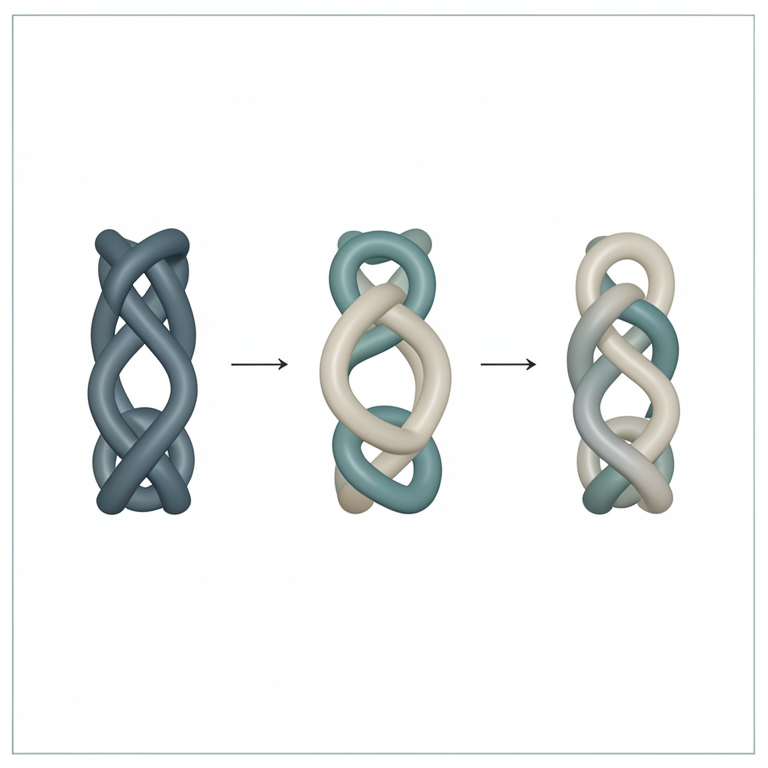
Positional Substitution Effect
AI-generated conceptual illustration (collagen triple helix model peptides). It explains the idea and does not show study data.
View source evidence ↗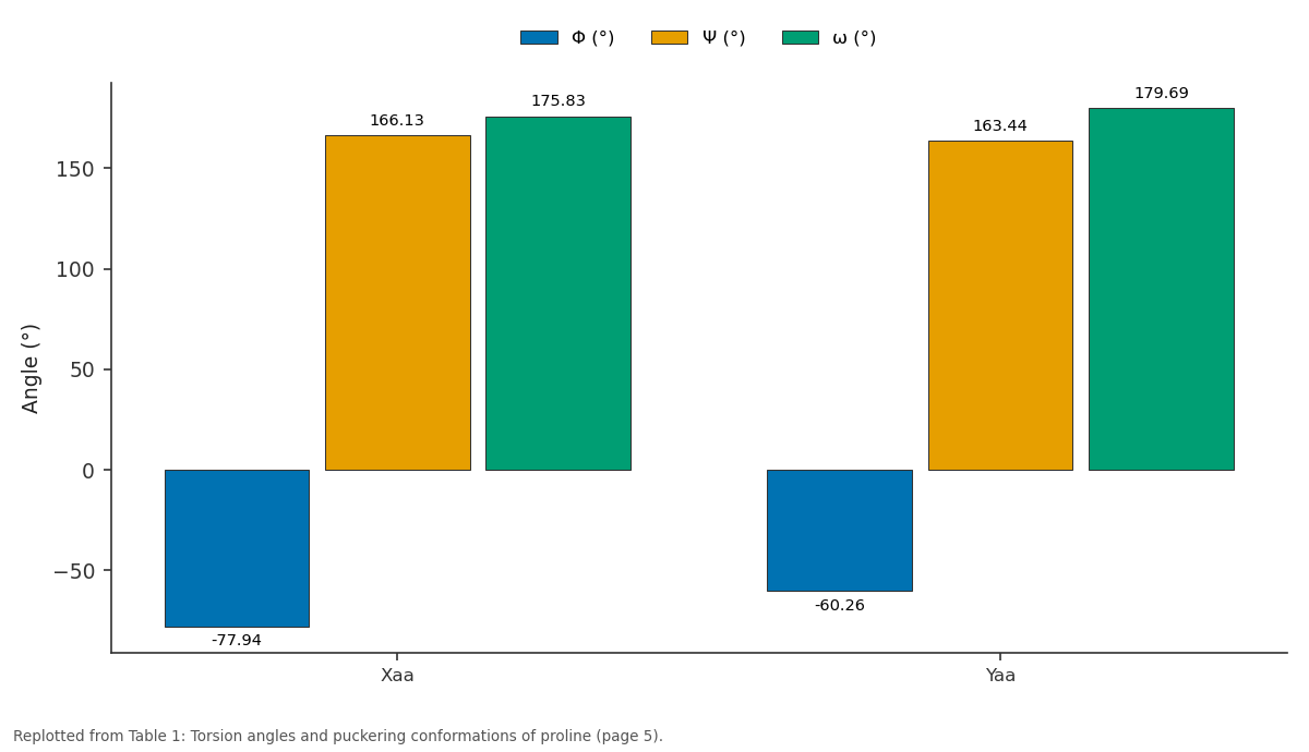
Φ (°) and Ψ (°) and more: Xaa vs Yaa
Replotted from Table 1 (page 5) of the original paper. Every bar is the paper's own table value.
View source evidence ↗Figures & tables
From the original paper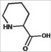 Figure 1
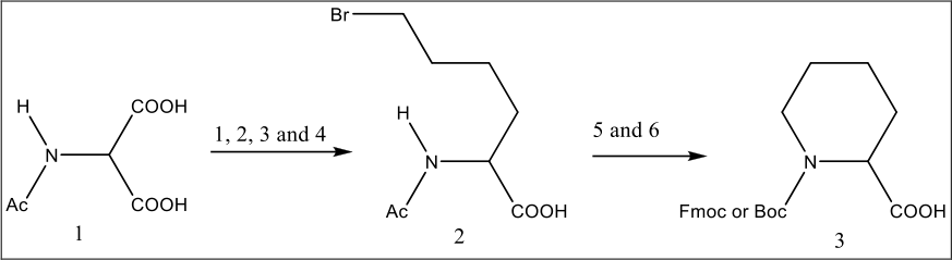 Figure 1
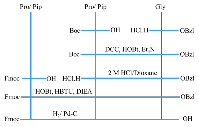 Figure 2
 Table 1
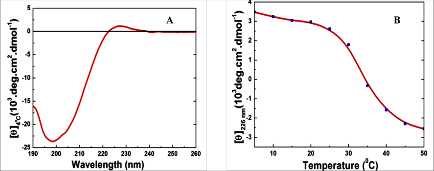 Figure 3
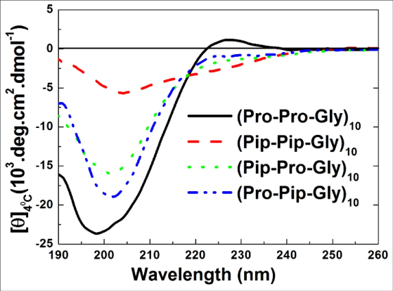 Figure 4
Text
4R-Flp-Gly) 10 , (Pro-4S-Flp-Gly) 10 , and (4S-Flp-4R-Gly) 10 to examine electronegative effects. (Shoulders & Raines, 2009) [31] Babu and Ganesh demonstrated that 4(R)-Amp collagen mimetics exhibit greater pH-dependent stability than Hyp, attributed to the protonation effects of the amino group.(Babu & Ganesh, 2001 [2] ; Research involving azetidine carboxylic acid (Aze) indicated that (Gly-Pro-Aze) forms triple helices analogous to poly(Gly-Pro-Pro), albeit with reduced stability. Both (G1y-Aze-Pro)n and poly(Gly-Aze-Aze) formed stable yet geometrically distinct helices, with Aze chains displaying increased flexibility. (Fowden & Richmond, 1963; Zagari et al., 1990) [7,38] These findings imply that modelling the effects of ring conformation entropy on angles and hydrophobicity, alongside pipecolic acid, could elucidate stability factors. Pipecolic acid (Pip), also referred to as (S)-Piperidine-2carboxylic acid, is a proline analogue characterized by a sixmembered piperidine ring. (Fig 1) The incorporation of Pip influences the structural properties of collagen by altering the torsion angles in model peptides, thereby affecting the hydration of the PPII helix. Pip is naturally present in legumes (Grobbelaar & Steward, 1953) [10] , Neurospora (Schweet et al., 1954), cerebrospinal fluid (Lam et al., 1984), blood (Gatfield et al., 1968), urine (Lam et al., 1984), and the rat brain (Nishio & Segawa, 1983) [20] ,
and it activates aminobutyric acid receptors in neonatal chicks (Takagi et al., 2003) [35] . It is an essential component of peptide mimetics found in immunosuppressants such as FK506 (Tanaka et al., 1987) [36] , bupivacaine, VX710, demethoxyrapamycin, and trapoxins. Pip has been utilized as a substitute for proline in structureactivity studies of peptides. The substitution of proteinogenic amino acids with cyclic imino acids has been employed in structure-activity investigations and for the development of peptidomimetics with enhanced pharmacological profiles. A pentapeptide containing Pip influences the thermodynamic properties of the interaction between peptidyl prolyl cis-trans isomerase Pin1 and its substrate.
Experimental 2.1 Synthesis of Pipecolic acid
Various methodologies are available for the synthesis of pipecolic acid (Pip), encompassing chemoenzymatic techniques (Nazabadioko et al., 1998) [18] , asymmetric induction (Roos & Dastlik, 2003) [29] , and catalytic processes (Ginesta et al., 2002). A photocatalytic transformation employing TiO₂ or CdS offers an environmentally friendly approach, yielding ammonia as a by-product, albeit with limited efficiency. (Myers et al., 1997) [17] In the chemoenzymatic pathway developed by Watanabe, diethyl acetamidomalonate undergoes alkylation with dibromobutane, facilitated by sodium ethoxide as the alkylating agent. (Watanabe et al., 2005) [37] The resultant compound is subsequently hydrolyzed using sodium hydroxide, followed by decarboxylation to produce a monoester. Further hydrolysis yields acetyl-DL-2-amino-6bromohexanoic acid. The racemic mixture is resolved using aminoacylase, with the L-enantiomer undergoing cyclization to form L-pipecolic acid, which is subsequently converted into derivatives such as Fmoc-L-Pip-OH, Boc-L-Pip-OH, and HCl•H-L-Pip-OBzl (Scheme 1).
Synthesis of tripeptides
Unless otherwise specified, all solvents and reagents were of reagent grade and utilized without further purification. 1-Hydroxybenzotriazole (HOBt), N,N'-Dicyclohexylcarbodiimide (DCC), O-(Benzotriazole-N,N,N',N'-tetramethyluronium) hexafluorophosphate (HBTU), N,N-Diisopropylethylamine (DIEA) were used to synthesis of tripeptide fragment (Figure 2). Flash chromatography was conducted using silica gel 60 (230-400 mesh) with the indicated solvents as eluents. All compounds were routinely analyzed using thin-layer chromatography (TLC) or high-performance liquid chromatography (HPLC). TLC was performed on aluminum-backed silica gel plates (Merck DC-Alufolien Kieselgel 60 F254), with spot visualization achieved through UV light exposure. Analytical HPLC was conducted on a Hitachi instrument equipped with a Chromolith Performance RP-18e column. The mobile phases employed were A: H2O with 0.1% TFA, and B: CH 3 CN with 0.1% TFA, utilizing a solvent gradient from A to B over 15 minutes, with detection at 220 nm and a flow rate of 2 mL/min. Fast atom bombardment (FAB)
mass spectra and high-resolution mass spectra (HRMS) were obtained using a JEOL JMS-SX 102A instrument. Nuclear magnetic resonance (NMR) spectra were recorded on a Bruker 500 MHz spectrometer. Unless otherwise noted, all NMR spectra were recorded in CDCl3 solutions with tetramethylsilane (TMS) as the internal standard. All 1H chemical shifts are reported in parts per million (s = singlet; d = doublet; t = triplet; m = multiplet).
Fmoc-L-Pro-L-Pip-Gly-OH
To a cooled solution of HCl. H-Gly-OBzl (5.5 g, 24 mmol) in DMF (60 mL), triethylamine (3.4 mL, 24 mmol), Boc-L-Pip-OH (1.29 g, 5.5 mmol), HOBt•H 2 O (3.67 g, 24 mmol), and DCC (6 g, 28.8 mmol) were added. The mixture was then stirred overnight at room temperature. After completion of the reaction, DMF was evaporated, and the residue was dissolved in ethyl acetate. The DCU was removed by filtration, and the filtrate was successively washed with 10% citric acid, 4% sodium bicarbonate, and brine. The ethyl acetate solution was dried over anhydrous MgSO 4 and concentrated to an oily residue, which was purified by silica gel chromatography using a mixture of chloroform and methanol (99:1) to yield Boc-L-Pip-Gly-OBzl (7.
Fmoc-L-Pip-L-Pro-Gly-OH
Boc-L-Pro-Gly-OBzl (365 g, 1 mmol) was dissolved in 2 M HCl/dioxane (10 mL) and the mixture was kept at room temperature for 2 h. The reaction was monitored using TLC.
After completion of the reaction, HCl/dioxane was evaporated. The residue was washed with diethyl ether and pumped to obtain HCl.H-L-Pro-Gly-OBzl as a white powder (300 mg, 100%). To a cooled solution of HCl.H-L-Pro-Gly-OBzl (300 mg, 1 mmol) in DMF (3 mL), Fmoc-L-Pip-OH (0.351 mg, 1 mmol), HOBt•H 2 O (153 mg, 1 mmol), HBTU (379 mg 1 mmol), and DIEA (0.50 mL, 3 mmol) were added and stirred for 4 h at room temperature. After completion of the reaction, DMF was evaporated, and the residue was dissolved in ethyl acetate and successively washed with 10% citric acid, 4% sodium bicarbonate and brine. The ethyl acetate solution was dried over anhydrous MgSO 4 and concentrated to an oily residue, which was purified by silica gel chromatography using a mixture of chloroform and methanol (99:1) to obtain Fmoc-L-Pip-L-Pro-Gly-OBzl (475 mg, 82%) as white foam, HPLC, rt Fmoc-L-Pip-L-Pro-Gly-OBzl (475 mg, 1 mmol) was dissolved in MeOH (5 mL). Pd-C (50 mg) was added, and the mixture was stirred overnight under a hydrogen atmosphere. The reaction was monitored using TLC and HPLC. After completion of the reaction, Pd-C was filtered off, and methanol was evaporated. The product was purified by crystallization using ether-petroleum ether (1:3 v/v) to yield Fmoc-L-Pip-L-Pro-Gly-OH (410 mg, 97%) as a white solid. HPLC, rt 6. Fmoc-L-Pip-L-Pip-Gly-OBzl (3.1 g, 5.1 mmol) was dissolved in methanol (30 mL). Pd-C (250 mg) was added, and the mixture was stirred under a hydrogen atmosphere for 10 h. The reaction was monitored using TLC and HPLC.
After completion of the reaction, Pd-C was filtered off, and methanol was evaporated. The product was purified by crystallization using ether-petroleum ether (1:3 v/v) to yield Fmoc-L-Pip-L-Pip-Gly-OH (2.4 g, 91%) as a white solid. HPLC, rt 9.26 min. HR-FAB MS, [M+H] + 520.2499 for C 29 H 34 O 6 N 3 (calcd. 520.2448). Fmoc-L-Pip-L-Pip-Gly-OBzl (3.1 g, 5.1 mmol) was dissolved in methanol (30 mL). Pd-C (250 mg) was added, and the mixture was stirred under a hydrogen atmosphere for 10 h. The reaction was monitored using TLC and HPLC.
After completion of the reaction, Pd-C was filtered off, and methanol was evaporated. The product was purified by crystallization using ether-petroleum ether (1:3 v/v) to yield Fmoc-L-Pip-L-Pip-Gly-OH (2.4 g, 91%) as a white solid. HPLC, rt 9.26 min. HR-FAB MS, [M+H] + 520.2499 for C 29 H 34 O 6 N 3 (calcd. 520.2448).
against nucleophiles, a requisite condition for solid-phase peptide synthesis (SPPS). Product cleavage occurs under mild conditions due to the high stability of trityl cations. Standard cleavage conditions include HOAc/TFE/DCM or diluted TFA in methylene chloride, although TFA vapor or HCl gas can also be directly applied to the dry bead. Due to these advantages, Barlos resin has been increasingly utilized for the synthesis of collagen model peptides. In this study, it served as a solid support for peptide synthesis. Fmoc derivatives of the tripeptide residues were used for coupling. This strategy employs Fmoc as the protective group and piperidine for deprotection. As the peptide chains grow, they have the potential to interact with each other; therefore, the surface concentration of peptides on the solid support was kept low. Using this strategy segments Fmoc-L-Pro-L-Pro-Gly-OH, Fmoc-Pip-Pip-Gly-OH, Fmoc-L-Pip-Pro-Gly-OH and Fmoc-Pro-Pip-Gly-OH were anchored on Barlos resin with to obtain tripeptide segments with low surface concentration (0.13-0.20 mmol/g). Manual peptide synthesis can be accomplished using a fritted-filter reaction vessel. Fmoc deprotection after each amino acid coupling was accomplished using 20% piperidine in DMF. An amino acid is coupled to the free N-terminal of the peptide using a coupling mixture such as the protected amino acid (3 eq), PyBOP (3 eq), HOBt (3 eq), and DIPEA (6 eq) in 1:1 DCM:DMF until the resin is negative for the free amine test.
Another popular set of coupling conditions is amino acid (4.4 eq), HBTU (4 eq), N, N-diisopropylethylamine (DIPEA) (8 eq), and DMF. In this case, only two equivalents of tripeptide residues were applied for coupling with one equivalent of HOBt, one equivalent of HBTU, and 2.9 equivalents of DIEA. The progress of the amino acid coupling reactions was monitored using p-chloranil. The pchloranil solution turns the resin beads dark black or blue in the presence of a primary amine if acetaldehyde is used as the solvent or in the presence of a secondary amine if acetone is used instead; the beads remain colorless or pale yellow otherwise. In addition, from time to time, a small part of the resin was removed, and the fragments were cleaved and analyzed by mass spectrometry. Matrix-assisted laser desorption onization (MALDI) time-of-flight (TOF) MS is particularly useful for verifying and improving peptide synthesis. After reaching the desired length the peptides were cleaved from the solid support by the mixture of AcOH/TFE/CH 2 Cl 2 (2:2:6). The peptides were precipitated using tert.-butylmethylether and finally purified by semi-preparative RP-HPLC. Peptides were characterized by HPLC and MALDI-TOF MS. [31] The incorporation of Pip residues into peptides results in two primary differences: (1) alterations in ring conformations and (2) variations in torsion angles. Proline, in particular, demonstrates distinct torsion angles and puckering at the Xaa and Yaa positions.
Results and Discussion
The conformational characteristics of proline residues in collagen are determined by their backbone torsion angles (Φ, Ψ, and ω) and the puckering mode of the pyrrolidine ring. The torsion angles, specifically Φ (rotation about the N-Cα bond), Ψ (rotation about the Cα-C bond), and ω (rotation about the peptide bond), restrict the orientation of the peptide backbones. Furthermore, the pyrrolidine ring of proline can assume distinct puckering modes, namely Cγendo ("Down") and Cγ-exo ("Up"). Within the collagen triple helix, proline residues at the Xaa position preferentially adopt the Cγ-endo conformation, whereas those at the Yaa position predominantly assume the Cγ-exo conformation. This alternating ring puckering is essential for stabilizing the triple helical structure by minimizing steric strain and facilitating favorable interchain interactions. The experimentally observed torsion angles and puckering modes of the proline residues at the Xaa and Yaa positions.
(Table 1) Pip demonstrates a preference for the chair conformation; however, its capacity for variation in the torsion angle remains undetermined. This indicates that while a single substitution at the Xaa and Yaa positions maintains similarity with the reference peptide, simultaneous substitutions at both positions result in a significant alteration in the peptide's properties. Peptides comprising 50% to 75% hydrophobic residues are typically insoluble or only partially soluble in aqueous solutions. In such instances, it is advisable to initially dissolve these peptides in a minimal volume of stronger solvents, such as DMF, acetonitrile, isopropyl alcohol, ethanol, acetic acid, 4-8 M GdnHCl or urea, DMSO (provided the sequence does not contain C, W, or M), and other similar organic solvents. Subsequently, the solution should be gradually introduced into a stirred aqueous buffer solution. In this context, (Pip-Pip-Gly)10 was dissolved in a minimal amount of TFE and then diluted with PBS to yield a solution in 4% TFE/PBS.
| Position | Φ (°) | Ψ (°) | ω (°) | Puckering |
|---|---|---|---|---|
| Xaa | -77.94 | 166.13 | 175.83 | Down |
| Yaa | -60.26 | 163.44 | 179.69 | Up |
Table text extracted from the original PDF; superscript letters are shown inline. The original table image is below.
View original table image
Conformational analysis
Various analytical techniques have been employed to study collagen and its model peptides, including X-ray crystallography (Berisio et al., 2000) [4] , electron microscopy (Olsen et al., 1971), sedimentation equilibrium methods (Sakakibara et al., 1968) [30] , NMR spectroscopy (Long et al., 1992) [15] , Raman (Renugopalakrishnan et al., 1985) [28] and infrared spectroscopy (Lazarev et al., 1978) [14] , differential scanning calorimetry (Slatter et al., 2003), fluorescence spectroscopy (Simon-Lukasik et al., 2003), and neutron scattering (Middendorf et al., 1995) [16] . Each of these methods provides unique structural and dynamic insights. Nevertheless, many of these techniques present experimental challenges and are not always suitable for large-scale or routine analyses. In contrast, Circular Dichroism (CD) spectroscopy has emerged as a rapid, sensitive, and versatile method that requires minimal sample quantities and facilitates systematic exploration of solvent conditions, pH, temperature, and cofactors. (Figure 3) Consequently, CD spectroscopy is the most widely utilized technique for examining the secondary structure and thermal stability of collagen and its model peptides. (Nishihara & Doty, 1958) [19] The reference model peptide, (Pro-Pro-Gly) 10 , exhibited characteristic circular dichroism (CD) spectra of collagen, marked by significant negative ellipticity at 200 nm and positive ellipticity at 225 nm. In contrast, peptides containing Pip did not display positive ellipticity, and the intensity of negative ellipticity was reduced compared to the reference model peptides. These findings are presented in.
Positive ellipticity is indicative of the right-handed superhelical conformation achieved by collagen model peptides, which also promotes triple helical association. The absence of positive ellipticity near 225 nm suggests a similar conformation in Pip-containing model peptides. All samples exhibit negative ellipticity, a characteristic feature of the polyproline-II (PPII) structure that indicates the left-handed helicity of the peptide. Notable differences are observed in the negative minima, including a shift in minima towards higher wavelengths and a reduction in ellipticity.
The observed shift in the ellipticity minima suggests that the incorporation of Pip alters the conformation from the PPII type of (Pro-Pro-Gly) 10 . This observation requires further analysis for confirmation. The reduction in the minima indicates a decrease in the degree of left-handed helicity.
The substitution of all Pip residues results in only residual helicity in the model peptide, as exemplified by (Pip-Pip-Gly) 10 . A notable feature of these CD spectra is the variation in the ellipticity pattern between (Pro-Pip-Gly) 10 and (Pip-Pro-Gly) 10 , clearly demonstrating that the two positions are not equivalent for Pip substitution. The (Pro-Pip-Gly) 10 exhibits deeper negative maxima compared to (Pip-Pro-Gly) 10 , further indicating the difference in conformational change due to positional variation. It is also evident that the deviation in conformation of (Pip-Pip-Gly) 10 from that of (Pro-Pro-Gly) 10 is more significantly influenced by Pip in the Xaa position than in the Yaa position. Additionally, the destabilization observed in (Pip-Pip-Gly) 10 is not merely an additive effect of the destabilization in (Pip-Pro-Gly) 10 and (Pro-Pip-Gly) 10 , but also involves additional interactions between two adjacent Pip residues.
Conclusion
In the investigation of collagen model peptides, the substitution of Pro with Pip in the sequence (Pro-Pro-Gly) 10 has demonstrated that the imino acid positions Xaa and Yaa contribute distinctly to the stability of the helix. Specifically, replacing Pip at the Xaa position resulted in a more pronounced negative impact on helicity compared to the Yaa position. Furthermore, when both positions were substituted concurrently, significant alterations were observed, including reduced solubility and changes in the circular dichroism (CD) spectral pattern.
Acknowledgement
AcknowledgementsThe authors express their gratitude to Professor Norikazu Nishino for his invaluable support in conducting the experiments.
Figures and tables from the paper
References
38 in the original paper- Astbury WT. The fourth Spiers memorial lecture. X-ray adventures among the proteins. Trans Faraday Soc. 1938;34(0):378-388.
- Babu IR, Ganesh KN. Enhanced triple helix stability of collagen peptides with 4R-aminoprolyl (Amp) residues: relative roles of electrostatic and hydrogen bonding effects. J Am Chem Soc. 2001;123(9):2079-2080.
- Berisio R, Vitagliano L, Mazzarella L, Zagari A. Crystal structure of the collagen triple helix model [(Pro-Pro-Gly)₁₀]₃. Protein Sci. 2002;11(2):262-270.
- Berisio R, Vitagliano L, Sorrentino G, Carotenuto L, Piccolo C, Mazzarella L, et al. Effects of microgravity on the crystal quality of a collagen-like polypeptide. Acta Crystallogr D. 2000;56(1):55-61.
- Cohen C, Bear RS. Helical polypeptide chain configuration in collagen. J Am Chem Soc. 1953;75(11):2783-2784.
- Eastoe JE. The amino acid composition of mammalian collagen and gelatin. Biochem J. 1955;61(4):589-595.
- Fowden L, Richmond MH. Replacement of proline by azetidine-2-carboxylic acid during biosynthesis of protein. Biochim Biophys Acta. 1963;71(C):459-461.
- Gatfield PD, Taller E, Hinton GG, Wallace AC, Abdelnour GM, Haust MD. Hyperpipecolatemia: a new metabolic disorder associated with neuropathy and hepatomegaly. Can Med Assoc J. 1968;99(25):12151219.
- Ginesta X, Pericàs MA, Riera A. Straightforward entry to the pipecolic acid nucleus: enantioselective synthesis of baikiain. Tetrahedron Lett. 2002;43(5):779-782.
- Grobbelaar N, Steward FC. Pipecolic acid in Phaseolus vulgaris: evidence on its derivation from lysine. J Am Chem Soc. 1953;75(17):4341-4343.
- Kenjiokuyama L, Nagarajan V, Kamitori S. 7/2-helical model for collagen: evidence from model peptides. Proc Indian Acad Sci Chem Sci. 1999;111(1):19-34.
- Kramer RZ, Vitagliano L, Bella J, Berisio R, Mazzarella L, Brodsky B, et al. X-ray crystallographic determination of a collagen-like peptide with the repeating sequence (Pro-Pro-Gly). J Mol Biol. 1998;280(4):623-638.
- Lam S, Azumaya H, Karmen A. High-performance liquid chromatography of amino acids in urine and cerebrospinal fluid. J Chromatogr A. 1984;302(C):2129.
- Lazarev YA, Lazareva AV, Shibnev VA, Esipova NG. Infrared spectra and structure of synthetic polytripeptides. Biopolymers. 1978;17(5):1197-1214.
- Long CG, Li MH, Baum J, Brodsky B. Nuclear magnetic resonance and circular dichroism studies of a triple-helical peptide with a glycine substitution. J Mol Biol. 1992;225(1):1-4.
- Middendorf HD, Hayward RL, Parker SF, Bradshaw J, Miller A. Vibrational neutron spectroscopy of collagen and model polypeptides. Biophys J. 1995;69(2):660673.
- Myers AG, Gleason JL, Yoon T, Kung DW. Highly practical methodology for the synthesis of D-and L-αamino acids, N-protected α-amino acids, and N-methylα-amino acids. J Am Chem Soc. 1997;119(4):656-673.
- Nazabadioko S, Pérez RJ, Brieva R, Gotor V. Chemoenzymatic synthesis of (S)-2-cyanopiperidine: a key intermediate in the route to (S)-pipecolic acid and 2-substituted piperidine alkaloids. Tetrahedron Asymmetry. 1998;9(9):1597-1604.
- Nishihara T, Doty P. The sonic fragmentation of collagen macromolecules. Proc Natl Acad Sci U S A. 1958;44(5):411-417.
- Nishio H, Segawa T. Determination of pipecolic acid in rat brain areas by high-performance liquid chromatography of dansyl derivatives with fluorimetric detection. Anal Biochem. 1983;135(2):312-317.
- Olsen BE, Berg RA, Sakakibara S, Kishida Y, Prockop DJ. The synthetic polytripeptides (Pro-Pro-Gly)₁₀ and (Pro-Pro-Gly)₂₀ form microcrystalline structures similar to those formed by collagen. J Mol Biol. 1971;57(3):301-318.
- Pauling L, Corey RB. The pleated sheet: a new layer configuration of polypeptide chains. Proc Natl Acad Sci U S A. 1951;37(5):251-256.
- Pauling L, Corey RB. The structure of synthetic polypeptides. Proc Natl Acad Sci U S A. 1951;37(5):241-250.
- Ramachandran GN, Kartha G. Structure of collagen. Nature. 1955;176(4482):593-595.
- Ramachandran GN, Kartha G. Studies on collagen -I. Structure of the collagen group of proteins. Proc Indian Acad Sci A. 1955;42(5):215-234.
- Ramshaw JAM, Shah NK, Brodsky B. Gly-X-Y tripeptide frequencies in collagen: a context for hostguest triple-helical peptides. J Struct Biol. 1998;122(12):86-91.
- Ranvier L. On the cellular elements of tendons and of loose connective tissue. J Cell Sci. 1870;s2-10(40):367380.
- Renugopalakrishnan V, Collette TW, Bhatnagar RS, Carreira LA. Low-frequency Raman spectra as a conformational probe for polypeptides and proteins. Macromolecules. 1985;18(9):1786-1788.
- Roos GHP, Dastlik KA. Highly diastereoselective alkylation of the Dellaria oxazinone template with bifunctional electrophiles. Synth Commun. 2003;33(13):2197-2208.
- Sakakibara S, Kishida Y, Kikuchi Y, Sakai R, Kakiuchi K. Synthesis of poly-(L-prolyl-L-prolylglycyl) of defined molecular weights. Bull Chem Soc Jpn. 1968;41(5):1273-1273.
- Schweet RS, Holden JT, Lowy PH. The metabolism of lysine in Neurospora. J Biol Chem. 1954;211(2):517529.
- Shoulders MD, Raines RT. Collagen structure and stability. Annu Rev Biochem. 2009;78:929-958.
- Simon-Lukasik KV, Persikov AV, Brodsky B, Ramshaw JAM, Laws WR, Ross JBA, et al. Fluorescence determination of tryptophan side-chain accessibility and dynamics in triple-helical collagenlike peptides. Biophys J. 2003;84(1):501-508.
- Slatter DA, Miles CA, Bailey AJ. Asymmetry in the triple helix of collagen-like heterotrimers confirms that external bonds stabilize collagen structure. J Mol Biol. 2003;329(1):175-183.
- Takagi T, Bungo T, Tachibana T, Saito ES, Saito S, Yamasaki I, et al. Intracerebroventricular administration of GABA-A and GABA-B receptor antagonists attenuate feeding and sleeping-like behavior induced by L-pipecolic acid in neonatal chicks. J Neurosci Res. 2003;73(2):270-275.
- Tanaka H, Kuroda A, Marusawa H, Hatanaka H, Kino T, Goto T, et al. Structure of FK506: a novel immunosuppressant isolated from Streptomyces. J Am Chem Soc. 1987;109(16):5031-5033.
- Watanabe LA, Haranaka S, Jose B, Yoshida M, Kato T, Moriguchi M, et al. An efficient access to both enantiomers of pipecolic acid. Tetrahedron Asymmetry. 2005;16(4):903-908.
- Zagari A, Némethy G, Heraga HA. The effect of the Lazetidine-2-carboxylic acid residue on protein conformation. II. Homopolymers and copolymers. Biopolymers. 1990;30(9-10):961-966.
Cited by 1
From OpenAlex, OpenCitations and reference lists read by Scinovex
- Reversal of helical conformation of pipecolic acid containing collagen model peptides with increase in temperature ↗
Sonu Ram Shankar, Bhavesh Kumar Shandilya · International Journal of Chemical Studies · 2025
Collagen is most abundant protein found in the animal kingdom. Its structure has been analysed with collagen model peptides. These peptides have (Xaa-Yaa-Gly) repeating units where Xaa and Yaa are proline (Pro) or its derivatives. Pipecolic acid (Pip) is ring homologue of proline. Substitution of Pipecolic acid in Xaa and Yaa positions lead to disruption of right-handed triple helix formation,