
Research article · 2025 · Full text
International Journal of Agriculture and Food ScienceIn vitro influence of physicochemical properties on the growth of Fusarium oxysporum f. sp. lentis, the causal agent of lentil wilt
Volume 7(9) · Pages 874–880 · DOI: 10.33545/2664844x.2025.v7.i9l.837 ↗
Research areasTraditional Chinese Medicine StudiesDiverse Approaches in Healthcare and Education StudiesEcology and Conservation Studies
Abstract
Author-writtenLentil (Lens culinaris Medik) is a major pulse crop in India, but its productivity is severely threatened by Fusarium wilt caused by Fusarium oxysporum f. sp. lentis. A field survey conducted during the Rabi season of 2024-25 in Satna district, Madhya Pradesh, revealed wilt incidence across different growth stages of lentil, with symptoms including stunted growth, chlorosis, vascular discoloration, and complete plant collapse in severe cases. Symptomatic plants were collected, and the pathogen was isolated, purified through single spore technique, and identified based on cultural and morphological characteristics. On PDA medium, colonies appeared initially white and fluffy, later turning pinkish-violet, with abundant aerial mycelium. Microscopy showed hyaline, septate, branched hyphae; unicellular oval to rod-shaped microconidia; slightly curved 3-4 septate macroconidia; and thick-walled chlamydospores borne terminally or intercalarily. Physicochemical studies revealed that PDA supported maximum radial growth, followed by CMA, while SDA was least favorable. Temperature assays showed optimum growth at 25 °C, with reduced growth at 10-15 °C and 30-35 °C. pH studies indicated that neutral to slightly alkaline conditions were most suitable, with highest growth at pH 8.0, while acidic pH levels (5.0-6.0) restricted growth. These findings confirm that Fusarium oxysporum f. sp. lentis best on PDA medium, at 25 °C, and under neutral to slightly alkaline conditions.
Summary
Source-checkedPlain-language reading of the full paper, prepared with AI and checked against the text. Each point links to the authors' own words.
Why it matters
- Lentil is a globally important pulse crop valued for its high protein content and nutritional versatility in human and animal diets. View evidence ↗
- Fusarium wilt, caused by the fungus Fusarium oxysporum f. sp. lentis, is the most destructive and widespread disease affecting lentil productivity. View evidence ↗
- Under favorable conditions, Fusarium wilt can lead to severe yield losses between 60% and 100%. View evidence ↗
The question
- The study aimed to investigate how different physicochemical properties influence the in vitro growth of Fusarium oxysporum f. sp. lentis. View evidence ↗
What they did
- A field survey was conducted in the Satna district of Madhya Pradesh during the 2024-25 Rabi season to assess wilt incidence. View evidence ↗
- The researchers tested three different culture media, Potato Dextrose Agar (PDA), Sabouraud Dextrose Agar (SDA), and Corn Meal Agar (CMA), to determine which best supported fungal growth. View evidence ↗
- Temperature effects were measured by incubating plates at six levels: 10, 15, 20, 25, 30, and 35 °C for up to 7 days after inoculation (DAI). View evidence ↗
- The effect of pH was studied using PDA medium adjusted to seven different levels ranging from 5.0 to 8.0. View evidence ↗
What they found
- PDA was the most favorable medium, supporting the highest radial growth of 3.280 cm at 3 DAI, 5.930 cm at 5 DAI, and 7.440 cm at 7 DAI. View evidence ↗
- The optimum temperature for growth was 25 °C, reaching 6.633 cm at 7 DAI, while the lowest growth occurred at 10 °C and 35 °C. View evidence ↗
- Neutral to slightly alkaline conditions (pH 7.0-8.0) are most favorable for the fungus, whereas acidic pH levels restrict its growth. View evidence ↗
What it means
- The authors conclude that understanding the ecological requirements of the pathogen will help in developing disease management strategies and identifying resistant lentil cultivars. View evidence ↗
Figures & tables
From the original paper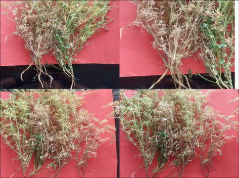 Figure 1
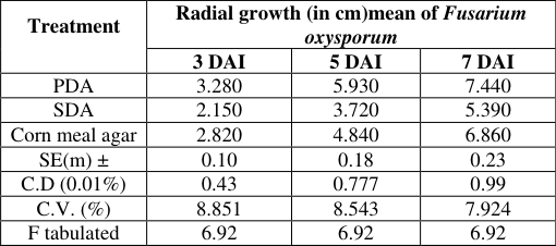 Table 1
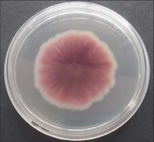 Figure 2
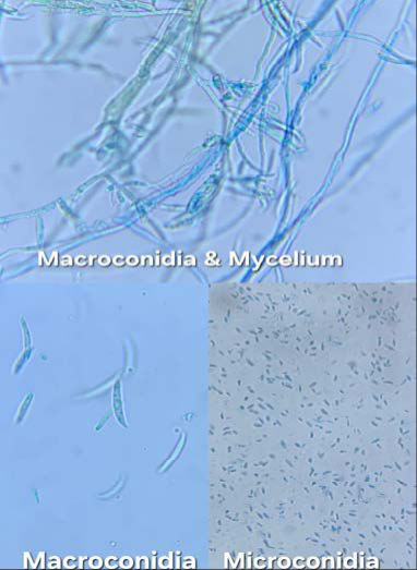 Figure 3
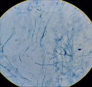 Figure 4
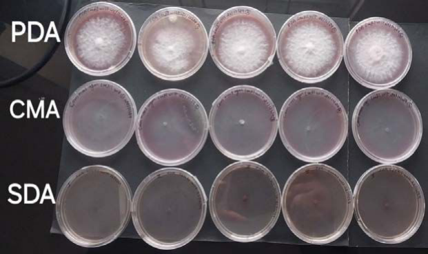 Figure 5
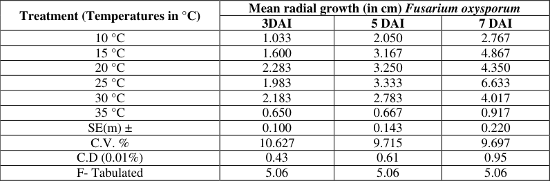 Table 2
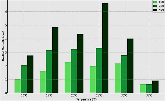 Figure 6
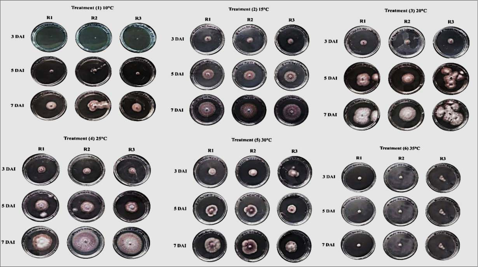 Figure 7
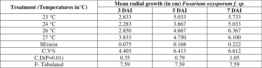 Table 3
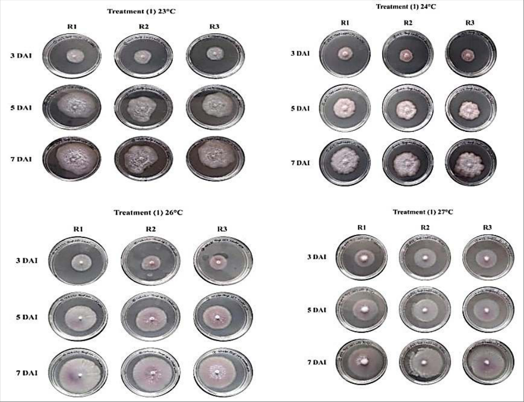 Figure 8
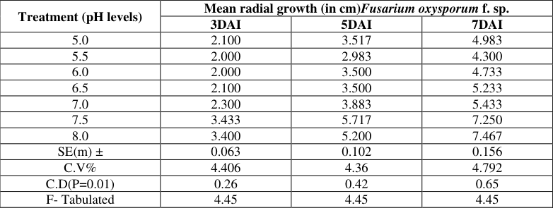 Table 4
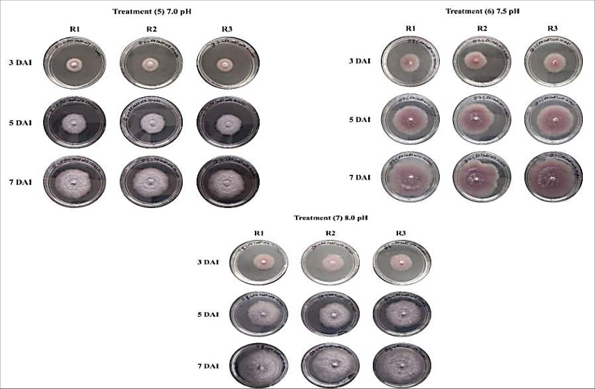 Figure 9
Introduction
Lentil (Lens culinaris Medik) is one of the earliest domesticated pulse crops and remains a vital component of global agriculture due to its high protein content, rich nutritional value, and multipurpose use in both human diets and animal feed (Sandhu & Singh, 2007; Zapata et al., 2004) [6,9] . In India, which contributes nearly 40% of global lentil production, the crop holds special importance as a primary protein source for the largely vegetarian population. However, lentil productivity is severely constrained by several fungal and bacterial diseases, include rust (Uromyces viciae-fabae), Ascochyta blight, Botrytis gray mold, Sclerotinia white mold, and Stemphylium blight. Among these, Fusarium wilt, caused by Fusarium oxysporum f. sp. lentis, is the most widespread and destructive disease, posing a significant threat to lentil productivity. Under favorableconditions, Fusarium wilt can cause yield losses ranging from 60% to 100% (Singh et al., 2007; Kumar et al., 2010) [6,4] , making it one of the most critical challenges in lentil farming. The disease progresses in stages, beginning with seed rot at the seedling stage, followed by drooping and wilting of upper leaves as the infection spreads. The most critical period for disease impact occurs between flowering and pod formation, leading to premature yellowing, plant death, and poor seed development. Infected seeds are often shriveled and exhibit low germination potential, ultimately causing a significant reduction in yield. Due to its severe impact on lentil productivity, Fusarium wilt remains a major concern for lentil growers worldwide (Grewal, 1988) [3] .
Materials and Methods
Materials and Methods
Survey of the Disease
A systematic survey was conducted during the Rabi season of 2024-25 to assess the lentil wilt caused by Fusarium oxysporum f. sp. lentis. A total of 10 fields were surveyed at intervals of 5-7 km across the fileds. In each field, lentil plants showing typical wilt
Selection of Suitable Culture Medium
Three culture media, viz., corn meal agar, Sabouraud dextrose agar (SDA) and potato dextrose agar (PDA) were used to select one for fast growth of the fungus. These culture media were prepared and sterilized as per the standard protocol and poured in 90mm polyethylene petri plates in laminar air flow. Three petri plates (R1, R2 and R3) with culture medium were inoculated with a single spore with culture medium were inoculated with discs of 5mm diameter cut from fresh culture of Fusarium oxysporum. Total three replicates of each culture media were maintained. All inoculated petri plates were incubated in a BOD incubator at 27±1°C for 7 days. Radial growth of the test fungus and spore formation were observed at 3, 5 and 7 Days After Inoculation (DAI). The data were tabulated to determine the best medium for fungal growth.
Effect of Temperatures on Mycelial Growth
Three replicates (R1, R2 and R3) with PDA culture medium were inoculated with a single spore from more than 7 days old culture were inoculated with discs of 5mm diameter cut from 3-5 days old culture of Fusarium oxysporum. temperature level were maintained. Inoculated petri plates were incubated at six different temperatures: 10 °C, 15 °C, 20 °C, 25 °C, 30 °C, and 35 °C up to 7 DAI. Twodimensional average radial growth was measured at 3, 5, and 7 days after inoculation (DAI) and tabulated for statistical analysis. When maximum radial fungal growth was found from any tested temperatures, two lower and two upper ranges of temperatures of maximum growth temperature were also tried to find out the optimum temperature. The experiment was arranged in a Completely Randomized Design (CRD) with three replications.
Effect of pH levels on Mycelial Growth
Potato Dextrose Agar (PDA) medium was prepared and divided into seven portions, each adjusted to a specific pH level ranging from 5.0 to 8.0 using 0.1 N NaOH or 0.1 N HCl prior to autoclaving. After sterilization, the pH-adjusted media were poured into Petri plates (20 ml per plate), with three replicates maintained for each pH level. Mycelial discs (5 mm diameter) were cut from actively growing fresh cultures and aseptically inoculated at the center of the plates. The inoculated plates were incubated at the optimum temperature of 26±1 °C for 7 days. Radial mycelial growth was recorded at 3, 5, and 7 days after inoculation (DAI), and the data were compiled and tabulated for statistical analysis.
Results and Discussion
Results and Discussion
Survey of the Disease
During the survey, lentil crops in different areas of Satna district were found at varying growth stages, ranging from 20-day-old seedlings to advanced flowering and pod-filling stages, due to differences in sowing time and prevailing weather conditions. whereas sowing in AKS University fields extended into November. Infected seedlings exhibited stunted growth, chlorosis, and poor root development, often with reduced or absent lateral roots. External foliar symptoms included yellowing and wilting of lower leaves, which later turned brown and necrotic. As the disease progressed, wilting advanced upward, ultimately leading to complete plant collapse. Typical root symptoms were characterized by dark brown to black discoloration of vascular tissues, often extending along the taproot. The terminal portions of the taproot and lateral roots appeared shriveled and necrotic. Severely infected plants frequently occurred in scattered patches across fields, reflecting the systemic and progressive nature of wilt caused by Fusarium oxysporum f. sp. lentis. Disease severity was observed to increase with plant age; in many cases, apical leaves of affected plants turned chlorotic while the rest of the plant became desiccated. Representative symptomatic plants from different growth stages (Fig. 1) were uprooted and collected in labeled polythene bags for further pathological studies.
Isolation and Identification of the Fungus (Fusarium oxysporum)
The fungus was isolated from wilt-infected lentil samples collected from different locations of Satna district, including Sohawal, Sherganj, and the AKS University fields. The isolates were purified using the single spore isolation technique and identified as Fusarium oxysporum f. sp. lentis based on their cultural and morphological characteristics. On PDA medium, the fungal colonies initially appeared white and fluffy, later developing pinkish to violet pigmentation with age (Fig. 2). Abundant aerial mycelium was also observed in some plates. The hyphae exhibited branching, which is a typical diagnostic feature of the species. Microscopic examination revealed that the vegetative mycelium was hyaline, septate, slender, and extensively branched, with septa frequently occurring near the branching points. The microconidia were hyaline, unicellular, slightly broad in the middle (oval-shaped), while some appeared short and rod-shaped (Fig. 3). The macroconidia were thin-walled, slightly curved, typically 3-4 septate, and possessed pointed ends (Fig. 4).
Chlamydospores were observed both terminally and intercalarily as thick-walled resting structures, occurring singly, in pairs, or occasionally in clusters. These morphological features confirmed the identity of the pathogen as Fusarium oxysporum f. sp. lentis.
Selection of Suitable Culture Medium
The results presented in Table 1, Figure 5, clearly indicate that PDA supported significantly higher radial growth of the fungus compared to SDA and CMA. The PDA supported the highest radial growth of the fungus at all observation intervals, recording (T1) 3.280 cm at 3 DAI, 5.930 cm at 5 DAI, and 7.440 cm at 7 DAI. Corn Meal Agar (CMA) also promoted substantial fungal growth, with radial growth measurements of (T3) 2.820 cm, 4.840 cm, and 6.860 cm at 3, 5, and 7 DAI, respectively. In contrast, Sabouraud Dextrose Agar (SDA) exhibited comparatively lower radial growth, recording (T2)2.150 cm, 3.720 cm, and 5.390 cm at the corresponding intervals. confirmed 25 °C as the optimum temperature for mycelial development (Table 3, Fig. 8). Statistical analysis showed these differences were highly significant. Similar findings have been reported by Chakrapani et al. (2023) [1] , Cruz et al. (2019) [2] , and Singh et al. (2011) [8] , who also observed optimum growth of Fusarium oxysporum at 25-27 °C, with reduced growth at both lower and higher extremes.
| Treatment (Temperatures in°C) | Mean radial growth (in cm) Fusarium oxysporum f. sp. | — | — |
|---|---|---|---|
| 3 DAI | 5 DAI | 7 DAI | |
| 23 °C | 2.833 | 5.033 | 5.733 |
| 24 °C | 2.283 | 3.667 | 5.033 |
| 26 °C | 2.850 | 4.667 | 6.367 |
| 27 °C | 3.833 | 4.750 | 6.100 |
| SE(m)± | 0.075 | 0.168 | 0.222 |
| C.V% | 4.403 | 6.413 | 6.612 |
| C.D(P=0.01) | 0.35 | 0.79 | 1.05 |
| F- Tabulated | 7.59 | 7.59 | 7.59 |
Table text extracted from the original PDF; superscript letters are shown inline. The original table image is below.
View original table image
| Treatment | Radial growth (in cm)mean of Fusarium oxysporum | — | — |
|---|---|---|---|
| 3 DAI | 5 DAI | 7 DAI | |
| PDA | 3.280 | 5.930 | 7.440 |
| SDA | 2.150 | 3.720 | 5.390 |
| Corn meal agar | 2.820 | 4.840 | 6.860 |
| SE(m) ± | 0.10 | 0.18 | 0.23 |
| C.D (0.01%) | 0.43 | 0.777 | 0.99 |
| C.V. (%) | 8.851 | 8.543 | 7.924 |
| F tabulated | 6.92 | 6.92 | 6.92 |
Table text extracted from the original PDF; superscript letters are shown inline. The original table image is below.
View original table image
Effect of pH on Growth of Fusarium oxysporum
The result presented in Table 4 shown in Figure 9 clearly demonstrate that different pH levels had a significant influence on the radial growth of Fusarium oxysporum f. sp. lentis at 3, 5, and 7 days after inoculation (DAI). The data clearly indicated that pH levels significantly affected the radial growth of the pathogen across all observation periods. At 3 DAI, the highest mean radial growth was observed at pH 7.5 (3.433 cm), followed closely by pH 8.0 (3.400 cm), while the lowest growth was recorded at pH 5.5 and 6.0 (2.000 cm each). At 5 DAI, maximum growth occurred at pH 7.5 (5.717 cm), followed by pH 8.0 (5.200 cm), whereas the least growth was recorded at pH 5.5 (2.983 cm). Similarly, at 7 DAI, the highest growth was recorded at pH 8.0 (7.467 cm), closely followed by pH 7.5 (7.250 cm), and the lowest at pH 5.5 (4.300 cm). The statistical analysis revealed that the differences among treatments were significant at P=0.01 level, as indicated by the calculated critical differences (C.D). These findings indicate that neutral to slightly alkaline conditions (pH 7.0-8.0) are most favorable for the growth of Fusarium oxysporum f. sp. lentis, while acidic pH considerably restricts its development. Similar observations were reported by Singh and Kumar (2016) [7] , who found that Fusarium oxysporum f. sp. lentis exhibited maximum growth and sporulation at pH 6.5-7.0, whereas acidic conditions significantly inhibited its growth (Indian Phytopathology).
| Treatment (pH levels) | Mean radial growth (in cm)Fusarium oxysporum f. sp. | — | — |
|---|---|---|---|
| 3DAI | 5DAI | 7DAI | |
| 5.0 | 2.100 | 3.517 | 4.983 |
| 5.5 | 2.000 | 2.983 | 4.300 |
| 6.0 | 2.000 | 3.500 | 4.733 |
| 6.5 | 2.100 | 3.500 | 5.233 |
| 7.0 | 2.300 | 3.883 | 5.433 |
| 7.5 | 3.433 | 5.717 | 7.250 |
| 8.0 | 3.400 | 5.200 | 7.467 |
| SE(m) ± | 0.063 | 0.102 | 0.156 |
| C.V% | 4.406 | 4.36 | 4.792 |
| C.D(P=0.01) | 0.26 | 0.42 | 0.65 |
| F- Tabulated | 4.45 | 4.45 | 4.45 |
Table text extracted from the original PDF; superscript letters are shown inline. The original table image is below.
View original table image
Conclusion
The present investigation confirmed that Fusarium oxysporum f. sp. lentis is a major causal agent of wilt in lentil crops across Satna district, Madhya Pradesh, during the Rabi season of 2024-25. Field surveys revealed typical wilt symptoms, including chlorosis, vascular discoloration, and plant collapse, with increasing incidence as crop growth advanced. Pathogen isolation and characterization through cultural and morphological features further validated its identity. The fungus produced white to pinkish-violet colonies on PDA with abundant aerial mycelium, while microscopic studies revealed septate hyphae, oval to rodshaped microconidia, slightly curved macroconidia, and thick-walled chlamydospores. Physiological assays indicated that PDA was the most suitable medium for rapid growth, while SDA showed the least support. Temperature trials demonstrated optimum mycelial development at 25 °C, with growth declining under both lower (10-15 °C) and higher (30-35 °C) regimes. Similarly, pH studies revealed that neutral to slightly alkaline conditions, particularly pH 7.5-8.0, favored maximum growth, while acidic conditions significantly restricted fungal proliferation. These findings provide valuable insights into the ecological requirements of F. oxysporum f. sp. lentis, which will be useful in devising effective disease management strategies and in identifying resistant cultivars for sustainable lentil production.
Figures and tables from the paper
| Treatment (Temperatures in °C) | Mean radial growth (in cm) Fusarium oxysporum | — | — |
|---|---|---|---|
| 3DAI | 5 DAI | 7 DAI | |
| 10 °C | 1.033 | 2.050 | 2.767 |
| 15 °C | 1.600 | 3.167 | 4.867 |
| 20 °C | 2.283 | 3.250 | 4.350 |
| 25 °C | 1.983 | 3.333 | 6.633 |
| 30 °C | 2.183 | 2.783 | 4.017 |
| 35 °C | 0.650 | 0.667 | 0.917 |
| SE(m) ± | 0.100 | 0.143 | 0.220 |
| C.V. % | 10.627 | 9.715 | 9.697 |
| C.D (0.01%) | 0.43 | 0.61 | 0.95 |
| F- Tabulated | 5.06 | 5.06 | 5.06 |
Table text extracted from the original PDF; superscript letters are shown inline. The original table image is below.
View original table image
References
9 in the original paper- Chakrapani S, Ramesh R, Kumar P. Effect of temperature on growth and sporulation of Fusarium f. sp. lentis under in vitro conditions. J Mycol Plant Pathol. 2023;53(2):145-150.
- Cruz JF, Silva RN, Oliveira AC. Influence of environmental factors on growth and pathogenicity of Fusarium oxysporum. Braz J Microbiol. 2019;50(3):617-625. doi:10.1007/s42770-019-00086-1
- Grewal JS. Diseases of pulse crops -An overview. Indian Phytopathol. 1988;41:1-14.
- Kumar S, Kumar J, Singh S, Ahmed S, Chaudhary R, Sarker A. Vascular wilt disease of lentil: A review. J Lentil Res. 2010;4:1-14.
- Sandhu JS, Singh S. History and origin. In: Yadav SS, McNeil DL, Stevenson PC, editors. Lentil: An Ancient Crop for Modern Times. Dordrecht: Springer Netherlands; 2007. p.1-9.
- Singh G, Chaudhary RG, Singh H. Screening of lentil genotypes for resistance against Fusarium wilt (Fusarium oxysporum f. sp. lentis). Indian J Pulses Res. 2007;20(1):67-69.
- Singh P, Kumar S. Effect of different media and pH on growth and sporulation of Fusarium oxysporum f. sp. lentis causing wilt of lentil. Indian Phytopathol. 2016;69(4 Suppl):80-82.
- Singh RP, Sharma A, Verma P. Effect of temperature and pH on growth and sporulation of Fusarium oxysporum f. sp. ciceris and lentis. Ann Plant Prot Sci. 2011;19(2):356-359.
- Zapata L, Peña-Chocarro L, Pérez-Jordá G, Stika HP. Early Neolithic agriculture in the Iberian Peninsula. J World Prehist. 2004;18(4):283-325.