
Research article · 2025 · Full text
International Journal of Agriculture and NutritionIn vitro evaluation of bioagents against Fusarium oxysporum F. sp. lentis causing wilt in lentil (Lens culinaris Medik)
Volume 7(8) · Pages 41–46 · DOI: 10.33545/26646064.2025.v7.i8a.266 ↗
Research areasGeochemistry and Geologic MappingGeological Modeling and AnalysisElectrical and Electromagnetic Research
Abstract
Author-writtenLentil (Lens culinaris Medik), a crucial pulse crop, is significantly affected by wilt caused by Fusarium oxysporum f. sp. lentis. This study investigates the occurrence, isolation, and in vitro management of this pathogen using three bioagents: Trichoderma viride, Neem oil, and Pseudomonas fluorescens. Surveys conducted in the Sohawal block of Satna district during the Rabi season of 2024–25 confirmed variable wilt incidence. The pathogen was isolated, purified. In vitro evaluations using poisoned food technique showed that all three bioagents inhibited fungal growth, with Trichoderma viride (T3) and Pseudomonas fluorescens (2%) demonstrating the highest efficacy. These findings suggest promising eco-friendly strategies for managing lentil wilt.
Summary
Source-checkedPlain-language reading of the full paper, prepared with AI and checked against the text. Each point links to the authors' own words.
Why it matters
- Lentil is an important pulse crop that suffers significant damage from wilt disease caused by the fungus Fusarium oxysporum f. sp. lentis. View evidence ↗
- Lentils are a vital protein source for the Indian population, especially those following vegetarian diets. View evidence ↗
- There is a need for eco-friendly management strategies to control lentil wilt. View evidence ↗
The question
- The study aimed to investigate the occurrence of the pathogen and evaluate the in vitro effectiveness of Trichoderma viride, Neem oil, and Pseudomonas fluorescens in managing it. View evidence ↗
What they did
- Disease surveys were conducted in the Sohawal block of Satna district during the 2024–25 Rabi season. View evidence ↗
- The pathogen was isolated by placing 5–6 mm pieces of infected roots and stems on Potato Dextrose Agar (PDA) and incubating them at 27 °C. View evidence ↗
- The in vitro test used 5 mm discs of 7-day-old fungal cultures on bioagent-amended PDA, with three replications per treatment and untreated controls. View evidence ↗
What they found
- At 7 days after inoculation (DAI), Pseudomonas fluorescens at 2% concentration resulted in the lowest radial growth of 6.013 cm, though this was lower than the 1% (8.338 cm) and 1.5% (8.063 cm) treatments. View evidence ↗
- Neem oil at a 1% concentration was the most effective dose, limiting fungal growth to 3.750 cm at 7 DAI compared to 4.163 cm in the control. View evidence ↗
- At 3 DAI, all Neem oil concentrations inhibited growth relative to the control (1.225 cm), with the 0.5% dose showing the lowest growth at 1.000 cm. View evidence ↗
- At 5 DAI, Trichoderma viride treatment T3 showed the lowest radial growth at 4.017 cm, compared to the control's 5.333 cm. View evidence ↗
What it means
- The authors concluded that Trichoderma viride (T3) provided the highest suppression, followed by 2% Pseudomonas fluorescens and 1% Neem oil. View evidence ↗
Visual summary
1 visualsIllustrations explain the paper; they do not show study data.
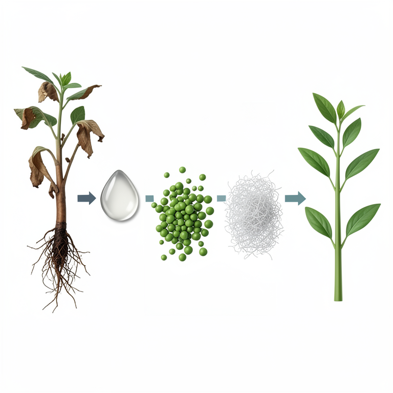
Comparison of Bioagent Efficacy
AI-generated conceptual illustration (lentil plants (Lens culinaris Medik) and Fusarium oxysporum fungi). It explains the idea and does not show study data.
View source evidence ↗Figures & tables
From the original paper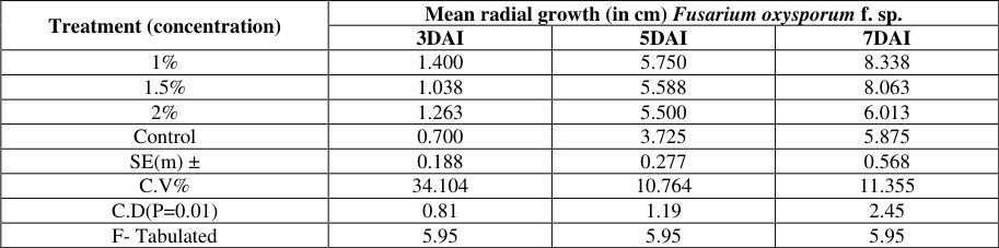 Table 3
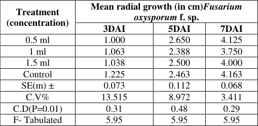 Table 3
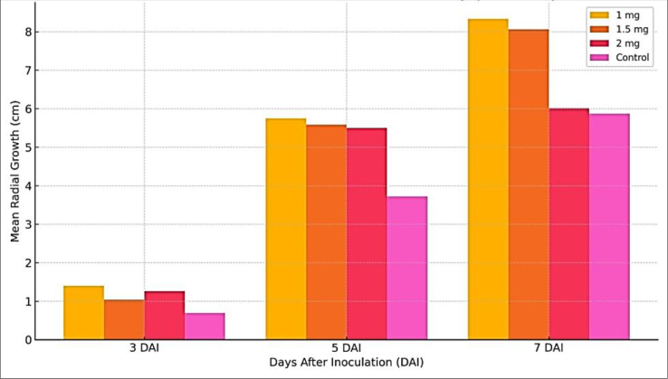 Figure 1
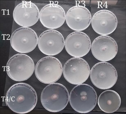 Figure 1
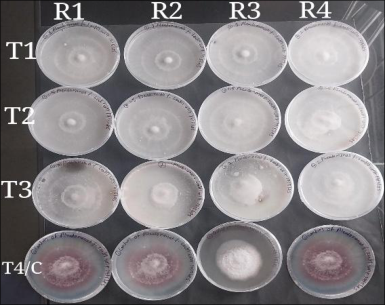 Figure 1
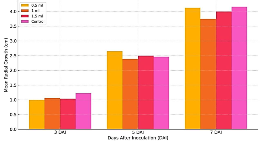 Figure 2
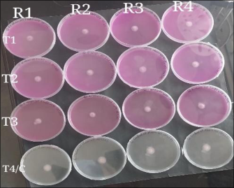 Figure 2
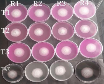 Figure 2
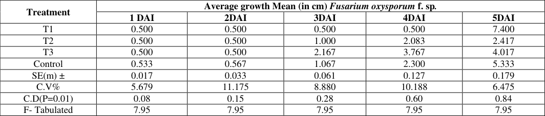 Table 3
Introduction
Lentil (Lens culinaris Medik) is among the earliest domesticated pulse crops and holds significant importance in global agriculture due to its high protein content and rich nutritional profile (Sandhu & Singh, 2007; Zapata et al., 2004) [8,12] . Pulses, including lentis, serve as a primary source of protein in Indian diets, particularly for the predominantly vegetarian population. However, India's pulse production has struggled to meet the increasing demand, necessitating enhanced cultivation strategies to bridge the supply gap. Lentil seeds are a nutrient-dense food source, comprising approximately 23.7% protein, 60% carbohydrates, and 11% water. In addition to macronutrients, lentils are rich in essential minerals such as calcium, iron, and niacin, making them a valuable component of a balanced diet. Beyond human consumption, lentil straw is widely utilized as a nutritious and costeffective animal feed, further reinforcing the crop's agricultural and economic significance. The present investigation explores the use of bioagents as an eco-friendly approach to manage lentil wilt.
Material methods Survey of disease incidence
A survey was conducted to record the occurrence and distribution of wilt of lentil in Sohawal block, Satna districts across one major lentil-growing block in central India -Madhya Pradesh, during the Rabi season of 2024-25. The number of fields in Sohawal block visited Satna district ranged from 10, with a distance of 5-7 km. A total of write numbers fields across Shohawal block of Satna districts were surveyed, and disease incidence, was recorded. In each field infected plant were randomly selected, and counted. Disease incidence was calculated based on the number of infected plants relative to the total number of plants. lentil plants displaying typical wilt symptoms were collected from the surveyed areas, packed in labelled polythene bags, and were brought to the laboratory for pathogen isolation.
Isolation and purification of fungus from infected roots and stem
Plants showing typical wilt symptoms were washed under running tap water and blot dried. Infected roots and stems were cut into pieces of 5-6 mm in size. After thorough washing in sterile distilled water the pieces were transferred using forceps onto sterilized Potato Dextrose Agar (PDA) medium in Petri dishes and incubated at 27 °C to obtain mycelium, conidia, conidiophores, and chlamydospores growth. After 48 hours of incubation, the growing mycelium and spores were observed and marked. Mycelium and spores from the margins of the resulting colonies were cut with a sterilized 5 mm cork borer and transferred to a Petri dish containing PDA for re-isolation of the pathogen. The cultures were then purified using the single spore isolation technique. Spores were collected from a sporulating fungal culture of 48 hrs old using a sterile loop and suspended in sterile distilled water in a test tube. Serial dilutions (10⁻ 1 to 10⁻⁵) of the spore suspension were prepared to reduce the spore concentration. A small drop of the diluted suspension was pipetted onto PDA medium in a Petri dish and spread gently using a sterile bent glass rod. The Petri dishes were incubated at an optimal temperature of 25-28 °C for 12-24 hours until germination was observed. Using a compound microscope, well-separated single germinating spores on the agar surface were located and transferred to fresh PDA plates. These plates were incubated at the appropriate temperature for fungal growth. Colony development was monitored, and the cultures were checked for purity by examining colony morphology and microscopic features to ensure separation of individual spores. The purified isolates were then transferred to PDA slants. The pure cultures of all isolates grown on PDA slants were stored at 8 °C for further studies.
In vitro evaluation of different bio-agents against the fungus
An in vitro study was conducted to assess the efficacy of different three Bio-agent: Trichoderma viride, Neem oil, Pseudomonas fluorescens against Fusarium oxysporum f. sp. lentis using the poisoned food technique with Potato Dextrose Agar (PDA) as the basal medium.
Bio-agents Tested and Their Concentrations: 1. Tricoderma viride 2. Neem Oil -0.5%, 1.0%, 1.5%, 2.0% (Neemanjali) 3. Pseudomonas fluorescens -1.0%, 1.5%, 2.0% (Multiplex)
Based on the active ingredient, the required amount of each bio-agents was weighted mixed thoroughly with autoclaved and cooled (40 °C) PDA in conical flasks to achieve the desired concentrations. Twenty milliter of boiagentsamended PDA was poured aseptically into 90 mm sterile glass Petri plates and allowed to solidify at room temperature. Each plate was then inoculated at the center with a 5 mm disc of actively growing Fusarium oxysporum f. sp. lentis culture (7-day-old), placed in an inverted position. Control plates (without any bio-agents) were also inoculated with the same culture disc to serve as untreated controls. Three replications were maintained for each treatment. Plates were incubated at optimum temperature and suitable pH for 7 days. After incubation, radial mycelium growth (in mm) of the pathogen was measured.
The percent inhibition of mycelium growth was calculated using the following formula
Where, C= growth of the test fungus in control plates (mm) T= growth of the test fungus in treated plates (mm)
Results and Discussion
Results and Discussion
Effect of Pseudomonas fluorescens on the Growth of
Effect of Pseudomonas fluorescens on the Growth of
Fusarium oxysporum f. sp. lentis
The radial growth of Fusarium oxysporum f. sp. lentis was significantly influenced by different concentrations of Pseudomonas fluorescens at 3, 5, and 7 days after inoculation (DAI). The results, as presented in Table 3.1, indicate a varied suppressive effect of the bacterial antagonist across different time intervals. At 3 DAI, the highest radial growth was observed in the 1% treatment (1.400 cm), followed by 1.5% (1.038 cm) and 2% (1.263 cm). Interestingly, the control (0.700 cm) recorded the lowest growth, likely due to slower initial colonization of the pathogen in the absence of bioagent competition. However, by 5 DAI, all treated plates exhibited increased growth compared to the control (3.725 cm), with the highest growth again at 1% (5.750 cm), followed closely by 1.5% (5.588 cm) and 2% (5.500 cm). This trend suggests that at early stages, P. fluorescens may not inhibit the pathogen significantly but exerts a stronger effect over time. By 7 DAI, a clear antagonistic effect was observed at the 2% concentration, which recorded the lowest radial growth (6.013 cm), significantly less than both 1% (8.338 cm), 1.5% (8.063 cm), and the control (5.875 cm). These findings imply that while lower concentrations may initially stimulate fungal growth due to the presence of additional nutrients or by competitive stress induction, higher concentrations exert an inhibitory influence as the bacterial population stabilizes and produces antifungal metabolites. These observations align with those reported by Raghuchander et al. (2004) [4] and Sharma and Sharma (2008) [10] , who demonstrated that P. fluorescens significantly inhibits Fusarium spp. in vitro through the production of antibiotics (such as pyoluteorin and 2,4-diacetylphloroglucinol), siderophores, and lytic enzymes. Gopalakrishnan et al. (2011) [3] also confirmed the suppressive effects of Pseudomonas species against soilborne pathogens including F. oxysporum in chickpea and lentil systems, suggesting that strain specificity and application dosage are key determinants of efficacy.
| Treatment | Average growth Mean (in cm) Fusarium oxysporum f. sp. | — | — | — | — |
|---|---|---|---|---|---|
| 1 DAI | 2DAI | 3DAI | 4DAI | 5DAI | |
| T1 | 0.500 | 0.500 | 0.500 | 0.500 | 7.400 |
| T2 | 0.500 | 0.500 | 1.000 | 2.083 | 2.417 |
| T3 | 0.500 | 0.500 | 2.167 | 3.767 | 4.017 |
| Control | 0.533 | 0.567 | 1.067 | 2.300 | 5.333 |
| SE(m) ± | 0.017 | 0.033 | 0.061 | 0.127 | 0.179 |
| C.V% | 5.679 | 11.175 | 8.880 | 10.188 | 6.475 |
| C.D(P=0.01) | 0.08 | 0.15 | 0.28 | 0.60 | 0.84 |
| F- Tabulated | 7.95 | 7.95 | 7.95 | 7.95 | 7.95 |
Table text extracted from the original PDF; superscript letters are shown inline. The original table image is below.
View original table image
| Treatment (concentration) | Mean radial growth (in cm)Fusarium oxysporum f. sp. | — | — |
|---|---|---|---|
| 3DAI | 5DAI | 7DAI | |
| 0.5 ml | 1.000 | 2.650 | 4.125 |
| 1 ml | 1.063 | 2.388 | 3.750 |
| 1.5 ml | 1.038 | 2.500 | 4.000 |
| Control | 1.225 | 2.463 | 4.163 |
| SE(m) ± | 0.073 | 0.112 | 0.068 |
| C.V% | 13.515 | 8.972 | 3.411 |
| C.D(P=0.01) | 0.31 | 0.48 | 0.29 |
| F- Tabulated | 5.95 | 5.95 | 5.95 |
Table text extracted from the original PDF; superscript letters are shown inline. The original table image is below.
View original table image
| Treatment (concentration) | Mean radial growth (in cm) Fusarium oxysporum f. sp. | — | — |
|---|---|---|---|
| 3DAI | 5DAI | 7DAI | |
| 1% | 1.400 | 5.750 | 8.338 |
| 1.5% | 1.038 | 5.588 | 8.063 |
| 2% | 1.263 | 5.500 | 6.013 |
| Control | 0.700 | 3.725 | 5.875 |
| SE(m) ± | 0.188 | 0.277 | 0.568 |
| C.V% | 34.104 | 10.764 | 11.355 |
| C.D(P=0.01) | 0.81 | 1.19 | 2.45 |
| F- Tabulated | 5.95 | 5.95 | 5.95 |
Table text extracted from the original PDF; superscript letters are shown inline. The original table image is below.
View original table image
Effect of Neem Oil on the Growth of Fusarium oxysporum f. sp. lentis
Neem oil treatments at 0.5%, 1.0%, and 1.5% concentrations exhibited a moderate inhibitory effect on F. oxysporum f. sp. lentis, with the highest suppression generally observed at the 1.0% concentration. At 3 DAI, all Neem oil treatments showed slight inhibition of fungal growth compared to the control (1.225 cm), with the lowest growth observed at 0.5% (1.000 cm). The difference, although minor, was statistically significant (CD = 0.31 at P = 0.01). By 5 DAI, 1% Neem oil recorded the least radial growth (2.388 cm), followed by 1.5% (2.500 cm) and 0.5% (2.650 cm), while the control remained at 2.463 cm. At 7 DAI, the 1% treatment continued to perform best, with growth limited to 3.750 cm, lower than the control (4.163 cm). These results suggest that Neem oil at 1% concentration exerts a consistent fungistatic effect on Fusarium growth. The antifungal action is likely due to azadirachtin and other limonoids, which interfere with fungal membrane function and enzyme activity. Similar inhibitory effects of Neem-based products have been reported by Patel et al. (2015) [6] and Manjunatha et al.
(2012) against Fusarium spp. and Rhizoctonia bataticola in legumes. Saxena and Pandey (2001) [9] also documented that Neem oil inhibits sporulation and conidial germination, contributing to disease suppression under laboratory conditions. However, it was also noted in these studies that excessive concentrations (>2%) may result in phytotoxicity or reduced antagonistic efficiency, reinforcing the effectiveness of the 1% dose observed in the current experiment. [2] and Papavizas (1985) [5] , who described the strong antagonistic potential of Trichoderma spp. against several phytopathogens through production of cell wall-degrading enzymes (chitinases, glucanases) and volatile inhibitors. Dubey and Patel (2001) [1] and Singh et al. (2010) [11] also reported similar trends in lentil and chickpea pathosystems, affirming Trichoderma as a potent biocontrol agent under both in vitro and field conditions.
Conclusion
The present study reveals the differential inhibitory effects of three bioagents on the growth of Fusarium oxysporum f. sp. lentis. Among the tested agents: 1. Trichoderma viride (T3) showed highest suppression 2. Pseudomonas fluorescens at 2% was also highly effective 3. Neem oil at 1% had moderate efficacy
Figures and tables from the paper
References
12 in the original paper- Dubey SC, Patel B. Biological management of lentil wilt caused by Fusarium oxysporum f. sp. lentis. Indian Phytopathol. 2001;54(3):306-309.
- Elad Y, Chet I, Henis Y. Parasitism of Trichoderma spp. on Rhizoctonia solani and Sclerotium rolfsii: scanning electron microscopy and fluorescence microscopy. Phytopathology. 1983;73(1):85-88. doi:10.1094/Phyto-73-85.
- Gopalakrishnan S, Upadhyaya HD, Vadlamudi S, Humayun P, Vijayabharathi R. Evaluation of bacteria isolated from rice rhizosphere for biological control of chickpea wilt caused by Fusarium oxysporum f. sp. ciceris. Arch Phytopathol Plant Prot. 2011;44(7):700714. doi:10.1080/03235400903464653.
- Manjunatha G, Naik MK, Mini C. Evaluation of plant extracts and bio-agents for the management of root rot disease of greengram incited by Rhizoctonia bataticola. Plant Arch. 2012;12(2):841-843.
- Papavizas GC. Trichoderma and Gliocladium: biology, ecology, and potential for biocontrol. Annu Rev Phytopathol. 1985;23:23-54. doi:10.1146/annurev.py.23.090185.000323.
- Patel A, Meena PD, Yadav RK. Antifungal activity of neem-based formulations against some important plant pathogenic fungi. Ann Plant Prot Sci. 2015;23(1):168170.
- Raghuchander T, Samiyappan R, Anand T. Evaluation of native strains of Pseudomonas fluorescens for the management of chickpea wilt. J Biol Control. 2004;18(2):121-127.
- Sandhu JS, Singh G. Lentil. In: Handbook of Agriculture. New Delhi: Indian Council of Agricultural Research; 2007. p. 947-951.
- Saxena A, Pandey RN. Neem for management of plant diseases. Indian J Plant Prot. 2001;29(1-2):47-52.
- Sharma JP, Sharma P. Effect of bioagents on management of wilt complex in chickpea. Indian Phytopathol. 2008;61(3):344-346.
- Singh D, Singh AK, Upadhyay JP. Biocontrol potential of Trichoderma viride and T. harzianum against Fusarium oxysporum f. sp. lentis causing lentil wilt. Indian Phytopathol. 2010;63(2):178-183.
- Zapata FJ, Axmann H, Roy RN. Fertilizer use by crop in India. Rome: Food and Agriculture Organization of the United Nations; 2004.