
Research article · 2025 · Full text
International Journal of Biology SciencesComparative evaluation of lateral flow assay and polymerase chain reaction in the detection of canine parvovirus
Volume 7(11) · Pages 40–42 · DOI: 10.33545/26649926.2025.v7.i11a.516 ↗
Research areasVirus-based gene therapy researchParvovirus B19 Infection StudiesAnimal Virus Infections Studies
Abstract
Author-writtenCanine parvovirus (CPV) infection poses a significant threat to the health and well-being of dogs worldwide. It is a small, non-enveloped virus consisting of linear, single-stranded negative-sense DNA of approximately 5 kb in length. The present study was conducted in Department of Veterinary Medicine, Veterinary College, Bengaluru to assess the efficacy of lateral flow assay (LFA) in comparison to the conventional polymerase chain reaction (PCR) in detecting CPV antigen in the faecal samples of CPV- suspected dogs under one year of age. CPV antigen was detected by LFA in 44 percent of the suspected dogs, whereas PCR detected CPV DNA in 72 percent. PCR targeting partial VP (Viral Protein)-2 gene, yielding the amplicon size of 630 base pairs (bp) was performed. The specificity and sensitivity of LFA was 100 and 61.1 percent, respectively, in comparison with the PCR. Although LFA allow patient side diagnosis PCR remains the most sensitive and reliable method for the confirmatory detection of CPV infection in clinical cases.
Summary
Source-checkedPlain-language reading of the full paper, prepared with AI and checked against the text. Each point links to the authors' own words.
Why it matters
- Canine parvovirus (CPV) 2 is a significant pathogen in dogs, causing myocarditis and hemorrhagic gastroenteritis. View evidence ↗
- CPV is a global virus with high mortality rates, reaching approximately 90% without treatment and 10% with treatment. View evidence ↗
- Lateral flow assay (LFA) and polymerase chain reaction (PCR) are the most common diagnostic techniques used in routine veterinary hospital laboratories. View evidence ↗
The question
- The study aimed to compare the efficacy of LFA and conventional PCR for detecting CPV antigen in fecal samples from suspected dogs under one year of age. View evidence ↗
What they did
- The study included 50 dogs under one year old at the Veterinary College in Bengaluru that showed symptoms like lethargy, diarrhea, vomiting, and anorexia. View evidence ↗
- The LFA was performed using the Canine Parvo Virus Ag Rapid Test kit from PetX (J&G Biotech Ltd.). View evidence ↗
- The PCR targeted the partial VP-2 capsid protein gene, producing an amplicon size of 630 base pairs. View evidence ↗
What they found
- LFA detected CPV antigen in 44% (22/50) of the samples, while 56% (28/50) were negative. View evidence ↗
- When compared to PCR, the LFA demonstrated 100% specificity but only 61.1% sensitivity. View evidence ↗
What it means
- The authors conclude that PCR remains the most specific and sensitive technique for confirming CPV infection in clinical cases. View evidence ↗
- While LFA is useful for point-of-care diagnosis, it has a higher rate of false negatives due to antibody neutralization or low viral shedding in early infection. View evidence ↗
Visual summary
1 visualsIllustrations explain the paper; they do not show study data.
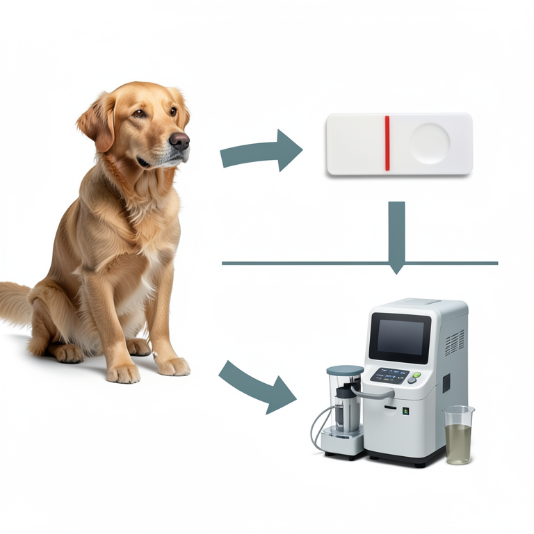
Diagnostic Comparison Concept
AI-generated conceptual illustration (Canine parvovirus in dog fecal samples). It explains the idea and does not show study data.
View source evidence ↗Figures & tables
From the original paper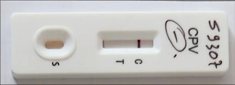 Figure 1
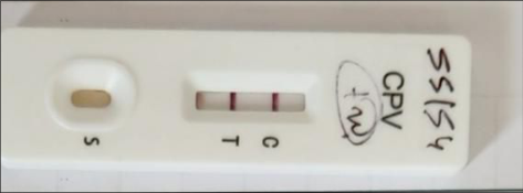 Figure 2
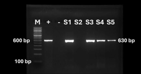 Figure 3
Introduction
Canine parvovirus (CPV) 2 is a major canine pathogen that causes hemorrhagic gastroenteritis and can also induce myocarditis in dogs. Identified in 1977, it has become a global enteric virus in canines, with high morbidity and mortality reported up to about 10 percent with treatment and 90 percent without treatment (Olaifa et al., 2025) [10] . Several diagnostic methods exist in research settings for CPV detection, including hemagglutination assays, immunofluorescence, enzyme linked immunosorbent assay (ELISA), polymerase chain reaction (PCR), lateral flow assay (LFA) and cell culture, but in routine veterinary hospital laboratories, the most commonly used techniques are the LFA and PCR (Faz et al., 2017) [4] . The positive LFA results were considered reliable, but negative results required PCR confirmation, reinforcing that clinical diagnosis alone was indecisive and that laboratory confirmation on faeces was essential with PCR (Khajeh-Kazerooni, 2020) [7] . The present compared the sensitivity and specificity of LFA and PCR.
Materials And Methods
The study population comprised of 50 dogs below one year of age, that were presented to the Department of Veterinary Medicine, Veterinary College, Bengaluru, exhibiting clinical manifestations suggestive of CPV infection, including anorexia, vomiting, diarrhoea and lethargy. All the faecal samples of the study population underwent LFA and PCR for the confirmation of CPV antigen. The manufacturer's guidelines were followed LFA, using LFA kits (Canine Parvo Virus Ag Rapid Test, PetX, J&G Biotech Ltd., London, England). Faecal samples were collected from dog's rectum using a sterile cotton swab and were added into the provided assay buffer tube and agitated. The test device was taken out from the package just prior to examination and placed it horizontally on the table and about two millilitres of assay buffer containing the sample was sucked into a dropper provided, and five drops of it was placed into the sample hole "S" of the test device. The result was interpreted within five to 10 minutes. Only clear control (C) line, with no test (T) line was reported as negative, whereas presence of both C line and T line was reported as positive. If no coloured line appeared in C zone, irrespective of T line, the test was declared invalid. The faecal samples were stored at -20℃ for further analysis. The extraction of viral DNA from faecal samples was conducted in accordance with the manufacturer's instructions using DNA extraction kit (DNeasy, QIAGEN, Germany). In a 1.5 mL microcentrifuge tube, 200 µL of the sample was mixed with 200 µL of the lysis buffer (Buffer ATL) and 20 µL of Proteinase K. The mixture was thoroughly vortexed to ensure complete lysis. Subsequently, 200 µL of absolute ethanol was added, and the solution was mixed again by vortexing. The entire lysate, including any precipitate, was then transferred to a DNeasy Mini spin column placed in a two mL collection tube and centrifuged at 8000 rpm for one minute. The flow-through and collection tube were discarded. The column was transferred to a new collection tube and 500 µL of Buffer AW1 was added before centrifugation at 8000 rpm for one minute. The flow-through and tube were again discarded and 500 µL of Buffer AW2 was added to the column, followed by centrifugation at 14,000 rpm for three minutes. Finally, the column was placed in a clean 1.5 mL microcentrifuge tube and 200 µL of Buffer AE was added directly to the membrane. After incubation at room temperature for one minute, DNA was eluted by centrifugation at 8000 rpm for one minute. The DNA from a known CPV-positive sample was extracted and used as the positive control. Nuclease free water was used as no template control (NTC). The quantity and purity of the extracted DNA was evaluated, before preparing the PCR reaction mix, using nucleic acid analyser (Genetix, Biotech Asia, India). The extracted DNA was stored at -80 ℃ till further analysis. Conventional PCR targeting the partial viral protein (VP)-2 capsid protein gene yielding amplicon size of 630 base pairs (bp) was performed as described by Buonavoglia et al. (2001), using the forward primer (CAGGTGATGAATTTGCTACA) and reverse primer (CATTTGGATAAACTGGTGGT) for the positions 3556-3575 and 4166-4185, respectively. The PCR was carried out in a final reaction volume of 25 μl. Each reaction mixture consisted of 12.5 μl of 2X PCR Master Mix, 5.5 μl of nuclease-free water, five μl of template DNA, one μL each of forward and reverse primers. The PCR amplification was performed under the following thermal cycling conditions, an initial denaturation at 95 ℃ for five minutes, followed by 35 cycles consisting of denaturation at 95 ℃ for 30 seconds, annealing at 56 ℃ for 30 seconds, and extension at 72 ℃ for one minute. This was followed by a final extension step at 72 ℃ for 10 minutes, and the reaction was then held at 4 ℃ until further analysis.
Ten microliters of the PCR products were loaded into their respective wells in the 1.5 percent agarose gel, along with 10 microlitre 100 base pair ladder in a single separate well. Electrophoresis device was done at 50 Volts capacity for 45 minutes and gel was analysed in a UV transilluminator for visualization of bands. The results were documented by using a gel documentation system (GELSTAN 1312, Mediccare Scientific Supplies, India).
Results and Discussion
Of the 50 samples analysed using LFA, 44 percent (22) tested positive (Fig. 2) for CPV antigen, while 56 percent (28) tested negative (Fig. 1). PCR (Fig. 3) detected CPV antigen in 72 percent (36) samples, whereas 28 percent ( 14) samples were negative. Compared to PCR, LFA had 100 percent specificity and 61.1 percent sensitivity. to the infection in naive or poorly immunized dogs through oronasal exposure (Ford et al. 2017) [5] .
Although LFA allow patient side diagnosis, likelihood of false negatives in LFA has been attributed to low faecal viral loads during early infection or the neutralization of antigen by antibodies in the intestinal lumen (Navarro, 2020; Tangolli et al., 2024) [9,13] . Sensitivity of LFA was improved substantially when tests were performed immediately after collection, thus negative LFA results did not exclude CPV, particularly with recent vaccination or stored samples (Kantere et al. 2015) [9] . Dogs tested too early in the course of infection might not yet shed detectable levels of virus, contributing to false-negative LFA outcomes, but clinical signs, including severity and prognosis, did not significantly differ between LFA-positive and LFA-negative dogs, except for a variation in defecation frequency (Proksch et al. 2015) [11] .
The most sensitive and reliable method for the confirmatory detection of CPV infection in clinical cases was PCR, as demonstrated in the present study (Navarro, 2020; Tangolli et al., 2024) [9,13] . [14] . PCR can also be used for detection of viral DNA from various tissues like heart and bone marrow, which is helpful in understanding the pathogenesis of the disease (Stancu et al., 2025) [12] .
Conclusion
Though LFA enables point-of-care diagnosis, the higher percentage of false negatives has been linked to reduced fecal viral shedding in the early stages of infection or antigen neutralization by antibodies. In contrast, PCR continues to be the most sensitive and specific technique for confirming CPV infection in clinical cases, as shown in the present study.
References
14 in the original paper- Buonavoglia C, Martella V, Pratelli A, Tempesta M, Cavalli A, Buonavoglia D, et al. Evidence for evolution of canine parvovirus type 2 in Italy. J Gen Virol. 2001 Dec;82(12):3021-3025.
- Decaro N, Elia G, Martella V, Campolo M, Desario C, Camero M, et al. A real-time PCR assay for rapid detection and quantitation of canine parvovirus type 2 DNA in the feces of dogs. Vet Microbiol. 2005 Jan;105(1):19-28.
- Desario C, Decaro N, Campolo M, Cavalli A, Cirone F, Elia G, et al. Canine parvovirus infection: which diagnostic test for virus? J Virol Methods. 2005 Sep;126(1):179-185.
- Faz M, Rojas M, Segura-Correa JC, Cob-Galera L, Echeverría JC, Larios HM. Reliability of clinical diagnosis and laboratory testing for canine parvovirus type 2 in Mexico. PLoS One. 2017 Nov;12(11):18-24.
- Ford J, McEndaffer L, Renshaw R, Molesan A, Kelly K. Parvovirus infection is associated with myocarditis and myocardial fibrosis in young dogs. Vet Pathol. 2017 Nov;54(6):964-971.
- Kantere M, Athanasiou LV, Chatzopoulos D, Martellos P, Kanellos T, Ikonomopoulos J, Polizopoulou Z. Evaluation of a rapid in-clinic test for the detection of canine parvovirus. J Small Anim Pract. 2015 Jun;56(6):389-393.
- Khajeh-Kazerooni S. Canine parvovirus infection: clinical features and diagnostic approaches. Vet Res Forum. 2020 Sep;11(3):241-248.
- Miranda C, Thompson G. Canine parvovirus: the worldwide occurrence of antigenic variants. J Gen Virol. 2016 Sep;97(9):2043-2057.
- Navarro C. Diagnosis of canine parvovirus infection: updates and pitfalls. Top Companion Anim Med. 2020 Jun;39:100-108.
- Olaifa S, Ajayi O, Akande F, Adebiyi A, Ahmed S. Clinical outcome of canine parvoviral enteritis: a systematic review and meta-analysis. Front Vet Sci. 2025 Feb;12:15-21.
- Proksch AL, Unterer S, Speck S, Truyen U, Hartmann K, Straubinger RK. Influence of clinical and laboratory variables on fecal antigen ELISA results in dogs with canine parvovirus infection. Vet J. 2015 Dec;204(3):304-308.
- Stancu A, Cârstina D, Bîrzan C, Gherghel I, Tănase A. Detection of canine parvovirus DNA in cardiac and hematopoietic tissues of infected dogs using PCR. Animals. 2025 Mar;15(3):512-521.
- Tangolli G, Patil S, Kumar A, Reddy P, Joseph J. Performance of rapid antigen tests versus PCR for canine parvovirus diagnosis in clinical practice. Vet Sci. 2024 Feb;11(2):87-94.
- Wang J, Liu L, Li R, Li J, Yang J, Xie Q, et al. Rapid and sensitive detection of canine parvovirus type 2 by recombinase polymerase amplification. Sci Rep. 2016 Feb;6:25-33.