
Research article · 2025 · Full text
International Journal of Agriculture and Food ScienceInfluence of nutrition, temperature, and pH on the mycelial growth and sclerotial development of Rhizoctonia solani Kühn, an incitant of rice sheath blight
Volume 7(11) · Pages 78–88 · DOI: 10.33545/2664844x.2025.v7.i11b.943 ↗
Research areasPlant Disease Resistance and GeneticsPlant-Microbe Interactions and ImmunityPlant and Fungal Interactions Research
Abstract
Author-writtenSheath blight of rice, caused by Rhizoctonia solani Kühn, poses a major threat to rice cultivation worldwide. This soil-borne fungus exhibits strong saprophytic growth and infects a wide range of host plants. Under favorable environmental conditions and with a high inoculum load, the disease severity increases significantly. Among the tested media, Potato Dextrose Agar, Czapek’s Dox Agar, and modified Czapek’s Agar were found to best support R. solani growth, each promoting maximum mycelial development (90 mm) and abundant sclerotia formation. Among the various carbon and nitrogen sources tested, sucrose, dextrose, and mannitol, along with ammonium nitrate and sodium nitrate, proved to be the most effective in promoting the growth and sclerotial formation of Rhizoctonia solani compared to other supplemented sources. Although the fungus was able to grow across a broad pH range, the highest mycelial growth and sclerotial production occurred at pH 5. Optimal growth and sclerotia formation were recorded at temperatures between 25 °C and 30 °C on Potato Dextrose Agar under in vitro conditions. These findings indicate that suitable nutrient sources, temperature, and pH serve as key edaphic factors influencing the growth and sclerotial development of R. solani.
Summary
Source-checkedPlain-language reading of the full paper, prepared with AI and checked against the text. Each point links to the authors' own words.
Why it matters
- Rice sheath blight, caused by the fungus Rhizoctonia solani, is a significant global threat to rice production. View evidence ↗
- Pests and diseases are estimated to cause a 35-40% loss in annual rice yields. View evidence ↗
- The pathogen is soil-borne and persists in the environment as mycelia or sclerotia. View evidence ↗
The question
- The study aimed to determine how different nutrient sources, temperature, and pH levels affect the mycelial growth and sclerotial production of R. solani. View evidence ↗
What they did
- Samples were collected from the southern Tamil Nadu districts of Tirunelveli, Thoothukudi, and Kanyakumari. View evidence ↗
- Pathogenicity was tested by inoculating 45-day-old rice plants with 1 g of rice hull-rice grain inoculum placed 3-4 cm above the water level. View evidence ↗
- The effect of temperature was tested using a range from 0 °C to 40 °C. View evidence ↗
- The effect of pH was evaluated across a range from 4.0 to 9.0. View evidence ↗
What they found
- Field surveys showed disease incidence between 32.58% and 44.05%, with the highest rate (44.05%) found in Thirupathisaram. View evidence ↗
- Isolate RS5 was the most virulent, causing 92.30% disease incidence under artificial inoculation. View evidence ↗
- The highest mycelial growth (90 mm) occurred on potato dextrose agar, Czapek’s dox agar, and modified Czapek’s dox agar, while King’s B medium supported the least growth (18.66 mm). View evidence ↗
- Sucrose, dextrose, and mannitol promoted the highest mycelial growth (90 mm), whereas carboxymethyl cellulose resulted in the lowest growth (54.30 mm). View evidence ↗
- Ammonium nitrate and sodium nitrate were the most effective nitrogen sources for growth (90 mm), while urea was the least effective (40.66 mm). View evidence ↗
- Maximum mycelial growth (90 mm) occurred at pH 5.0, with the minimum growth (76 mm) observed at pH 8.0. View evidence ↗
- Optimal growth (90 mm) and maximum sclerotial formation occurred at 25 °C and 30 °C, while no sclerotia formed at 5 °C and 10 °C. View evidence ↗
What it means
- The authors recommend nutrient management, altering soil pH, and forecasting seasonal changes to help rice crops avoid sheath blight. View evidence ↗
- The authors state that further research is needed on the survival of sclerotia in the field and the pathogenicity of different anastomosis groups. View evidence ↗
Visual summary
2 visualsIllustrations explain the paper; charts are replotted from the paper's own table values.
*Mycelial growth of the pathogen(mm): Potato dextrose agar... vs King`s B agar medium vs Nutrient agar medium vs Ken Knight`s agar medium vs Oat meal agar medium
Replotted from Table 4 (page 8) of the original paper. Every bar is the paper's own table value.
View source evidence ↗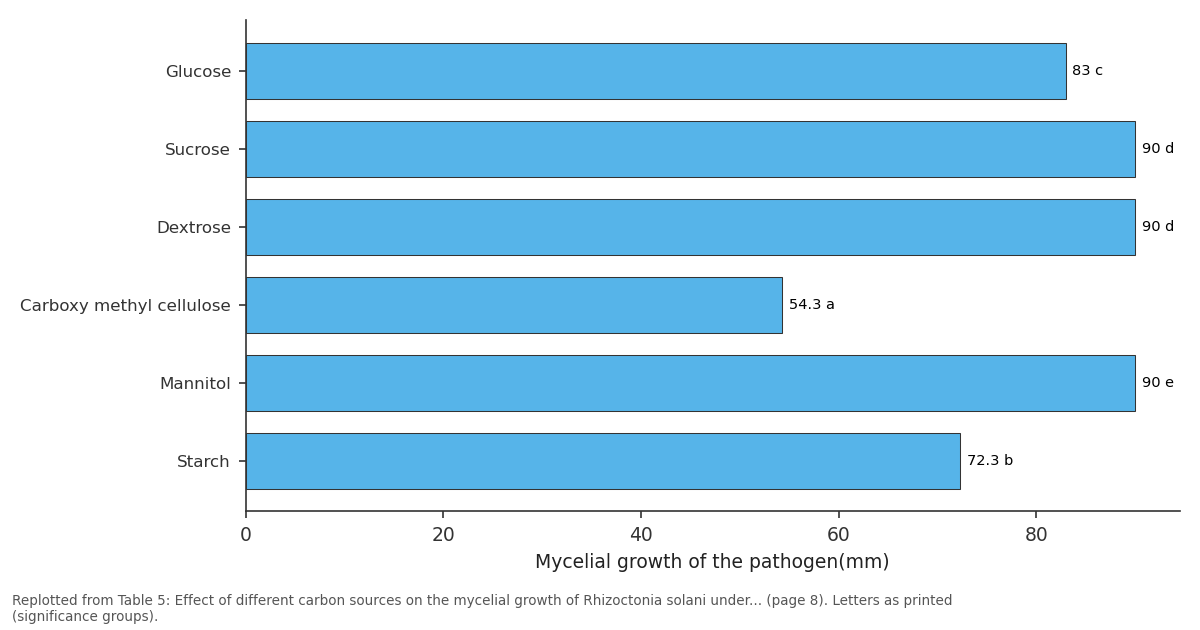
*Mycelial growth of the pathogen(mm): Glucose vs Sucrose vs Dextrose vs Carboxy methyl cellulose vs Mannitol
Replotted from Table 5 (page 8) of the original paper. Every bar is the paper's own table value.
View source evidence ↗Figures & tables
From the original paper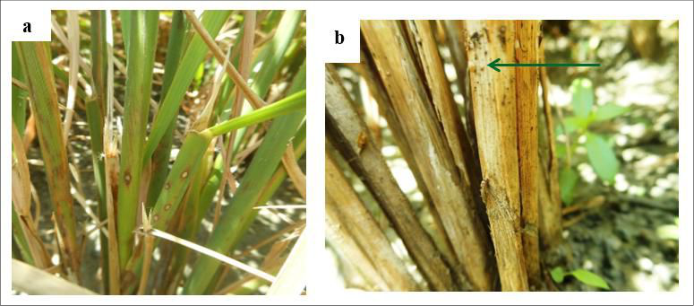 Figure 1
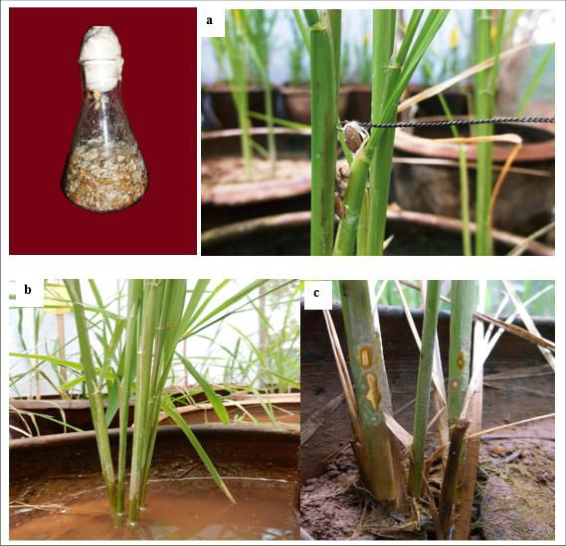 Figure 2
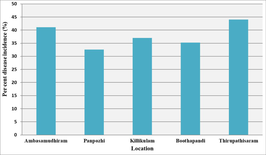 Figure 3
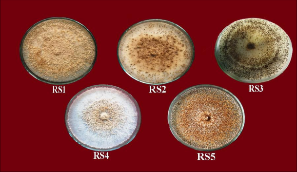 Figure 4
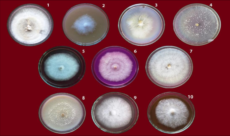 Figure 5
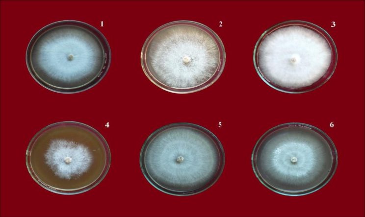 Figure 6
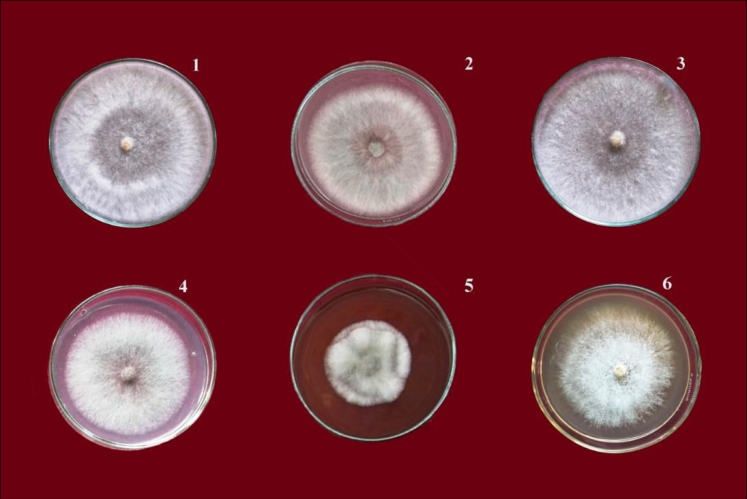 Figure 7
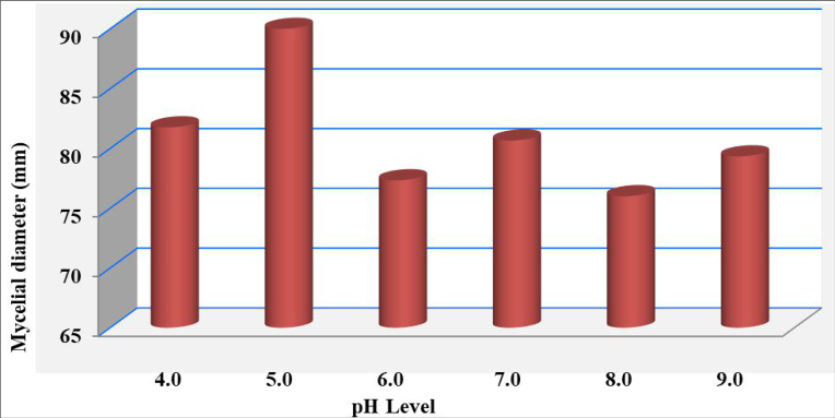 Figure 8
 Table 1
 Table 2
 Table 3
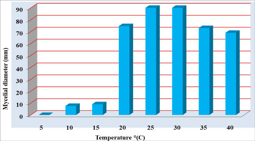 Figure 9
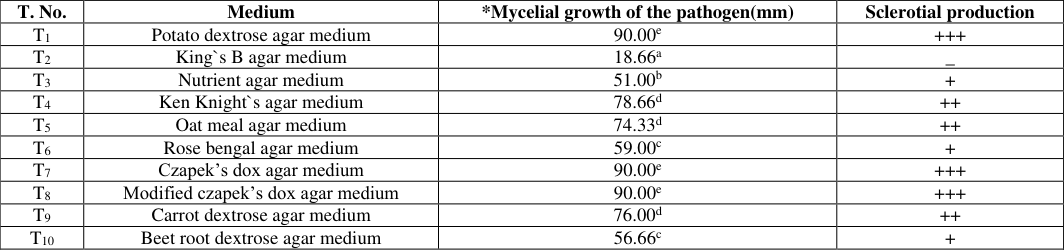 Table 4
 Table 5
 Table 6
 Table 7
 Table 8
Introduction
Rice (Oryza sativa L.) is one of the most important staple food crops widely consumed across Asian countries. India ranks as the world's second-largest rice producer, cultivating approximately 42, 949.80 thousand hectares and yielding about 112, 905 thousand tonnes annually (India Stat, 2022-23). The crop, however, is vulnerable to numerous diseases caused by fungi, bacteria, viruses, phytoplasmas, and nematodes. Collectively, pests and diseases account for an estimated 35-40% loss in annual rice yield [1] . Among these, sheath blight, caused by Rhizoctonia solani Kühn, has emerged as a rapidly increasing and serious threat to rice production across India. The disease primarily infects on leaf sheath. Initially, the symptoms appear on leaf sheath as1 to 3 cm long, elliptical or oval to irregular, greenish gray spots with brown margin at or above the water line. The tillering stage of the crop is more vulnerable for the infection to take place (Fig. 1a). Under conducive environmental conditions, the disease spreads faster even to the panicles and results in poor filling of the grains. Turaidar [2] reported that sheath blight pathogen during severe stage of infection and disease development forms brown sclerotia, which detach from the affected plant part and serves as an inoculum (Fig. 1b). The pathogen Rhizoctonia solani is soil-borne in nature and can survive for extended periods either as mycelia or sclerotia in the soil or on infected rice stubbles [3] . The severity of sheath blight largely depends on environmental factors, the genetic makeup of the rice cultivar, and the virulence of the pathogen. The disease primarily affects the leaf sheath and panicle, leading to substantial yield reductions. To date, no rice cultivars have been identified with complete genetic resistance to R. solani, and all existing varieties exhibit varying degrees of susceptibility to the pathogen [4] . Initially, the colonies on the media are nearly white and the colonies changes to shades of brown where the brown pigment, the matured colonies produces sclerotia appears to be the ideal diagnostic character [1] . Colony colour ranges from creamy white to light brown. Mycelial growth and sclerotial size significantly varies for each isolate [5] . Among the twelve isolates grown on PDA medium at 28±2ºC, five isolates formed macro-sclerotia which were dark brown but varied among their size and weight of sclerotia [6] . The maximum mycelial growth of the sheath blight pathogen was recorded on various solid media, including Potato Dextrose Agar (PDA), Czapek's Dox Agar (CDA), and Rose Bengal Agar (RBA) [7] . Additionally, studies on the morphological characteristics and sclerotial variability of Rhizoctonia solani isolates were conducted using four solid media-Potato Dextrose Agar, Czapek's Dox Agar, Corn Meal Agar and Water Agar-which revealed notable differences in growth patterns and sclerotia formation among isolates [8] . Nitrogen availability plays a crucial role in the growth and disease dynamics of plants. Nitrogen deficiency leads to stunted and abnormal plant development, while excessive nitrogen levels significantly enhance the incidence and severity of various diseases [9] . According to [10] , the effect of different media on the growth of Rhizoctonia solani was evaluated using five synthetic broths, among which trypto soyo (TS) broth supported the best fungal growth. This superior growth is attributed to the rich composition of the TS medium, which provides abundant nitrogen, carbohydrates, and essential minerals. Moreover, nutrient composition-particularly carbon and nitrogen sourcesstrongly influences the production of antimicrobial metabolites by fungi [11] . Among the nitrogen sources tested, sodium nitrate was found to promote higher mycelial growth and sclerotial density in R. solani. The dark pigmentation of the sclerotia and hyphae on sodium nitrate media indicated enhanced melanin biosynthesis [12] . Furthermore, elevated nitrate levels in the medium were associated with increased disease severity compared to normal or low nitrate concentrations [13] . It has been reported that most basidiomycete fungi, including R. solani, can grow across a wide range of pH and temperature conditions, although many bioactive compounds remain stable only within a narrow pH range [14] . The fungal growth of R. solani is thus strongly influenced by both temperature and pH. Findings from [10] indicated optimal mycelial growth at 30 °C and peak metabolic activity at 25 °C under pH 6 conditions. Similarly, [15] confirmed 30 °C as the optimal temperature for both mycelial growth and sclerotial production of R. solani, while [16] observed that isolates of R. solani (AG 1-1B) from lettuce exhibited maximum growth between pH 5 and 6. In the present study, the cultural and morphological variations in mycelial and sclerotial characteristics were examined to determine the effects of different nutrient sources, temperature, and pH levels on the growth and sclerotial production of R. solani.
Materials and methods
Materials and methods
Survey and disease assessment
A random field survey was carried out to evaluate the prevalence of rice sheath blight disease caused by Rhizoctonia solani across major rice-growing regions of the southern districts of Tamil Nadu, namely Tirunelveli, Thoothukudi, and Kanyakumari. In each selected field, five plots measuring 1 × 1 m were demarcated for observation. One plot was established at the center of the field, while the remaining four plots were randomly distributed at different locations, ensuring that border rows were excluded to avoid edge effects. The incidence of sheath blight was determined by counting the number of infected plants in relation to the total number of plants within each 1 m² plot. For each locality, observations were recorded from three fields, and the mean disease incidence was calculated. The percentage of sheath blight incidence was expressed as percent disease incidence (PDI) using the following formula:
Isolation of pathogen
Rice sheath blight samples were collected from different rice-growing districts of southern Tamil Nadu, namely Tirunelveli, Thoothukudi, and Kanyakumari. For the isolation of Rhizoctonia solani, the infected sheath portions were cut into small segments and thoroughly washed with distilled water. The tissue pieces were then surface-sterilized using a 5% sodium hypochlorite solution for 3-5 minutes, followed by repeated rinsing with sterile distilled water to remove any traces of the disinfectant. The sterilized tissue segments were subsequently inoculated onto acidified Potato Dextrose Agar (PDA) medium and incubated at room temperature (28±2 °C) for seven days. Emerging fungal colonies were carefully sub-cultured onto fresh PDA plates to obtain pure cultures. Identification of the pathogen was based on characteristic mycelial morphology observed on PDA and conidial features examined microscopically using standard diagnostic keys.
To maintain pure cultures, hyphal tip transfers were performed aseptically, and the resulting isolates were preserved on PDA slants using the single hyphal tip method [17] . The stock cultures were stored at 4 °C under refrigerated conditions for long-term preservation.
Pathogenicity test
Pathogenicity of Rhizoctonia solani was confirmed by fulfilling Koch's postulates. To assess the virulence of different isolates, each was cultured on a rice hull-rice grain medium prepared by mixing rice hulls (300 g), rice grains (100 g), and distilled water (200 ml). The mixture was dispensed into 500 ml conical flasks and sterilized by autoclaving at 121 °C for 20 minutes. After cooling, a 5 mm agar disc taken from the margin of a five-day-old R. solani culture grown on Potato Dextrose Agar (PDA) was aseptically transferred to each flask. The inoculated flasks were then incubated for two weeks at room temperature (27±2 °C) to allow sufficient fungal growth and colonization of the substrate.
For pathogenicity testing, 45-day-old rice plants were inoculated with R. solani by placing approximately 1 g of the prepared rice hull-rice grain inoculum between the stem and the basal leaf sheath of each tiller, at a height of 3-4 cm above the water level (as shown in Fig. 2). The development of sheath blight symptoms was monitored, and the percentage of disease incidence in plants was determined using the following formula:
Disease incidence (%) = Number of infected tillers Total number of tillers × 100
Mycelial dry weight and sclerotial production of different isolates of R. solani under In vitro A 5 mm mycelial disc from each Rhizoctonia solani isolate was individually inoculated into separate conical flasks containing 100 ml of potato dextrose broth. The cultures were incubated at room temperature (28±2 °C) for seven and fifteen days to measure mycelial dry weight and sclerotial formation, respectively. After incubation, the mycelium was collected on filter paper and oven-dried at 70 °C for 24 hours. The dry weight of the fungal biomass was then recorded. Sclerotial production was assessed after 15 days of incubation [7] .
Growth characters of R. solani on different solid media under In vitro
To evaluate the growth of Rhizoctonia solani on various solid media, different culture media-potato dextrose agar [18] , King's B medium [19] , nutrient agar [20] , Ken Knight's agar [21] , oat meal agar [22] , rose bengal agar [23] , Czapek's dox agar [24] , modified Czapek's dox agar [25] , carrot dextrose agar [26] , and beet root dextrose agar [26] were employed in the study. Each medium was prepared separately, sterilized by autoclaving at 1.4 kg/cm² pressure for 20 minutes, and allowed to cool. A medium containing only agar dissolved in 1000 ml of distilled water was solidified and used as a control. Five-millimeter mycelial discs were excised from five-day-old cultures using a sterile cork borer and placed at the center of sterile Petri plates containing each type of medium. The plates were incubated at room temperature (28±2 °C) for seven days. Observations on the mycelial growth were made after three days of inoculation, and the colony morphology and growth characteristics of the pathogen on each medium were recorded separately.
Effect of Different Carbon and Nitrogen Sources on Mycelial Growth of Rhizoctonia solani under in vitro Conditions
Czapek's Dox medium, which normally contains sucrose as the carbon source and sodium nitrate as the nitrogen source, was modified by substituting these components with various alternatives. The carbon sources tested included glucose, sucrose, dextrose, carboxymethyl cellulose, mannitol, and starch, while the nitrogen sources evaluated were ammonium nitrate, potassium nitrate, sodium nitrate, ammonium sulphate, urea, and peptone. The prepared media were sterilized, poured into sterile Petri plates, and allowed to solidify. Each plate was inoculated at the center with a 5 mm disc taken from a five-day-old culture of the pathogen. The plates were incubated at room temperature (28±2 °C).
Mycelial growth diameter was measured on the fifth day, while sclerotial formation was assessed on the tenth day after inoculation. All treatments were performed in triplicate [10] .
Effect of Different pH Levels on Mycelial Growth of R. solani under In vitro Conditions
Potato dextrose agar (PDA) medium (100 ml) was prepared and dispensed into 250 ml conical flasks. The pH of each flask was adjusted to different levels (4.0, 5.0, 6.0, 7.0, 8.0, and 9.0) using 0.1 N HCl or 0.1 N NaOH with the help of a pH meter. The media were sterilized at 1.4 kg/cm² pressure for 20 minutes and then poured into sterile Petri dishes (15 ml per plate). After solidification, each plate was inoculated with a 5 mm PDA culture disc of actively growing R. solani under aseptic conditions. Plates were incubated at room temperature (28±2 °C) for seven days. Each pH treatment was replicated three times. The colony diameter and sclerotial production were recorded at the end of the incubation period [10] .
Effect of Different Temperatures on Mycelial Growth of R. solani under in vitro Conditions
The influence of temperature on mycelial growth and sclerotial production was studied using PDA medium. Sterilized, molten PDA was poured into sterile Petri dishes, allowed to solidify, and inoculated at the center with a 5 mm mycelial disc from an actively growing culture of R. solani. The inoculated plates were incubated at temperatures of 0, 5, 10, 15, 20, 25, 30, 35, and 40 °C. Each temperature treatment included three replications. Observations on mycelial growth diameter and sclerotial formation were recorded after incubation [27] .
Results
Results
Survey and Disease Assessment
A field survey was undertaken to evaluate the incidence of rice sheath blight in the districts of Tirunelveli, Thoothukudi, and Kanyakumari (Tamil Nadu). Among the surveyed locations, Ambasamudhiram, Panpozhi, Killikulam, Boothapandi, and Thirupathisaram exhibited the highest disease incidence. The overall sheath blight incidence ranged from 32.58% to 44.05%, with the maximum incidence (44.05%) recorded at Thirupathisaram in Kanyakumari district, followed by 41.14% at Ambasamudhiram in Tirunelveli district. The lowest incidence (32.58%) occurred at Panpozhi in Tirunelveli district (Table 1; Fig. 3).
| S. No. | Location | District | Variety | Per cent disease incidence (%)* |
|---|---|---|---|---|
| 1 | Ambasamudhiram | Tirunelveli | ASD 16 | 41.14d (39.89) |
| 2 | Panpozhi | Tirunelveli | ASD 16 | 32.58a (34.80) |
| 3 | Killikulam | Thoothukudi | ADT 45 | 37.03c (37.48) |
| 4 | Boothapandi | Kanyakumari | BPT 5204 | 35.23b (36.41) |
| 5 | Thirupathisaram | Kanyakumari | TPS 5 | 44.05e (41.58) |
Table text extracted from the original PDF; superscript letters are shown inline. The original table image is below.
View original table image
Isolation of the Pathogen
Five Rhizoctonia solani isolates were selected to study their virulence. Infected rice plants showing sheath blight symptoms were collected, and the pathogen was purified using single hyphal tip and single sclerotial isolation techniques. Pure cultures were maintained on sterile PDA slants at 4 °C for further experimentation (Fig. 4).
Pathogenicity of the Isolates
A pot culture experiment was performed to determine the virulence of the five R. solani isolates obtained from the surveyed districts. Among them, isolate RS5 (collected from Thirupathisaram, Kanyakumari district) showed the highest disease incidence (92.30%) under artificial inoculation, followed by RS1 (90%). The remaining isolates showed disease incidence ranging from 60.03% to 72.89% (Table 2). RS5 was identified as the most virulent isolate, producing characteristic greyish, water-soaked lesions with dark brown margins on leaf sheaths above the water level (Fig. 2). The pathogen was re-isolated to confirm pathogenicity, and its cultural characteristics were compared with the original isolate. The virulent isolate RS5 was used for all subsequent studies.
| S. No. | Location | Isolates | Per cent disease incidence* |
|---|---|---|---|
| 1 | Ambasamudhiram | RS1 | 90.00d (71.56) |
| 2 | Panpozhi | RS2 | 61.73b (51.78) |
| 3 | Killikulam | RS3 | 72.89c (58.62) |
| 4 | Boothapandi | RS4 | 60.03a (50.94) |
| 5 | Thirupathisaram | RS5 | 92.30e (73.89) |
Table text extracted from the original PDF; superscript letters are shown inline. The original table image is below.
View original table image
Variability in Mycelial Dry Weight and Sclerotial Production
The five isolates were compared for their ability to produce mycelial biomass and sclerotia. The virulent isolate RS5 recorded the highest mycelial dry weight (562.33 mg), followed by RS1 (556.33 mg). Sclerotial formation was observed in all isolates after 15 days of incubation on PDA, with RS5 and RS1 exhibiting the highest sclerotial production (Table 3).
| S. No | Isolates | Mycelial dry weight (mg)* | Sclerotial production |
|---|---|---|---|
| 1 | RS1 | 556.33d | +++ |
| 2 | RS2 | 416.33b | ++ |
| 3 | RS3 | 469.00c | ++ |
| 4 | RS4 | 395.00a | ++ |
| 5 | RS5 | 562.33e | +++ |
Table text extracted from the original PDF; superscript letters are shown inline. The original table image is below.
View original table image
Cultural Characteristics of R. solani Growth on Different Solid Media
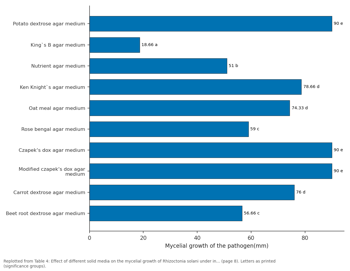
Mycelial growth of R. solani varied significantly among the ten solid media tested, ranging from 18.66 mm to 90 mm. Maximum growth (90 mm) was obtained on potato dextrose agar, Czapek's dox agar, and modified Czapek's dox agar, followed by Ken Knight's agar (78.66 mm). The least growth (18.66 mm) occurred on King's B medium.
Similarly, these three media also supported maximum sclerotial production (Table 4; Fig. 5).
| T. No. | Medium | *Mycelial growth of the pathogen(mm) | Sclerotial production |
|---|---|---|---|
| T1 | Potato dextrose agar medium | 90.00e | +++ |
| T2 | King`s B agar medium | 18.66a | _ |
| T3 | Nutrient agar medium | 51.00b | + |
| T4 | Ken Knight`s agar medium | 78.66d | ++ |
| T5 | Oat meal agar medium | 74.33d | ++ |
| T6 | Rose bengal agar medium | 59.00c | + |
| T7 | Czapek’s dox agar medium | 90.00e | +++ |
| T8 | Modified czapek’s dox agar medium | 90.00e | +++ |
| T9 | Carrot dextrose agar medium | 76.00d | ++ |
| T10 | Beet root dextrose agar medium | 56.66c | + |
Table text extracted from the original PDF; superscript letters are shown inline. The original table image is below.
View original table image
Effect of Different Carbon Sources
When six different carbon sources were tested, sucrose, dextrose, and mannitol supported the highest mycelial growth (90 mm) in Czapek's dox agar, followed by glucose (83 mm). The lowest growth (54.30 mm) was observed in carboxymethyl cellulose-supplemented medium. Sclerotial formation was also most abundant in sucrose, dextrose, and mannitol treatments (Table 5; Fig. 6).
| T. No. | Carbon Source | *Mycelial growth of the pathogen(mm) | Sclerotial production |
|---|---|---|---|
| T1 | Glucose | 83.00c | +++ |
| T2 | Sucrose | 90.00d | +++ |
| T3 | Dextrose | 90.00d | +++ |
| T4 | Carboxy methyl cellulose | 54.30a | + |
| T5 | Mannitol | 90.00e | +++ |
| T6 | Starch | 72.30b | ++ |
Table text extracted from the original PDF; superscript letters are shown inline. The original table image is below.
View original table image
Effect of Different Nitrogen Sources
Among six nitrogen sources evaluated, ammonium nitrate and sodium nitrate promoted the highest mycelial growth (90 mm) and sclerotial production, followed by ammonium sulphate (70 mm). Urea-supported media recorded the lowest growth (40.66 mm) (Table 6; Fig. 7).
| T. No. | Nitrogen Source | *Mycelial growth(mm) | Sclerotial production |
|---|---|---|---|
| T1 | Ammonium nitrate | 90.00c | +++ |
| T2 | Potassium nitrate | 62.00b | ++ |
| T3 | Sodium nitrate | 90.00c | +++ |
| T4 | Ammonium sulphate | 70.00b | ++ |
| T5 | Urea | 40.66a | + |
| T6 | Peptone | 67.66b | _ |
Table text extracted from the original PDF; superscript letters are shown inline. The original table image is below.
View original table image
Effect of pH on Mycelial Growth
The influence of pH on R. solani growth revealed maximum colony diameter at pH 5.0 (90 mm), followed by pH 4.0 (81.76 mm). Minimum growth (76 mm) occurred at pH 8.0. Optimal sclerotial production was observed between pH 4.0 and 5.0 (Table 7; Fig. 8).
| T. No. | PH Level | *Mycelial growth (mm) | Sclerotial production |
|---|---|---|---|
| T1 | 4.0 | 81.76a | +++ |
| T2 | 5.0 | 90.00b | +++ |
| T3 | 6.0 | 77.33a | + |
| T4 | 7.0 | 80.66a | ++ |
| T5 | 8.0 | 76.00a | + |
| T6 | 9.0 | 79.33a | + |
Table text extracted from the original PDF; superscript letters are shown inline. The original table image is below.
View original table image
Effect of Temperature on Mycelial Growth
Temperature markedly influenced mycelial growth and sclerotial development on PDA. The fungus grew optimally at 25 °C and 30 °C (90 mm growth), followed by 20 °C (74.66 mm). Maximum sclerotial formation also occurred at 25 °C and 30 °C, while no sclerotia developed at 5 °C and 10 °C (Table 8; Fig. 9). Each value represents the mean of three replications. Data in parentheses indicate arcsine-transformed values. Treatment means were compared using Duncan's Multiple Range Test (DMRT). In each column, means followed by the same letter(s) do not differ significantly at P = 0.05. Each value represents the mean of three replications. Data in parentheses indicate arcsine-transformed values. Treatment means were compared using Duncan's Multiple Range Test (DMRT). In each column, means followed by the same letter(s) do not differ significantly at P = 0.05.
| T. No. | Temperature (ºC) | *Mycelial growth (mm) | Sclerotial production |
|---|---|---|---|
| T1 | 5 | 0.00a | 0.00 |
| T2 | 10 | 7.60b | 0.00 |
| T3 | 15 | 9.00b | - |
| T4 | 20 | 74.66d | + |
| T5 | 25 | 90.00e | +++ |
| T6 | 30 | 90.00e | +++ |
| T7 | 35 | 73.00d | ++ |
| T8 | 40 | 69.00c | ++ |
Table text extracted from the original PDF; superscript letters are shown inline. The original table image is below.
View original table image
Discussion
Discussion
Survey
A field survey carried out in three districts-Tirunelveli, Thoothukudi, and Kanyakumari-revealed that the incidence of rice sheath blight ranged from 32.58% to 44.05%. According to [28] , pathogenic variation among Rhizoctonia solani isolates is influenced by their distribution across different climatic regions. Consequently, the highest disease incidence of 44.05% recorded at Thirupathisaram (Kanyakumari district), followed by 41.14% at Ambasamudhiram (Tirunelveli district), could be attributed to the presence of distinct pathogenic isolates adapted to varied climatic conditions. Variations in sheath blight severity may arise from differences in isolate virulence, geographic origin, and rice cultivar susceptibility. Similar findings were reported by [29] , who observed that the virulence of R. solani isolates varied depending on the resistance level of rice cultivars. The disease has been shown to cause up to 50% yield reduction in susceptible cultivars under field conditions [30] .
Symptomatology
In the present investigation, infected rice plants exhibited greyish, water-soaked lesions on the leaf sheaths near or slightly above the water level. These lesions gradually enlarged, developing dark brown margins and bleached, greyish-white centers. Comparable symptoms were reported by [31] who described circular to oblong, greenish-grey, water-soaked spots that expanded into grey lesions with purple-brown borders, characteristic of R. solani infection.
Morphological Characters of the Isolates
Marked differences were observed among the isolates of R. solani with respect to colony colour, mycelial growth, and sclerotial production. The isolate collected from Thirupathisaram (Kanyakumari district) exhibited the highest mycelial dry weight and sclerotial yield. Similar variations among isolates were previously reported by several researchers [32][33][34] . Basu [33] stated that the number and size of sclerotia are directly proportional to isolate virulence, explaining the higher disease incidence observed in isolates RS5 and RS1, both of which produced abundant sclerotia.
Virulence of the Isolates
A pot culture experiment involving five R. solani isolates demonstrated that isolate RS5 was the most virulent, producing the highest disease incidence, followed by RS1. The virulent RS5 isolate was collected from Thirupathisaram, a conventional and intensively ricecultivated area where sheath blight occurs regularly. Comparable findings on pathogenic variability among R. solani isolates were reported by several researchers (31, 35, 34) who observed that the isolate RS-21 (Tamil Nadu) exhibited the highest virulence index (7.33), followed by RS-18 (Haryana). Dath [36] further reported that less aggressive isolates induced mild or resistant reactions, even in susceptible rice cultivars. Accordingly, isolate RS4, which showed the lowest disease incidence, was considered less virulent than the other isolates tested.
Cultural Characteristics of R. solani
Growth on Different Solid Media Among the various media tested, potato dextrose agar (PDA), Czapek's dox agar, and modified Czapek's dox agar supported the maximum mycelial growth and sclerotial production of R. solani. These findings are consistent with those of [8,37] , who reported maximum growth and sclerotial yield on PDA, followed by Czapek's dox agar. Similarly, [15,38] observed the highest number of sclerotia on Czapek's dox agar medium.
Growth on Different Carbon and Nitrogen Sources
Among the carbon sources tested, media containing sucrose, dextrose, and mannitol supported the highest mycelial growth and sclerotial formation. Similar results were reported by [7,39] , who found abundant growth and sclerotial development of R. solani in sucrose-amended media. [40] also noted that sucrose and glucose supported maximum mycelial growth, followed by mannitol. Regarding nitrogen sources, media supplemented with ammonium nitrate and sodium nitrate exhibited the highest growth and sclerotial production. Previous reports corroborate these results, indicating that sodium nitrate enhances both mycelial growth and sclerotial density [10,12] . Likewise, Pounraj (2014) found that ammonium nitrate yielded the highest mean mycelial growth of S. oryzae.
Effect of pH on Mycelial Growth
In this study, R. solani exhibited maximum growth at pH 5, followed by pH 4. [39] observed that R. solani isolates could grow across a pH range of 4-9, with optimal mycelial growth at pH 5.6 and maximum sclerotial production at pH 5. Similarly, [16] reported that isolates of R. solani AG 1-1B grew best at pH 5-6. The ability of the pathogen to grow over a wide pH range (4-9) aligns with the general observation that basidiomycetous fungi tolerate broad pH and temperature variations, even though certain bioactive compounds remain stable only within narrow pH limits [14] . Moreover, R. solani can modify the pH of its growth environment to a more favorable range, thereby sustaining successful colonization [41] .
Effect of Temperature on Mycelial Growth
Temperature had a pronounced effect on the growth and sclerotial production of R. solani on PDA. Growth was negligible at lower temperatures but increased progressively up to 30 °C, after which it declined sharply. Optimal growth and sclerotial development occurred between 25 °C and 30 °C, while no sclerotia were formed at 5 °C and 10 °C. These findings concur with previous studies. For instance, [42] reported the highest colony diameter (7.5 cm) at 25 °C after seven days of incubation, followed by 5.8 cm at 30 °C, with no growth observed at 40 °C. similarly [43] noted a substantial increase in colony diameter (from 8.6 mm to 50.7 mm) as temperature rose from 5 °C to 30 °C. Likewise, [10] identified 30 °C as the optimal temperature for R. solani growth. Lalan [15] emphasized temperature as a critical factor influencing fungal growth, sclerotial production, and consequently virulence. Since virulence correlates with sclerotial formation, temperature likely plays a major role in disease severity across climatic regions. At extreme low temperatures (15 °C and 35 °C), poor fungal growth was observed [44] . Additionally, [45] confirmed that all R. solani isolates exhibited excellent sclerotial formation at 30 °C, with optimal production between 25 °C and 35 °C and minimal production at 10 °C-15 °C.
Conclusion
Sheath blight disease is a serious threat in rice growing regions of world. The standard disease management strategies for Rhizoctonia solani were not easily adopted due to the anastomosis phenomenon among the isolates at each niche. The precautionary measures viz., forecasting the seasonal change for cropping, nutrient managements and altering the soil pH would be highly preferable for escaping rice crop from sheath blight incidence. Even though the in vitro studies do not correlate unconditionally with the biological niche, the results afford a rough understanding of the behaviour and growth of the pathogen in soil. The study proved the factors influencing the growth of R. solani under laboratory condition that have an effect on germination of sclerotia In vitro. Further research, on the pathogenicity of different anastomosis groups and the survival of sclerotia under field conditions would provide a greater understanding of the biology of this plant pathogen.
Figures and tables from the paper
References
41 in the original paper- Srinivas P, Ratan V, Patel AP, Madhavi GB. Review on banded leaf and sheath blight of rice caused by Rhizoctonia solani Kühn. 2013.
- Turaidar V, Reddy M, Anantapur R, Krupa K, Dalawai N, Deepak C, et al. Rice sheath blight: major disease in rice. Int J Curr Microbiol Appl Sci. 2018;7:976-988.
- Singh RS, Sinha A. Influence of soil nutrition on the effectivity of Pseudomonas fluorescens on rice sheath blight. 2005.
- Reddy BP, Reddy M, Kumar K. In vitro antagonistic potential of Pseudomonas fluorescens isolates and their metabolites against rice sheath blight pathogen Rhizoctonia solani. 2010.
- Balne PK, Barik MR, Sharma S, Basu S. Development of a loop-mediated isothermal amplification assay targeting the mpb64 gene for diagnosis of intraocular tuberculosis. J Clin Microbiol. 2013;51:3839-3840.
- Adhipathi P, Singh V, Meena SC. Virulence diversity of Rhizoctonia solani causing sheath blight disease in rice and its host-pathogen interaction. Bioscan. 2013;8:949-952.
- Indian Phytopathol. 1980;33.
- Hussain AH, Awan S, Iqbal SM, Anees MA, Ali SA. Variability among the potato sclerotial isolates of Rhizoctonia solani of mountainous region, GilgitBaltistan, Pakistan. 2014.
- Agrios GN. Plant pathology. 5th ed. Amsterdam: Elsevier; 2005.
- Muhsin TM, Selman MS. In vitro optimization of growth and bioactivity of antibacterial metabolite produced by Rhizoctonia solani Kühn. J Basrah Res (Sci). 2013;39.
- El-Banna NM. Effect of carbon source on the antimicrobial activity of Corynebacterium kutscheri and Corynebacterium xerosis. Afr J Biotechnol. 2006;5.
- Ellil AHA. Biochemical changes and pathogenicity variations related to esterase polymorphism for morphological traits of Rhizoctonia solani. Int J Agric Biol. 2005;7:555-559.
- Mohamed MH, Gado E, El-Deeb S, Mostafa M. Effect of nitrate levels as fertilizer or fungal nutrition on aggressiveness of Rhizoctonia solani on faba bean. Eur J Adv Res Biol Life Sci. 2014;2.
- Vahidi H, Kobarfard F, Namjoyan F. Effect of cultivation conditions on growth and antifungal activity of Mycena leptocephala. Afr J Biotechnol. 2004;3:606609.
- Sharma LS, Goswami SG, Nagrale D. Culture and physiological variability in Rhizoctonia solani responsible for foliar lesions on aerial parts of soybean. 2013.
- Grosch R, Kofoet A. Influence of temperature, pH and inoculum density on bottom rot on lettuce caused by Rhizoctonia solani. J Plant Dis Prot. 2003;366-378.
- Rangaswami G, Mahadevan A. Diseases of crop plants in India. New Delhi: PHI Learning Pvt. Ltd.; 1998.
- Ainsworth GC. A taxonomic review with keys: Ascomycetes and fungi imperfecti. In: The fungi -an advanced treatise. New York: Academic Press; 1973.
- King EO, Ward MK, Raney DE. Two simple media for the demonstration of pyocyanin and fluorescin. J Lab Clin Med. 1954;44:301-307.
- Kumar RR, Jadeja VJ. Isolation of actinomycetes: a complete approach. 2016. International Journal of Agriculture and Food Science https://www.agriculturaljournals.com
- Booth C. Fungal culture media. In: Methods in microbiology. Vol. 4. Amsterdam: Elsevier; 1971. p. 49-94.
- Martin JP. Use of acid, rose bengal, and streptomycin in the plate method for estimating soil fungi. Soil Sci. 1950;69:215-232.
- Dox AW. The intracellular enzymes of Penicillium and Aspergillus with special reference to those of Penicillium camemberti. US Dep Agric Bur Anim Ind. 1910;120.
- Manoharan P. Studies on the sheath blight of rice (Oryza sativa L.) caused by Rhizoctonia solani with special reference to biological control [MSc (Agri) thesis]. Coimbatore: Tamil Nadu Agric Univ; 1999.
- Pandey K, Vishwakarma S. Growth, sporulation and colony characters of Alternaria alternata on different vegetable-based media. J Mycol Plant Pathol (India). 1998;28.
- Khanzada MA, Rajput AQ, Shahzad S. Effect of medium, temperature, light and inorganic fertilizers on In vitro growth and sporulation of Lasiodiplodia theobromae isolated from mango. Pak J Bot. 2006;38:885-890.
- Goswami SK, Singh V, Kashyap PL. Population genetic structure of Rhizoctonia solani AG1IA from rice fields in North India. Phytoparasitica. 2017;45:299316.
- Sunder S, Singh R, Dodan D. Standardization of inoculation methods and management of sheath blight of rice. Indian J Plant Pathol. 2003;21:92-96.
- Basu A, Podder M, Sengupta PK. Variability and anastomosis among the rice isolates of Rhizoctonia solani. Indian Phytopathol. 2004;57:70-72.
- Lal M, Singh V, Kandhari J, Sharma P, Kumar V, Murti S. Diversity analysis of Rhizoctonia solani causing sheath blight of rice in India. Afr J Biotechnol. 2014;13.
- Manimekalai M. Biological control of stem rot of Crossandra infundibuliformis caused by Rhizoctonia solani Kühn [MSc (Agri) thesis]. Coimbatore: Tamil Nadu Agric Univ; 2012.
- Dath A. A better criterion in rating the reaction of rice cultivars against sheath blight. 1985.
- Prasad BN, Kumar MR. Scanning electron microscopic studies on mycoparasitic activity of Trichoderma spp. against Rhizoctonia solani, incitant of sheath blight of rice. 2013.
- Tiwari A, Khare M. Conditions inducing imperfect and perfect stages in Rhizoctonia solani causing diseases of mungbean. 2002.
- Ritchie F, Bain R, McQuilken M. Effects of nutrient status, temperature and pH on mycelial growth, sclerotial production and germination of Rhizoctonia solani from potato. J Plant Pathol. 2009;91:589-596.
- Patil L, Shinde V, Ghawade R, Wavare S. Physiological and nutritional studies of Botryodiplodia theobromae Pat. causing die-back disease of mango. 2006.
- Parmeter JR. Rhizoctonia solani: biology and pathology. Berkeley: Univ California Press; 2023.
- Attaullah S, Husain I, Ali F, Khan A, Khan G. Comparison of different techniques for the isolation of Rhizoctonia solani from infected potato tubers. Greener J Agric Sci. 2012;2:212-219.
- Goswami B, Rahaman M, Hoque A, Bhuiyan K, Mian I. Variations in different isolates of Rhizoctonia solani based on temperature and pH. Bangladesh J Agric Res. 2011;36:389-396.
- Kwee LT, Chong NC, Lan CC. Etiology and control of durian foliar blight and dieback caused by Rhizoctonia solani. Ann Appl Biol. 1987;111:301-307.
- Dubey S, Dwivedi R. Evaluation of fungicides against the web blight disease of groundnut. 1992.
Cited by 1
From OpenAlex, OpenCitations and reference lists read by Scinovex
- Laboratory studies on Rhizoctonia bataticola and Fusarium oxysporum causing collar/ root rot in teak nursery seedlings ↗
Sandip Kedare, Mina D. Koche, R. B. Kothikar · International Journal of Plant Pathology and Microbiology · 2026
Teak (Tectona grandis) is the most important commercial timber species grown all over the world. From Vidarbha region ‘Alapalli teak’ and Paratwada teak’ are the most famous all over the country for the good quality timber. Even though, it is a hardy tree species, it is affected by complex diseases and insect fauna. Among diseases, nursery diseases are very important