
Research article · 2025 · Full text
International Journal of Biology SciencesDiseases infecting Honey bees
Volume 7(8) · Pages 224–228 · DOI: 10.33545/26649926.2025.v7.i8c.485 ↗
Research areasInsect and Pesticide ResearchBee Products Chemical AnalysisHealthcare and Venom Research
Abstract
Author-writtenHoneybees, vital pollinators essential for ecosystem health, are vulnerable to various bacterial, fungal, and viral diseases that pose a threat to honeybee colonies. As many as 60% and 90% of plant species depend upon pollinators to reproduce (Kremen et al. 2007). Since many agricultural crops depend on pollinators, bees play a vital role in the ecology, economy, and food production across all countries (Garcia-Anaya et al. 2016; Hamadache et al. 2017). Apart from pollination services honey bees also provide honey, beeswax, royal jelly, propolis and bee venom (Tej et al. 2017, Greenleaf and Kremen, 2006, Bosch and Kemp, 2002). Similar to other living organisms, honeybees and their products are susceptible to harmful diseases, pests, and pesticides. Successful beekeeping requires regular and timely monitoring of any factors that harm bee colonies. Honeybee diseases have been recognized as one of the primary biotic factors hindering successful beekeeping practices. Bee diseases cause considerable loss for beekeepers resulting in absconding of colonies, reduction in honey and beeswax production. Perez-Sato et al. (2009) stated that the major constraint faced by the beekeepers all over the world is pests and diseases. It is evident that therefore, both beginning and advanced beekeepers should learn to recognize and control bee diseases. Diseases of honey bees are generally classified into brood diseases and adult diseases. Brood diseases are caused by bacteria (American foul brood and European foul brood); virus (sac brood disease) and fungi (chalk brood and stone brood diseases). Adult diseases are mainly caused by protozoa (nosema disease) (Abrol, 1997).
Summary
Source-checkedPlain-language reading of the full paper, prepared with AI and checked against the text. Each point links to the authors' own words.
Why it matters
- Honey bees are critical for global food production and ecology because 60% to 90% of plant species rely on pollinators for reproduction. View evidence ↗
- Beyond pollination, honey bees provide valuable commercial products including honey, beeswax, royal jelly, propolis, and bee venom. View evidence ↗
- Diseases are a primary biological obstacle to beekeeping, leading to colony loss and reduced production of honey and wax. View evidence ↗
The question
- The paper aims to provide information to help beekeepers recognize and control various honey bee diseases. View evidence ↗
What they did
- The review categorizes honey bee diseases into brood diseases (bacterial, viral, and fungal) and adult diseases (primarily protozoan). View evidence ↗
What they found
- American Foulbrood (AFB) is caused by Paenibacillus larvae, a bacterium whose spores can remain dormant for 50 years or more. View evidence ↗
- AFB is highly infectious; as few as ten spores consumed by larvae can cause a lethal infection. View evidence ↗
- European Foulbrood (EFB), caused by Melissococcus plutonius, typically affects larvae before they are capped. View evidence ↗
- Chalkbrood, caused by the fungus Ascosphaera apis, reduces the number of emerging worker bees and weakens the colony. View evidence ↗
- The Thai Sacbrood Virus (TSBV) is a severe threat in Asia, with colony losses exceeding 95% in several countries. View evidence ↗
- Nosema disease, caused by microsporidia, affects adult bees and leads to diarrhea and population decline. View evidence ↗
- Antibiotic use for AFB is problematic because it does not kill spores, leaves residues in honey, reduces bee longevity, and faces increasing bacterial resistance. View evidence ↗
What it means
- For Nosema, the authors suggest that the most effective management is reducing stress through proper ventilation and protection from cold and humidity. View evidence ↗
Visual summary
1 visualsIllustrations explain the paper; they do not show study data.
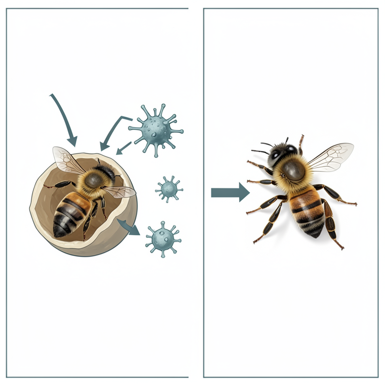
Honey Bee Disease Taxonomy
AI-generated conceptual illustration (Honey bees (Apis mellifera) in various life stages). It explains the idea and does not show study data.
View source evidence ↗Introduction
Introduction
Bacterial diseases
An earliest report of bacterial disease was given by Aristotle (384-322 B.C.) in his Book IX of History of Animals. Although several diseases have been observed in honey bee brood, American Foul Brood (AFB) and European Foul Brood (EFB) are considered a global threat to honey bees (Smith et al., 2014; Goulson et al., 2015) [54,31] . The name ''foulbrood" was given to the honey bee disease characterized by a foul odour emitted from brood infected with these pathogens (Schirach, 1769) [52] . Cheshire and Cheyne (1885) [20] further described the symptoms of the disease in the late 19th century and recognized that the cause of the disease was Bacillus alvei. However, White in 1912 described that there are actually two honey bee brood diseases, namely American foulbrood caused by the spore-forming bacterium Paenibacillus larvae (Genersch et al., 2005) [29] and European foulbrood caused by the bacterium Melissococcus plutonius (Bailey, 1956) [8] . American Foul Brood is predominantly found in tropical and subtropical regions. It was first documented by White in 1907 and subsequently recognized as a serious disease affecting various regions across the globe (Bailey and Ball, 1991) [7] .
Consequently, beekeepers and extension specialists must be familiar with the symptoms of this disease and understand how to manage it if necessary. AFB disease is caused by Paenibacillus larvae, which is a gram-positive, sporeforming, facultative anaerobic bacterium (Ash et al., 1994) [6] . It was originally described as Bacillus larvae, and It causes substantial economic losses to beekeepers. The spores of P. larvae are extremely infectious, but colonies differ in their resistance to AFB outbreaks. Honeybee workers are effective for the early identification of AFB (Lindstrom et al., 2005) [41] .
Symptomatology and control
In the early phase of American Foulbrood (AFB) infection, symptoms include isolated capped cells with no brood emergence, visible on the comb. The affected cells typically show dark, sunken, or perforated cappings with punctures made by the investigating adult bees, unlike healthy brood cells which are slightly protruding and fully closed. Infected larvae are brown in color (White, 1907) [60] , and an accompanying foul smell is produced, which is why this disease is also known as "Stinking disease." In advanced cases, the brood displays the pepper box symptom an irregular, patchy brood pattern on the comb resembling a pepperbox, caused by the disease killing and digesting larvae in their cells (Shimanuki, 1997) [53] . The highly resilient endospores represent the sole infectious form of this organism. These spores are infectious exclusively to larvae; adult bees do not become infected upon ingestion of P. larvae spores (Hitchcock et al., 1979; Wilson, 1971) [36, 64] . Larvae exhibit the highest susceptibility to infection during the initial larval stages, specifically 12-36 hours postegg hatching. Within this timeframe, oral consumption of approximately ten spores or fewer through contaminated larval food is sufficient to trigger a lethal infection (Bamrick and Rothenbuhler, 1961; Woodrow and Holst, 1942) [12,65] . To control the disease, oxytetracycline hydrochloride (OTC) or sulfathiazole help prevent multiplication of the agent, though they do not kill the spores. Consequently, multiplication can resume soon after treatment, necessitating repeated treatments at progressively shorter intervals. However, prolonged use of antibiotics poses several issues: (i) antibiotics do not target infectious spores and only alleviate clinical symptoms, thereby concealing the disease without curing AFB; (ii) chemical residues may remain in honey, affecting its quality and safety for human consumption (Lodesani and Costa, 2005; Martel et al., 2006) [42,43] ; (iii) administration of antibiotics to larvae and adult bees may impact the vitality of the brood as well as the longevity of the bees (Peng et al., 1992); and (iv) resistance to OTC and sulfathiazole in P. larvae has become prevalent (Evans, 2003; Piccini and Zunino, 2001; Negash, 2022) [25,50,67] , making new alternatives increasingly necessary (Alippi et al., 2005; Peng et al., 1996; Williams et al., 1998) [48,63] .
European foulbrood disease (EFB)
European foulbrood (EFB) is a bacterial infection that affects honey bee larvae before they reach the capped stage. The disease is characterized by the presence of dead and dying larvae, which may exhibit curled upward postures and can appear brown or yellow, as well as melted, desiccated, or rubbery. European foulbrood has long been reported from the United Kingdom (Cheshire and Cheyne, 1885) [20] and later from the United States of America (White, 1907) [60] in A. mellifera. Subsequently, the disease was documented worldwide, causing serious problems resulting in economic losses to beekeepers. The disease has been observed in many countries including North and South America, Europe, Japan, Australia, India, China, and South Africa (Matheson, 1993) [45] . Wilkins and colleagues reported in 2007 that EFB is the most widespread bacterial brood disease in Great Britain. Incidence of the disease has steadily increased in Switzerland since the late 1990s (Roetschi et al., 2008) [51] .
Symptomatology and control
The causative agent, Melissococcus plutonius, is consumed by honey bee larvae, where it competes for nourishment within the larvae. If the bacterium outcompetes the larva, the larva dies before the cell is sealed. Conversely, if the larvae have adequate food resources, they may develop into healthy adults. G. F. White is credited with first identifying the bacterium responsible for European foulbrood in 1908, initially naming it Bacillus Y, which he later renamed Bacillus pluton (Bailey, 1983) [9] . A patchy brood pattern arises when some larvae die in their cells due to disease, while others survive and become capped, resulting in a spotty or "shotgun" appearance of the capped brood. To manage bacterial diseases, beekeepers utilize a range of antibiotics, including tetracycline, chloramphenicol, sulphonamides, and glycosides. These antibiotics have been associated with residue problems in honey, thereby reducing its quality in the global market. Oxytetracycline hydrochloride (OTC) is an antibiotic acting as a bacteriostatic agent that inhibits the growth of M. plutonius and is used in many countries (Thompson and Brown, 2001) [56] . Since the 1950s, American beekeepers have applied OTC to prevent both European foulbrood (EFB) and American foulbrood (AFB). Although resistance to OTC in Paenibacillus larvae has been documented (Miyagi et al., 2000) [46] , no resistance to OTC has been reported in M. plutonius; research in the UK confirmed susceptibility of this bacterium to OTC (Waite et al., 2003). In Australia, where OTC is also applied against EFB, M. plutonius isolates remain sensitive (Hornitzky and Smith, 1999) [37] . In Great Britain, EFB-infected colonies are treated with OTC or the shook swarm method, whereby bees are transferred to new comb foundation and infected combs discarded; severely infected colonies may be destroyed (Wilkins et al., 2007) [62] . Combining the shook swarm method with OTC reduces the recurrence of clinical symptoms at the colony level (Waite et al., 2003) [58] . Requeening may further assist disease control by interrupting the brood cycle and introducing a more prolific queen (Shimanuki, 1997) [53] .
Fungal disease
Chalkbrood and stonebrood are two fungal diseases that affect honey bee brood. Chalkbrood, caused by Ascosphaera apis, is a prevalent and widespread disease that can significantly decrease the number of emerging worker bees, thereby impacting overall colony productivity. Although the fungus rarely causes the death of infected colonies, it weakens them, resulting in lower honey yields and increased vulnerability to other pests and diseases affecting bees. Engles et al. (2004) [24] also reported that during winter months, fungal diseases weakened honeybee colonies, which led to the development of European foulbrood.
Infected young larvae typically do not exhibit visible signs of illness but die upon being sealed in their cells as pupae. Worker bees often uncap the cells containing deceased larvae, making the mummified brood distinctly visible. Sometimes, they remove the mummified larvae and place them on the hive floor or at the hive entrance. According to Bailey (1967) [10] , honey bee larvae are particularly vulnerable to chalkbrood. A wide array of fungicides has been evaluated for their effectiveness in managing chalkbrood (Heath, 1982; Davis and Ward, 2003) [34,22] . Hornitzky (1999) [37] identified certain chemicals that appeared effective in inhibiting fungal growth, whether in culture or within bee colonies. Unfortunately, none of the tested compounds provided sufficient control to combat the disease. Stonebrood disease, caused by Aspergillus spp., is rarely observed, and consequently, its impact on colony health is not well understood (Jensen et al., 2013) [38] .
Protozoan disease Nosema disease
Nosematosis, caused by microsporidia from the genus Nosema, is recognized as one of the well-documented opportunistic infections affecting honey bees (Broadrup et al., 2019) [15] . To date, three distinct microsporidian species linked to honey bee infections have been identified within the genus Nosema. These infections are collectively referred to as nosematosis, regardless of whether they are caused by Nosema apis (Fries, 1993) [27] , Nosema ceranae (Fries et al., 1993) [27] , or Nosema neumanni (Chemurot et al., 2017) [18] . The first two species have spread globally and are responsible for observable symptoms such as diarrhea and a decline in bee populations within the hive (Higes et al., 2006; Klee et al., 2007) [35,39] . In contrast, N. neumanni has been recently identified in Ugandan bees, with its effects on host bees yet to be fully documented. Nosema disease is typically considered one of the most harmful diseases affecting adult bees, impacting workers, queens, and drones alike. Severely affected worker bees lose their ability to fly and may be observed crawling near the hive entrance or trembling atop the frames. Such bees exhibit signs of accelerated aging: their lifespan is significantly shortened, and their hypopharyngeal glands deteriorate, leading to a rapid decline in colony strength. Unfortunately, there is currently no reliable field diagnostic symptom that allows identification of diseased workers without killing them, nor can infected queens be easily recognized by beekeepers (FAO, 2006). However, in cases of severe infection, it may be possible to distinguish healthy from diseased bees by the swollen and shiny appearance of the abdomen in infected workers. The most effective management strategy for Nosema focuses on maintaining colony strength and minimizing stressors. Adequate ventilation and protection against cold and humidity are crucial for colonies and apiaries. Moreover, providing bees with regular opportunities to forage facilitates defecation, which helps reduce the spread of spores within the colony.
References
66 in the original paper- Abrol DP. Honey Bee Diseases and their Management. Kalyani Publishers, Ludhiyana; 1997. 112 p.
- Abrol DP. Honey Bee Diseases and their Management. Kalyani Publishers, Ludhiyana; 1997. 112 p.
- Alippi AM, Albo GN, Reynaldi FJ, De Giusti MR. In vitro and in vivo susceptibility of the honeybee bacterial pathogen Paenibacillus larvae subsp. larvae to the antibiotic tylosin. Vet Microbiol. 2005;109:47-55.
- Anderson DL, Gibbs AJ. Transpuparial transmission of Kashmir bee virus and sacbrood virus in the honey bee (Apis mellifera). Ann Appl Biol. 1989;114:1-7.
- Aristotle. History of Animals, Book IX. Translated by Crossware R. London: Georg Bell; 1907. 262 p. International Journal of Biology Sciences https://www.biologyjournal.net
- Ash C, Priest FG, Collins MD. Molecular identification of rRNA group 3 bacilli (Ash, Farrow, Wallbanks and Collins) using a PCR probe test: proposal for the creation of a new genus Paenibacillus. Antonie Van Leeuwenhoek. 1993/1994;64:253-260.
- Bailey L, Ball BV. Honey Bee Pathology. 2nd ed. Academic Press, London; 1991.
- Bailey L. Etiology of European foulbrood; a disease of the larval honey bee. Nature. 1956;178:3.
- Bailey L. Melissococcus pluton, the cause of European foulbrood of honey bees (Apis spp.). Bee World. 1983;64(3):107-117.
- Bailey L. The effect of temperature on the pathogenicity of the fungus Ascosphaera apis for larvae of the honey bee Apis mellifera. In: Van der Laan PA, editor. Insect Pathology and Microbial Control. North Holland Publishing Co.; 1967. p. 162-167.
- Bailey L. The multiplication and spread of sacbrood virus of bees. Ann Appl Biol. 1969;63:483-491.
- Bamrick JF, Rothenbuhler WC. Resistance to American foulbrood in honey bees. IV. The relationship between larval age at inoculation and mortality in a resistant and a susceptible line. J Insect Pathol. 1961;3:381-390.
- Beaurepaire A, Piot N, Doublet V, Antunez K, Campbell E, Chantawannakul P, et al. Diversity and global distribution of viruses of the western honey bee Apis mellifera. Insects. 2020;11:239.
- Bosch J, Kemp WP. Developing and establishing bee species as crop pollinators: The example of Osmia spp. (Hymenoptera: Megachilidae) and fruit trees. Bulletin of Entomological Research. 2002;92:3-16.
- Broadrup RL, Mayack CH, Schick SJ, Eppley EJ, White HK, Macherone A. Honey bee (Apis mellifera) exposomes and dysregulated metabolic pathways associated with Nosema ceranae infection. PLoS ONE. 2019;14:e0213249.
- Chagas DB, Monteiro FL, Hübner SDO, Lima MD, Fischer G. Viruses that affect Apis mellifera and their occurrence in Brazil. Ciência Rural. 2019;49.
- Chagas DB, Monteiro FL, Hübner SDO, Lima MD, Fischer G. Viruses that affect Apis mellifera and their occurrence in Brazil. Ciência Rural. 2019;49.
- Chemurot M, De Smet L, Brunain M, De Rycke R, de Graaf DC. Nosema neumanni n. sp. (Microsporidia, Nosematidae), a new microsporidian parasite of honeybees, Apis mellifera in Uganda. Eur J Protistol. 2017;61:13-19.
- Chen Y, Pettis JS, Collins A, Feldlaufer MF. Prevalence and transmission of honeybee viruses. Appl Environ Microbiol. 2006;72:606-611.
- Cheshire FR, Cheyne WW. The pathogenic history and the history under cultivation of a new Bacillus (B. alvei), the cause of the disease of the hive bee hitherto known as Foul brood. J. R. Microsc. Soc. 1885;5:581601.
- Choe SE, Nguyen LT, Noh JH, Kweon CH, Reddy KE, Koh HB, Kang SW. Analysis of the complete genome sequence of two Korean sacbrood viruses in the honey bee, Apis mellifera. Virology. 2012;432:155-161.
- Davis C, Ward W. Control of chalkbrood disease with natural products: a report for the RIRDC. Publication No. 03/107, Kingston, ACT, Australia; 2003.
- De Miranda J, Gauthier L, Ribiere M, Chen Y. Honey bee viruses and their effect on bee and colony health. Challenges and sustainable solutions, honey bee colony health. 2012. p. 71-102.
- Engles W, Nicholson GJ, Hertle R, Winkelmann G. Tyramine functions as a toxin in honey bee larvae during Varroa transmitted infection by Melissococcus pluton. FEMS Microbiol Lett. 2004;234:49.
- Evans JD. Diverse origins of tetracycline resistance in the honey bee bacterial pathogen Paenibacillus larvae. J Invertebr Pathol. 2003;83:46-50.
- FAO. Honey bee diseases and pests: a practical guide. 2006 (E).
- Fries I. Nosema apis A parasite in the honey bee colony. Bee World. 1993;74:5-19.
- García-Anaya MC, Romo-Chacón A, Zamudio-Flores PB, Ríos-Velasco C, Acosta-Muñiz CH. Detection of viruses in colonies of honey bees (Apis mellifera L.) in the state of Chihuahua, Mexico. Journal of Apicultural Research. 2016;55(3):240-242.
- Genersch E, Ashiralieva A, Fries I. Strain and genotype specific differences in virulence of Paenibacillus larvae subsp. larvae, a bacterial pathogen causing American foul brood disease in honey bees. Applied Environmental Microbiology. 2005;71:7551-7555.
- Ghosh RC, Ball BV, Willcocks MM, Carter MJ. The nucleotide sequence of sacbrood virus of the honey bee: an insect picorna-like virus. J Gen Virol. 1999;80:15411549.
- Goulson D, Nicholls E, Botias TC, Rotheray EL. Combined stress from parasites, pesticides and lack of flowers drives bee declines. Science. 2015;347:6229.
- Greenleaf SS, Kremen C. Wild bee species increase tomato production and respond differently to surrounding land use in Northern California. Biological Conservation. 2006;133:81-87.
- Hamadache M, Benkortbi O, Hanini S, Amrane A. QSAR modeling in ecotoxicological risk assessment: Application to the prediction of acute contact toxicity of pesticides on bees (Apis mellifera L.). Environmental Science and Pollution Research. 2017;25(1):896-907.
- Heath LAF. Development of chalk brood in a honey bee colony; chalkbrood pathogens: a review. Bee World. 1982;63(3):119-135.
- Higes M, Martín R, Meana A. Nosema ceranae a New Microsporidian Parasite in Honeybees in Europe. J Invertebr Pathol. 2006;92:93-95.
- Hitchcock JD, Stoner A, Wilson WT, Menapace DM. Pathogenicity of Bacillus pulvifaciens to honeybee larvae of various ages (Hymenoptera: Apidae). J Kansas Entomol Soc. 1979;52:238-246.
- Hornitzky MAZ, Smith LA. Sensitivity of Australian Melissococcus plutonius isolates to oxytetracycline hydrochloride. Austr J Exp Agric. 1999;39:881-883.
- Jensen AB, Aronstein K, Flores JM, Vojvodic S, Palacio MA, Spivak M. Standard methods for fungal brood disease research. J Apic Res. 2013;52(1):1-20.
- Klee J, Besana AM, Genersch E, Gisder S, Nanetti A, Tam DQ, et al. Widespread dispersal of the microsporidian Nosema ceranae, an emergent pathogen of the western honey bee, Apis mellifera. J Invertebr Pathol. 2007;96:1-10.
- Kremen C, Williams NM, Aizen MA, Gemmill-Herren B, LeBuhn G, Minckley R, Packer L, et al. Pollination and other ecosystem services produced by mobile International Journal of Biology Sciences https://www.biologyjournal.net organisms: a conceptual framework for the effects of land-use change. Ecology Letters. 2007;10(4):299-314.
- Lindstrom A, Fries I. Sampling of adult bees for detection of American foulbrood (Paenibacilluslarvae subsp. larvae) spores in honey bee (Apis mellifera) colonies. J Apic Res. 2005;44:82-86.
- Lodesani M, Costa M. Limits of chemotherapy in beekeeping: development of resistance and the problem of residues. Bee World. 2005;86:102-109.
- Martel AC, Zeggane S, Drajnudel P, Faucon JP, Aubert M. Tetracycline residues in honey after hive treatment. Food Addit Contam. 2006;23:265-273.
- Martin SJ, Highfield AC, Brettell L, Villalobos EM, Budge GE, Powell M, et al. Global honey bee viral landscape altered by a parasitic mite. Science. 2012;336:1304-1306.
- Matheson A. World Bee Health Report. Bee World. 1993.
- Miyagi T, Peng CYS, Chuang RY, Mussen EC, Spivak MS, Doi RH. Verification of oxytetracycline-resistant American foulbrood pathogen Paenibacillus larvae in the United States. J Invertebr Pathol. 2000;75:95-96.
- Peng CYS, Mussen EC, Fong A, Cheng P, Wong G, Montague MA. Laboratory and field studies on the effect of the antibiotic tylosin on honey bee Apis mellifera L. (Hymenoptera: Apidae) development and prevention of American foulbrood disease. J Invertebr Pathol. 1996;67:65-71.
- Peng CYS, Mussen EC, Fong A, Montague MA, Tyler T. Effects of chlortetracycline on honey bee worker larvae reared in vitro. J Invertebr Pathol. 1992;60:127133.
- Perez-Sato JA, Chaline N, Martin SJ, Hughes WOH, Ratneiks PL. Multi-level selection for hygienic behaviour in honey bees. Heredity. 2009;102(6):609615.
- Piccini C, Zunino P. American foulbrood in Uruguay: isolation of Paenibacillus larvae larvae from larvae with clinical symptoms and adult honeybees and susceptibility to oxytetracycline. J Invertebr Pathol. 2001;78:176-177.
- Roetschi A, Berthoud H, Kuhn R, Imdorf A. Infection rate based on quantitative real-time PCR of Melissococcus plutonius, the causal agent of European foulbrood, in honeybee colonies before and after apiary sanitation. Apidologie. 2008;39:362-371.
- Shimanuki H. Bacteria. In: Morse RA, Flottum K, editors. Honey Bee Pests, Predators, and Diseases. 3rd ed. A. I. Root Co.; 1997.
- Smith KM, Lob EH, Rostal MK, Zambrana-Torrelio CM, Mendiola L, Daszak P. Pathogens, Pests, and Economics: Drivers of Honey Bee Colony Declines and Losses. EcoHealth. 2014;10(4):434-445.
- Tej MK, Aruna R, Mishra G, Srinivasan MR. Beekeeping in India. Industrial Entomology. 2017;1:3566.
- Thompson HM, Brown MA. Is contact colony treatment with antibiotics an effective control for European foulbrood? Bee World. 2001;82:130-138.
- Ullah A, Gajger IT, Majoros A, Dar SA, Khan S, Kalimullah, et al. Saudi Journal of Biological Sciences. 2021;28:523-530.
- Waite R, Brown M, Thompson H, Bew M. Controlling European foulbrood with the shook swarm method and oxytetracycline in the UK. Apidologie. 2003;34:569575.
- Wei R, Cao L, Feng Y, Chen Y, Chen G, Zheng H. Sacbrood virus: a growing threat to honeybees and wild pollinators. Viruses. 2022;14:1871.
- White GF. The Cause of American Foul Brood. U.S. Department of Agriculture, Bur. Entomol., Circ. 1907;94:110.
- White GF. The Cause of European Foul Brood. Circ. U.S. Bur. Entomol. 1912;157:p810.
- Wilkins S, Brown M, Andrew A, Cuthbertson GS. The incidence of honey bee pests and diseases in England and Wales. Pest Manag Sci. 2007;63:1062-1068.
- Williams JR, Peng CYS, Chuang RY, Doi RH, Mussen EC. The inhibitory effect of azadirachtin on Bacillus subtilis, Escherichia coli, and Paenibacillus larvae, the causative agent of American foulbrood in the honeybee, Apis mellifera L. J Invertebr Pathol. 1998;72:252-257.
- Wilson WT. Resistance to American foulbrood in honey bees XI. Fate of Bacillus larvae spores ingested by adults. J Invertebr Pathol. 1971;17:247-255.
- Woodrow AW, Holst EC. The mechanism of colony resistance to American foulbrood. J Econ Entomol. 1942;35:327-330.
- Yoo MS, Noh JH, Yoon BS, Reddy KE, Kweon CH, Jung SC, Kang SW. Reverse transcription loopmediated isothermal amplification for sensitive and rapid detection of Korean sacbrood virus. J Virol Methods. 2012;186(1-2):147-151.
- Negash D. Demonstration and participatory evaluation of apiculture technologies in Sidama National Regional state, Ethiopia. Int. J. Agric. Extension. Social. Dev. 2022;5(2):12-16.