
Research article · 2025 · Full text
International Journal of Biology SciencesIdentification and characterization of proteins in testis and epididymal sperms of Rattus norvegicus
Volume 7(8) · Pages 208–223 · DOI: 10.33545/26649926.2025.v7.i8c.479 ↗
Research areasSperm and Testicular Function
Abstract
Author-writtenThe present study was conducted to identify and characterize proteins in the testis and sperm membrane abstract of male Rattus norvegcius. Male albino rats were dissected to collect spermatozoa from the testis and epididymis. Sperm membrane extract (SME) was prepared using deoxycholate detergent (DOC) (Naz et al 1986) [1]. Quantitative and qualitative analysis of total protein from testis and epididymal sperm membrane using was carried out by the method described by Lowry et al 1951 [2] and SDS-PAGE (Laemmli 1970) [3]. It can be concluded for the present study that the protein content was observed to be highest in caput and lowest in the cauda epididymis due to the maximum and minimum secretory activities respectively in these regions. The SDS-PAGE profile revealed 13 bands corresponding to molecular weight of 17, 20, 24, 35, 44, 45, 47, 55, 60, 80, 95, 115 and 133 kDa in testis; 20 bands corresponding to molecular weights 17, 20, 24, 26, 28, 33,35, 44, 45, 47, 55, 60, 64, 72, 80, 95, 115, 133, 135 and 170 kDa in caput;19 bands corresponding to molecular weights 17, 20, 24, 26, 28, 33,35, 44, 45, 47, 55, 60, 72, 80, 95, 115, 133, 135 and 170 kDa in corpus and 17 bands corresponding to molecular weights 17, 20, 24, 26, 33,35, 44, 45, 47, 55, 60, 72, 80, 95, 115, 133 and 135 kDa in the cauda epididymal sperm membrane extract.
Summary
Source-checkedPlain-language reading of the full paper, prepared with AI and checked against the text. Each point links to the authors' own words.
Why it matters
- Male fertility depends on both the production of sperm in the testes and their subsequent maturation as they move through the epididymis. View evidence ↗
- There is limited existing data comparing the protein profiles of sperm membranes across the testis and various regions of the epididymis in rats. View evidence ↗
The question
- The study aimed to identify and characterize the proteins found in the testis and epididymal sperm membrane extracts of male Rattus norvegicus. View evidence ↗
What they did
- The researchers used twenty sexually mature albino rats weighing between 130 and 150 grams. View evidence ↗
- The epididymis was divided into three distinct regions for analysis: the caput (head), corpus (body), and cauda (tail). View evidence ↗
- Sperm membrane extracts were prepared using deoxycholate detergent and then analyzed using SDS-PAGE to separate the proteins. View evidence ↗
- Total protein was quantitatively and qualitatively analyzed using the Lowry method. View evidence ↗
What they found
- Several protein bands (26, 28, 33, 64, 72, 135, and 170 kDa) were absent in the testis but appeared in the epididymal regions, suggesting they were acquired during maturation. View evidence ↗
- A 64 kDa protein was found only in the caput region and was absent in the corpus and cauda. View evidence ↗
- Proteins with molecular weights of 28 and 170 kDa were present in the caput and corpus but absent in the cauda, indicating they may have been reabsorbed. View evidence ↗
What it means
- The authors conclude that protein content is highest in the caput due to high secretory activity at the start of maturation and lowest in the cauda, where mature sperm are stored. View evidence ↗
Visual summary
2 visualsIllustrations explain the paper; charts are replotted from the paper's own table values.
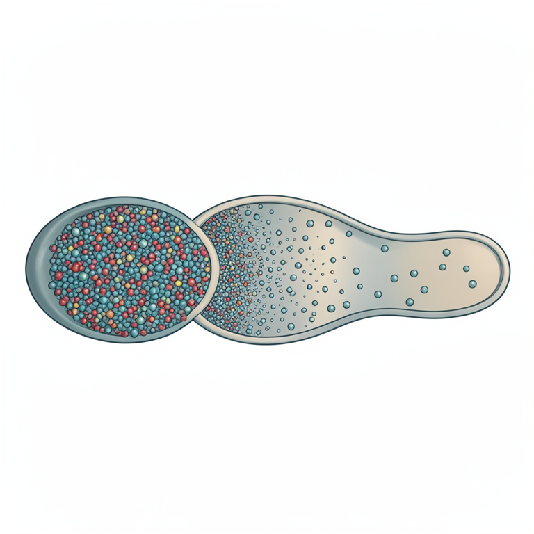
Epididymal Protein Gradient
AI-generated conceptual illustration (Rattus norvegicus epididymis and sperm). It explains the idea and does not show study data.
View source evidence ↗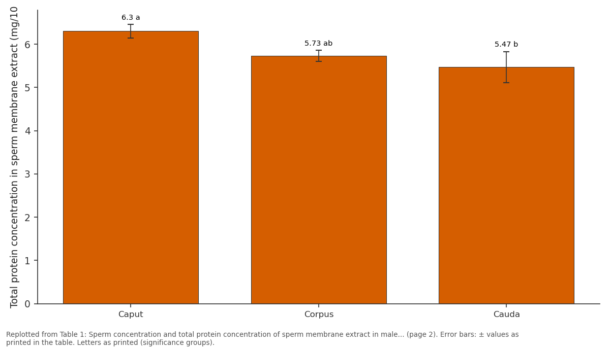
Total protein concentration in sperm...: Caput vs Corpus vs Cauda
Replotted from Table 1 (page 2) of the original paper. Every bar is the paper's own table value.
View source evidence ↗Figures & tables
From the original paper Table 1
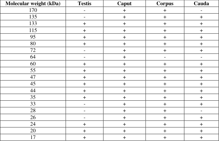 Table 2
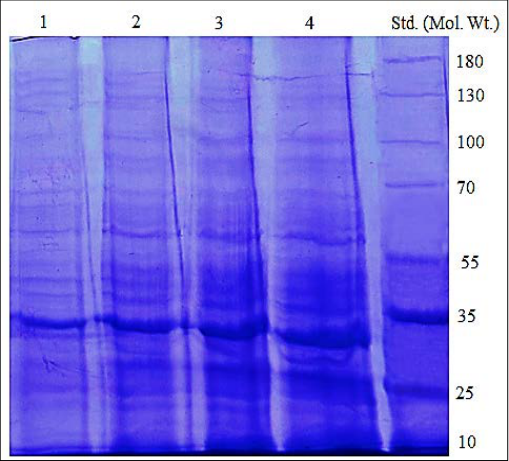 Figure 1
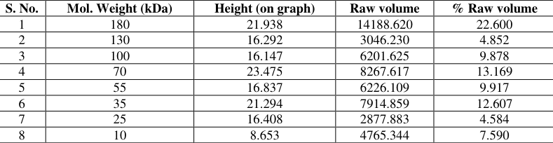 Table 3
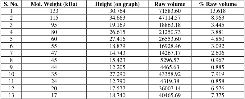 Table 4
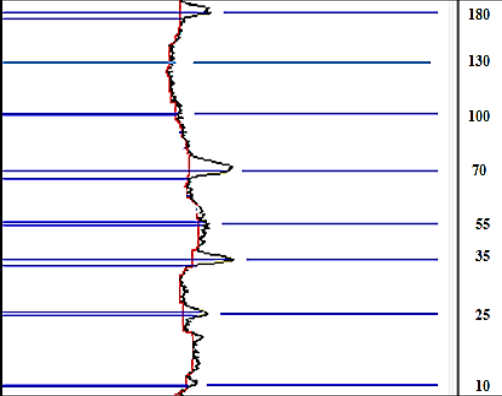 Figure 2
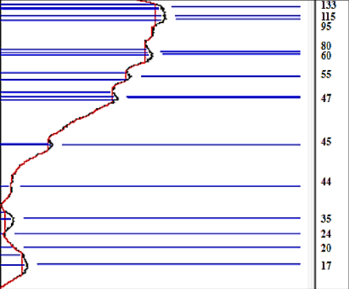 Figure 3
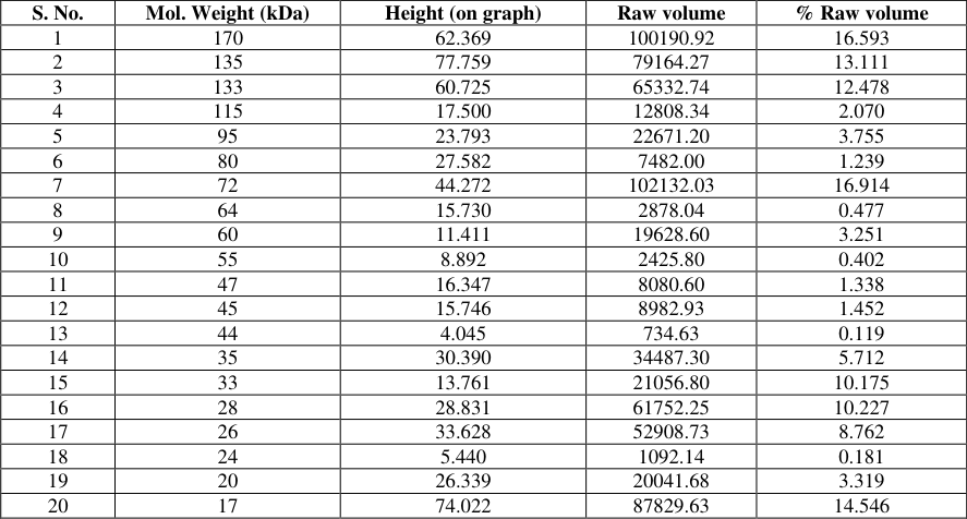 Table 5
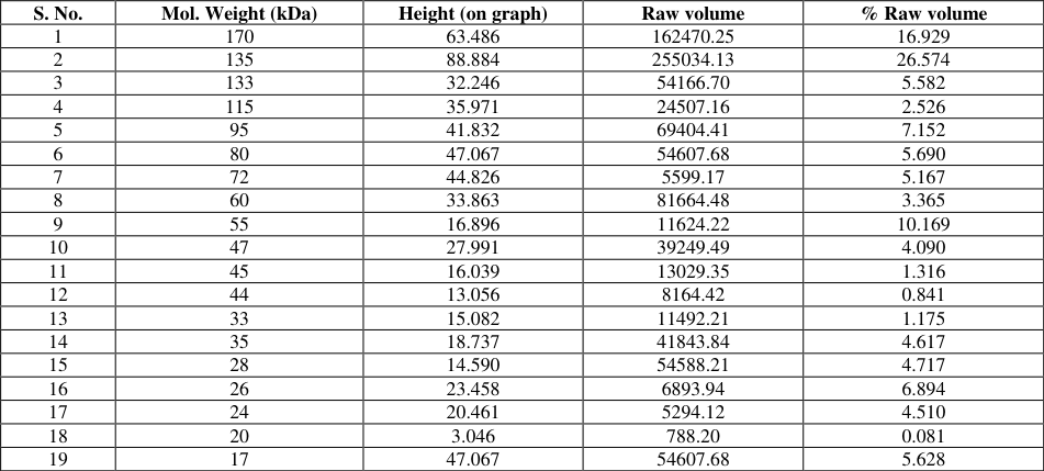 Table 6
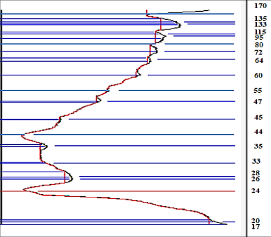 Figure 4
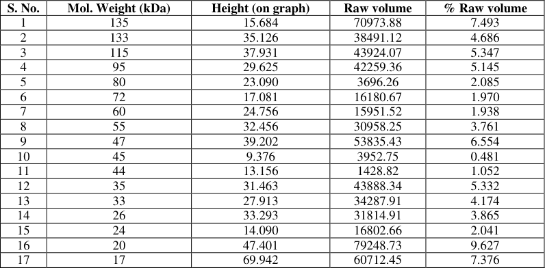 Table 7
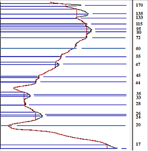 Figure 5
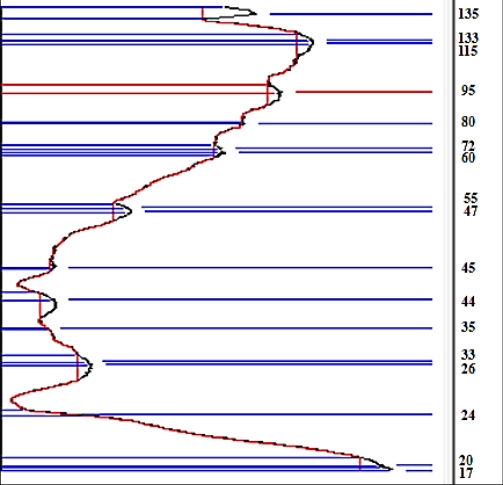 Figure 6
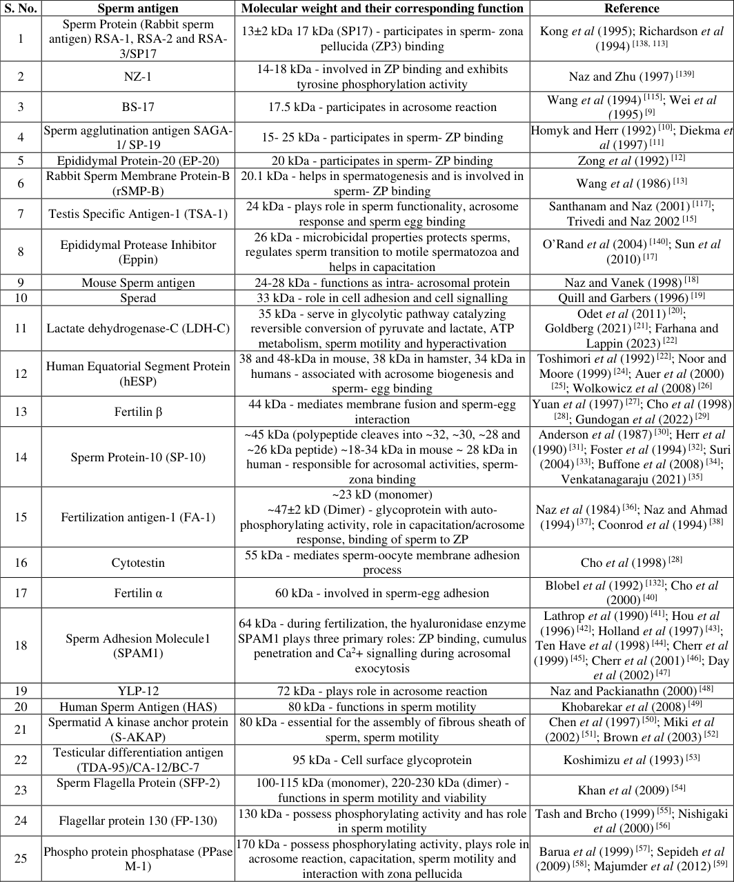 Table 8
Introduction
Male fertility relies not only on the production of spermatozoa in the testes but also on their structural and functional maturation during their passage through the epididymis. Sperm released from the testes are immature and lack fertilizing ability. As they transit through the caput, corpus and cauda regions of the epididymis, they progressively acquire motility, fertilizing competence and membrane stability (Cooper 1996; Robaire and Hinton, 2015) [134,141] . This transformation is driven by the specialized environment of the epididymis, which provides region-specific secretions and reabsorptive activities (Turner 1991; Cornwall 2009) [142, 136] . Proteins play a central role in this maturation process. Epididymal secretions and membrane remodeling alter the sperm protein landscape, with some proteins being removed while others are acquired or modified post-translationally (Belleannée et al 2012; Cooper and Yeung 2006) [131,135] . Many sperm proteins are directly involved in key reproductive events, such as motility regulation, zona pellucida recognition, acrosome reaction and sperm-egg fusion (Naz and Zhu 1997; O'Rand et al 2004) [139,140] . Characterized examples include SP-17, Fertilin and SPAM1, which participate in zona binding, sperm-egg adhesion, and enzymatic activities crucial for fertilization (Kong et [138,132,133] . Rodents, particularly Rattus norvegicus, are frequently used as models for reproductive studies due to their physiological resemblance to higher mammals in sperm maturation processes (Jones and Lopez 2004) [137] . Although individual sperm proteins have been identified in different species, limited information is available on the comparative protein profile of sperm membranes across testis and different epididymal regions in rats. Understanding these molecular variations is essential for clarifying the mechanisms that regulate sperm maturation and fertility. The present study was therefore undertaken to identify and characterize proteins in the testis and epididymal sperm membrane extracts of male Rattus norvegicus. By combining biochemical protein estimation with SDS-PAGE analysis, this work aims to provide insights into region-specific protein dynamics and their potential roles in sperm maturation.
Materials and Methods
Materials and Methods
Procurement and maintenance of animals
Twenty sexually mature albino rats weighing between 130-150 g were procured from Disease Free Animal House at Lala Lajpat Rai, University of Veterinary and Animal Sciences (LUVAS), Hisar, a registered animal breeding centre after getting permission from Institutional Animal Ethics Committee (IAEC). The animals were acclimatized to laboratory conditions for 10 days before beginning of experiments. The rats were kept in polypropylene cages having wheat straw as bedding. The animals were fed a standard pelleted diet and water was provided in glass water bottle feeder with a rubber cork. During the experiment, standard laboratory conditions were maintained including ambient temperature (22-25 °C) and 12-hour light: 12-hour dark cycle. The rats were maintained according to the Indian Committee for the purpose of Control and Supervision of Experiments with animals (CPCSEA) standards.
Quantitative and qualitative analysis of total protein from epididymal sperm membrane 2.2.1 Dissection of Rats
Sexually mature adult male rats (n=20) were weighed, anaesthetized in CO2 chamber and then dissected under hygienic conditions to take out testes and epididymii from both the sides. The organs were transferred into Phosphate buffer saline (PBS) kept at 37°C in petri dishes. The epididymis was split into three regions: caput (head), corpus (body) and cauda (tail) and each region was suspended in 1ml PBS. Each region of the epididymis was given a small incision and the epididymal fluid containing spermatozoa was collected. The various morphological parameters were analysed using epididymal fluid. The epididymal fluid containing spermatozoa was collected and centrifuged at 3000 rpm for 10 minutes to get spermatozoa in the pellet and the epididymal fluid (supernatant) was collected in separate vials. From the spermatozoa, sperm membrane extract (SME) was extracted using deoxycholate detergent and buffer and the fluid was processed for the partial purification of proteins with ammonium sulphate (NH4)2SO4.
Partial purification of epididymal fluid proteins
A saturated solution of ammonium sulphate (NH 4 ) 2 SO 4 was prepared by dissolving 7.67 g of ammonium sulphate in 10 ml of distilled water. 4 ml of ammonium sulphate was added to 1ml of epididymal fluid with continuous stirring with glass rod. The solution was kept overnight at 4°C. Precipitates of proteins were formed which were then separated by performing centrifugation at 10,000 g for 10 min at 4 °C. The precipitated proteins were then dissolved in PBS (pH 7.4) and dialysed against respective buffer to remove traces of ammonium sulphate.
Extraction of testis and epididymal sperm membrane proteins
The sperm membrane proteins of testicular and epididymal fluid were extracted with sodium dodecyl sulphate (SDS) (Jones et al 1983) and Deoxycholate (DOC) (Naz et al 1986). The fluid from testis and epididymis containing spermatozoa was centrifuged at 3000 rpm for 10 min. The pellet containing the spermatozoa was then given two PBS (pH 7.4) washes. The sperms were then suspended in 1 ml of 15 mM DOC/ 2% SDS in 62.5 mM Tris-HCl (pH 8.0) containing protease inhibitors (10 mM aprotinin, 1mM phenyl methyl sulphonyl fluoride, 25mM benzidine). Following this the sperm suspension was sonicated for 3 × 20 sec at 20 Watts in a cold ice bath. It was then centrifuged at 16,000 g for 40 minutes at 4 °C. The supernatant containing the sperm membrane extract (SME) was collected in aliquots and stored at -20 °C till further use.
Quantitative analysis of proteins (Lowry et al 1951)
Protein estimation was carried out by the method of Lowry et al 1951 in SME. The protein content was expressed as mg/10 9 spermatozoa.
Where; O.D is optical density [3] For the purpose of separating sperm membrane proteins, the extracted SME was subjected to sodium dodecyl sulphatepolyacrylamide gel electrophoresis (SDS-PAGE) (Laemmli 1970) [3] .
Qualitative analysis of proteins by Sodium dodecyl sulphate-Polyacrylamide gel electrophoresis (SDS-PAGE) (Laemmli 1970)
Qualitative analysis of proteins by Sodium dodecyl sulphate-Polyacrylamide gel electrophoresis (SDS-PAGE) (Laemmli 1970)
Statistical analysis
Values were calculated as Mean ± S.E. Significance of difference was determined between different parameters at 5% level of significance by one way-ANOVA followed by Duncan Test using SPSS 16.0 version software.
Results
Results
Quantitative analysis of total protein content in epididymal sperm membrane extract
The protein content in the caput, corpus and cauda sperm membrane extract was recorded to be 6.30±0.16, 5.73±0.16 and 5.47±0.36 mg /10 9 spermatozoa respectively as shown in (Table 1). Significant difference was observed in the protein content in all the three different regions of epididymis. According to the values obtained it was observed that the protein content was highest in caput and consecutively decreased in corpus and was found to be lowest in the cauda epididymis. The results obtained could be due to the maximum secretory activities in the caput (head) region remarking beginning of the process of sperm maturation and minimum secretory activities in cauda (tail) region depicting the presence of mature spermatozoa in this region. The concentration of sperm in the caput, corpus and cauda region of epididymis was 85.4±1.86, 55.13±1.42 and 125.1±2.69 × 10 6 /ml respectively (Table 1). Sperm concentration was observed to be highest in the cauda region as it is the storage site for mature spermatozoa. 1). An increase in intensity of bands has been observed from caput to cauda, high intensity bands were recorded in cauda region, consequently lower intensity bands in corpus and lowest intensity bands were observed in the caput region. Although the number of protein bands recorded were higher in caput region but their intensity was low. The trend for peak height, raw volume and % raw volume was not same for all the proteins in standard protein ladder, testis, caput, corpus and cauda sperm membrane extract as shown in the tables 3-7 and graphs for variation in protein intensity between the different epididymal regions are shown in figure 23456.
| Regions of Epididymis | Sperm concentration (× 106 spermatozoa/ml) | Total protein concentration in sperm membrane extract (mg/109 spermatozoa) |
|---|---|---|
| Caput | 85.4±1.86a | 6.30±0.16a |
| Corpus | 55.13±1.42b | 5.73±0.13ab |
| Cauda | 125.1±2.69c | 5.47±0.36b |
Table text extracted from the original PDF; superscript letters are shown inline. The original table image is below.
View original table image
The difference in band patterns on gel indicates that some proteins are lost and some are acquired during the spermatozoa's journey through the epididymis. The protein bands of molecular weight 26, 28, 33, 64, 72, 135 and 170 kDa were absent in the SME of testis but were present in the SME of different regions of epididymis. These proteins are thought to be acquired by the spermatozoa during epididymal maturation. A 64 kDa protein was absent in corpus and cauda region but were present in the caput region of epididymis depicting that possibly these proteins were absorbed during their transition through the epididymis. The protein bands with molecular weight 28 and 170 kDa were absent in cauda region but were present in caput and corpus region of epididymis indicating that these proteins were probably reabsorbed during their transition (Table 2). The presence or absence of certain protein bands in different regions of the epididymis is because, during the sperm's passage through the epididymis, its surface membrane undergoes sequential protein modifications. [138,113] 2 NZ-1 14-18 kDa -involved in ZP binding and exhibits tyrosine phosphorylation activity Naz and Zhu (1997) [139] 3 BS-17 17.5 kDa -participates in acrosome reaction Wang et al (1994) [115] ; Wei et al (1995) [9] 4 Sperm agglutination antigen SAGA-1/ SP-19 15-25 kDa -participates in sperm-ZP binding Homyk and Herr (1992) [10] ; Diekma et al (1997) [11] 5
| Molecular weight (kDa) | Testis | Caput | Corpus | Cauda |
|---|---|---|---|---|
| 170 | - | + | + | - |
| 135 | - | + | + | + |
| 133 | + | + | + | + |
| 115 | + | + | + | + |
| 95 | + | + | + | + |
| 80 | + | + | + | + |
| 72 | - | + | + | + |
| 64 | - | + | - | - |
| 60 | + | + | + | + |
| 55 | + | + | + | + |
| 47 | + | + | + | + |
| 45 | + | + | + | + |
| 44 | + | + | + | + |
| 35 | + | + | + | + |
| 33 | - | + | + | + |
| 28 | - | + | + | - |
| 26 | - | + | + | + |
| 24 | + | + | + | + |
| 20 | + | + | + | + |
| 17 | + | + | + | + |
Table text extracted from the original PDF; superscript letters are shown inline. The original table image is below.
View original table image
Epididymal Protein-20 (EP-20) 20 kDa -participates in sperm-ZP binding Zong et al (1992) [12] 6 Rabbit Sperm Membrane Protein-B (rSMP-B) 20.1 kDa -helps in spermatogenesis and is involved in sperm-ZP binding Wang et al (1986) [13] 7
Testis Specific Antigen-1 (TSA-1) 24 kDa -plays role in sperm functionality, acrosome response and sperm egg binding Santhanam and Naz (2001) [117] ; Trivedi and Naz 2002 [15] 8 Epididymal Protease Inhibitor (Eppin)
26 kDa -microbicidal properties protects sperms, regulates sperm transition to motile spermatozoa and helps in capacitation O'Rand et al (2004) [140] ; Sun et al (2010) [17] 9 Mouse Sperm antigen 24-28 kDa -functions as intra-acrosomal protein Naz and Vanek (1998) [18] Sperad 33 kDa -role in cell adhesion and cell signalling Quill and Garbers (1996) [19] Lactate dehydrogenase-C (LDH-C)
35 kDa -serve in glycolytic pathway catalyzing reversible conversion of pyruvate and lactate, ATP metabolism, sperm motility and hyperactivation
Odet et al (2011) [20] ; Goldberg (2021) [21] ; Farhana and Lappin (2023) [22] Human Equatorial Segment Protein (hESP)
38 and 48-kDa in mouse, 38 kDa in hamster, 34 kDa in humans -associated with acrosome biogenesis and sperm-egg binding
Toshimori et al (1992) [22] ; Noor and Moore (1999) [24] ; Auer et al (2000) [25] ; Wolkowicz et al (2008) [26] Fertilin β 44 kDa -mediates membrane fusion and sperm-egg interaction
Yuan et al (1997) [27] ; Cho et al (1998) [28] ; Gundogan et al (2022) [29] Sperm Protein-10 (SP-10) ~45 kDa (polypeptide cleaves into ~32, ~30, ~28 and ~26 kDa peptide) ~18-34 kDa in mouse ~ 28 kDa in human -responsible for acrosomal activities, spermzona binding
Anderson et al (1987) [30] ; Herr et al (1990) [31] ; Foster et al (1994) [32] ; Suri (2004) [33] ; Buffone et al (2008) [34] ; Venkatanagaraju (2021) [35] Fertilization antigen-1 (FA-1) ~23 kD (monomer) ~47±2 kD (Dimer) -glycoprotein with autophosphorylating activity, role in capacitation/acrosome response, binding of sperm to ZP Naz et al (1984) [36] ; Naz and Ahmad (1994) [37] ; Coonrod et al (1994) [38] Cytotestin 55 kDa -mediates sperm-oocyte membrane adhesion process Cho et al (1998) [28] Fertilin α 60 kDa -involved in sperm-egg adhesion Blobel et al (1992) [132] ; Cho et al (2000) [40] Sperm Adhesion Molecule1 (SPAM1) 64 kDa -during fertilization, the hyaluronidase enzyme SPAM1 plays three primary roles: ZP binding, cumulus penetration and Ca 2 + signalling during acrosomal exocytosis Lathrop et al (1990) [41] ; Hou et al (1996) [42] ; Holland et al (1997) [43] ; Ten Have et al (1998) [44] ; Cherr et al (1999) [45] ; Cherr et al (2001) [46] ; Day et al (2002) [47] YLP-12 72 kDa -plays role in acrosome reaction Naz and Packianathn (2000) [48] Human Sperm Antigen (HAS) 80 kDa -functions in sperm motility Khobarekar et al (2008) [49] Spermatid A kinase anchor protein (S-AKAP) 80 kDa -essential for the assembly of fibrous sheath of sperm, sperm motility Chen et al (1997) [50] ; Miki et al (2002) [51] ; Brown et al (2003) [52] Testicular differentiation antigen (TDA-95)/CA-12/BC-7 95 kDa -Cell surface glycoprotein Koshimizu et al (1993) [53] Sperm Flagella Protein (SFP-2) 100-115 kDa (monomer), 220-230 kDa (dimer)functions in sperm motility and viability Khan et al (2009) [54] Flagellar protein 130 (FP-130)
130 kDa -possess phosphorylating activity and has role in sperm motility Tash and Brcho (1999) [55] ; Nishigaki et al (2000) [56] Phospho protein phosphatase (PPase M-1) 170 kDa -possess phosphorylating activity, plays role in acrosome reaction, capacitation, sperm motility and interaction with zona pellucida Barua et al (1999) [57] ; Sepideh et al (2009) [58] ; Majumder et al (2012) [59] 4. Discussion 4.1 Quantitative analysis of total protein content in testicular and epididymal sperm membrane extract The total protein content in the sperm membrane extract was found to decrease when the spermatozoa passes from caput to cauda epididymis. The current experiment's result coincides with the results of previous researches. Several workers have reported changes in the protein concentration of spermatozoa during their transit from caput to cauda region of the epididymis (Lavon et al 1971, Brooks and Higgins 1980, Olson and Danzo 1981) [60,61,62] .
A spermatozoon can only become fertile when it has undergone a series of post-testicular alterations in both the male and female reproductive tracts. In order to acquire motility and the capacity to fertilize, the sperm goes through a number of biochemical and functional changes as it passes through the epididymis (Jones and Lopez 2004 [63] , Cooper and Yeung 2006 [135] , Robaire et al 2006 [135] , Robaire and Hinton 2015) [141] . After the seminiferous tubules produce sperm, the tubule's lumen releases the sperm, which then travels to the rete testis and eventually the efferent ducts via the flow of fluids (Hess et [67,68,69] . The sperm are then retained until ejaculation after passing through the caput (head), corpus (body) and cauda (tail) regions of the epididymis (Robaire et al 2006) [135] . Therefore, the epididymis performs three key roles: storage, transit and most importantly sperm maturation.
Each component of the epididymis serves a different physiological purpose. The sperm in each area have unique structural characteristics. The maturation of sperm is carried out by the caput and corpus, while the cauda epididymis serves as a reservoir for sperm (Cornwall 2009, Belleannée et al 2012) [136,131] . The epididymal epithelium plays a role in absorption, secretion, synthesis and creates an environment that is appropriate for acquiring sperm motility and its fertilizing ability (Turner 1991, Hinton et al 1995) [73] . This has also been reported by several other workers that epididymis can change the composition of luminal fluid through its secretion (Kirchhoff 1998 [75,76,77,78] and all the major changes in the protein are due to epididymal secretions ( [134,84,135,86,87] . The epididymis secretes proteins in a highly regulated and regionalized manner from different regions of the epididymis. The secretory activity of the head epididymal region is 6-8 times greater than that of the tail region in boar. Caput contributes 83%, corpus contributes 16% and cauda contributes mere 1% to the overall epididymal secretions (Dacheux et al 2005) [78] . Maximum secretory activities are observed in caput region representing the beginning of maturation process of spermatozoa in this region. Some of the synthesized and secreted proteins from the epididymis become associated with the sperm plasma membrane (Yeung et al 1997, Cooper 1998, Holland and Nixon 1998) [89,90,91] . The proteins in the plasma membrane are embedded partially or completely in the lipid bilayer and can protrude out from their cytoplasmic surface and have different characteristics depending on their functions (Etemadi 1989) [92] . These membrane proteins can be selectively extracted using various detergents such as SDS and DOC (Ahuja et al 1985, Kinger et al 1989) [93,94] . These detergents are of varying polarities; the detergents with different polarities selectively extract proteins of different nature and are embedded to different extents in the lipid bilayer of plasma membrane (Sundhey et al 1992) [95] .
Qualitative analysis of total protein from testis, caput, corpus and cauda epididymal sperm membrane by Sodium dodecyl sulphate-Polyacrylamide gel electrophoresis (SDS-PAGE)
Prior to ejaculation, functionally mature sperm cells are stored in the cauda epididymis (Cornwall 2009) [136] . The cauda's epithelial cells release substances that support the luminal environment intended to keep sperm in quiescent stage during storage (Robaire et al 2006) [135] . Upon ejaculation, sperm exit this quiescent state and their metabolic activity increases by 3-5 times (Jones 1999) [97] . Sperm maturation is a crucial process that takes place during epididymal transit and is necessary for normal male fertility. During this period, sperm go through a number of maturational changes, but most significantly, they gain motility through interaction with epididymosomes which are released by the epididymal epithelium and plays role in sperm competency (Topfer-Petersen 2000, Sullivan and Saez 2013, Gervasi and Visconti 2017) [98,99,100] . Epididymosomes are known to contain various proteins from different functional classes. Furthermore, the various locations of the epididymis have distinct protein compositions of epididymosomes (Girouard et al 2011) [101] . Sperm come into direct contact with the materials in the epididymal lumen environment during the maturation phase (Cornwall 2009) [136] . The sperm maturation is believed to be the result of modification of pre-existing sperm components and absorption of extracellular substances from epididymal secretions and incorporation of epididymal fluid peptides (Olson and Orgebin-Crist 1982) [102] . Testicular spermatozoa released into the seminiferous tubules are not yet mature and do not possess fertilizing capacity. They become mature and acquire the potential to fertilize due to its interaction with epididymal secretory proteins. The majority of the testicular sperm surface proteins are gradually lost or altered in immature gametes, while new transient or permanent proteins are added to well-organized sperm membrane protein domains. It has been shown that these alterations are necessary for male gametes to acquire motility and fertility. The epididymal microenvironment surrounding the spermatozoa is maintained by active secretion and reabsorption throughout the tract and by the presence of a continuous blood barrier formed by tight junctions in the epithelial cells of epididymis (Pardyak et al 2020, Gibb et al 2021) [103,104] . The interaction may result in removing, masking or unmasking pre-existing surface compounds (Voglmayr et al 1982) [105] . Sequential protein modifications occur on the sperm surface during their transit through the epididymis: immature gametes progressively loose or modify most of their testicular surface proteins and gain new transient or permanent proteins in well-organized sperm membrane protein domains. These stages have been demonstrated to be essential for acquisition of motility and fertility by the male gametes. Disruption in the proteins in any of the stage will ultimately affect the process of fertilization (Belleannée et al 2011, Dacheux and Dacheux 2014) [106,107] . Evidences from earlier studies have demonstrated that specific secretory proteins in the epididymis interact with the sperm surface to influence sperm maturation. The epididymis synthesizes and secretes various proteins some of which becomes associated with the sperm plasma membrane and some epididymal glycosidases are absorbed in the plasma membrane which are having role in fertilization (Vernon et al 1982, Hammerstedt and Parks 1987, Jones 1998, Gervasi and Visconti 2017) [107,108,109,110] . The different protein bands that appeared on the acrylamide gels correspond to different sperm proteins. Many of these proteins have been characterized by different researchers that support the present study. Some of the characterized proteins are as follows:
A protein band corresponding to molecular weight ranging between ~14-18 kDa is of NZ-1. The testis-specific proteins were isolated from human and mouse sperm antigens. The sperm antigens are involved in ZP binding and exhibits tyrosine phosphorylation activity. Mice immunized with a recombinant form of the NZ-1 antigen showed decreased rates of reproduction (Naz and Zhu 1997) [139] . A protein band corresponding to molecular weight ~17 kDa is of sperm Protein (SP-17). Sperm specific proteins namely RSA-1, RSA-2 and RSA-3/SP17 with low molecular weight ~13±2 kDa have been identified from rabbit sperm (O'Rand and Porter 1982) [110] . Cross-reactions between mouse, baboon and human sperm were observed with monoclonal antibodies against RSA antigens. These antibodies prevented human spermatozoa from penetrating hamster oocytes that were zona-free (O'Rand and Irons 1984) [111] . The most significant antigen among RSA-1, RSA-2 and RSA-3/SP17 is SP-17 with molecular weight corresponding to ~17 kDa, shown on the surface of sperm following an acrosome reaction in human, rabbit and mouse. The protein is localized in the equatorial section and participates in zona pellucida (ZP) binding in vitro (Kong et al 1995, Richardson et al 1994) [113] . Studies conducted in vitro demonstrated SP-17's selectivity for ZP3. Antibody titres were increased in both male and female mice immunized with a chimeric peptide including SP-17 and T-cell epitope. The peptide's immunogenic properties were validated by increase in antibody titre in both the sexes. A protein band corresponding to molecular weight ~17.5 kDa is of BS-17. It is one of the antigens that were found in infertile women's serum samples. A 758 bp cDNA fragment was obtained from the positive clone when polyclonal anti-BS-17 antibodies were used to probe the human testis λgt11 cDNA expression library. The protein possibly participates in acrosome reaction (Wang et al 1994, Wei et al 1995) [115,9] . Human sperm were unable to penetrate and fertilize zonafree hamster eggs when polyclonal antibodies targeting the BS-17 antigen were present (Wei et al 1994). Given that BS-17 and calpastatin had a 99.7% homology, it was hypothesized that the anti-BS-17 antibodies were responsible for destabilising the calpastatin-calpain complex. Following this destabilisation, calpain may cause the sperm acrosome reaction prematurely, before the sperm reach the ovum, which would diminish the sperm's capacity to fertilize the ovum. Sperm agglutination antigen or SP-19 corresponding to molecular weight ~ (15-25 kDa) was first identified and isolated from human spermatozoa. The presence of SAGA-1 on the entire sperm surface was demonstrated by immunolocalization using electron microscopy, which also indicated the reactivity of S-19 monoclonal antibody to human sperm surface (Homyk and Herr 1992) [10] . Sperm agglutination/immobilization and prevention of penetration in zona-free hamster ova were the outcomes of in vitro incubation of antibodies produced against SAGA-1 (Diekman et al 1997) [11] . A protein band corresponding to molecular weight ~20 kDa is similar to Epididymal Protein-20 (EP-20). It is a glycoprotein that was identified from rabbit cauda epididymal fluid. Polyclonal antibodies specific to EP-20 were used to perform immune localization of the protein, demonstrating its presence in the testis and epididymis. The protein is testis specific as strong staining was only seen in the human testes whereas germ cells, interstitial cells and nine other tissues remained unstained.
In addition to immobilising and agglutinating human sperm, polyclonal antibodies produced against EP-20 prevented human sperm from penetrating zona-free hamster eggs in vitro (Zong et al 1992, Nixon et al 2002) [12,76] . Rabbit Sperm Membrane Protein-B (rSMP-B) -A protein corresponding to molecular weight ~20.1 kDa is of rSMP-B, extracted from the tail of a rabbit sperm (Wang et al 1986) [13] . Using polyclonal antibodies against rSMP-B, the immunolocalization of rSMP-B on spermatozoa and somatic cells was examined. The findings implied that during spermatogenesis, germ cells produced this antigen. It was shown that monoclonal antibodies produced against rSMP-B could prevent human sperm from penetrating and fertilizing zona-free hamster eggs in vitro. When male rabbits were immunized with rSMP-B protein, spermatogenesis was blocked, leading to azoospermia. Additionally, it was discovered that 83.3% of immunized female rats showed infertility following mating when they were immunized with the rSMP-B 230 peptide (Kamada et al 1999) [116] . Testis Specific Antigen-1 (TSA-1) -A protein corresponding to molecular weight ~24 kDa is of TSA-1. Human and mouse sperm express this protein. The expression of TSA-1 in testis was detected using the northern blot technique. Acrosomal, equatorial, midpiece and tail regions of human sperm were identified by antibody (Santhanam and Naz 2001) [117] . In vitro tests revealed that sperm egg binding and acrosome response were both blocked by anti-recombinant TSA-1 antibodies (Trivedi and Naz 2002) [15] . These results suggested that a protein unique to the testis and sperm plays a role in sperm funtionality. A protein band corresponding to molecular weight ~26 kDa is of Epididymal Protease Inhibitor (Eppin) which is an epididymal protein (O'Rand et al 2004) [140] and was used to immunize male monkeys. 78% of the monkeys that had high anti-Eppin antibody titres following immunization lost their ability to reproduce, while 71% of the monkeys were able to conceive again after the immunization was stopped. Subsequent researches revealed that active immunization with Eppin reduced animal populations to a maximum of 90%. A recombinant version of Eppin was also tested in an attempt to increase its sensitivity. In contrast to native protein, immunization with recombinant protein only produced a 70% decrease in fertility (Sun et al 2010) [17] . A protein band corresponding to molecular weight ~24-28 kDa is of mouse sperm antigen. The protein was reported in testis, corpus and cauda and shows 60% homology with human SP-10 at protein level and expresses at the stage of post-meiotic germ cells in mouse and functions as intraacrosomal protein (Naz and Vanek 1998) [18] . A protein band corresponding to molecular weight ~33.3 kDa is of Sperad. An extensive family of potential cell adhesion molecules known as biliary glycoproteins bears a close resemblance to the sperad. The protein is first expressed by haploid spermatid and is confined to the plasma membrane covering the acrosome along with the role in cell adhesion/signaling. Sperad is lost from the sperm cells when the acrosome reaction is induced, but its molecular weight does not appear to change (Quill and Garbers 1996) [19] . A protein band in the gel corresponding to molecular weight ~35 kDa is of LDH-C data obtained from National Center for Biotechnology Information (NCBI). LDH-C protein family members characteristically are distributed in tissue and cell in specific patterns and serve as the terminal enzyme of glycolysis, catalyzing reversible oxidation-reduction between pyruvate and lactate (Farhana and Lappin 2023) [22] . They are present as tetramers and only one family member, LDH-C is found abundant in spermatocytes, spermatids and spermatozoa. Modest amounts of LDH-C are also found in oocytes. Lactate dehydrogenase-C gene (Ldhc) gene is localized to human chromosome 11. The gene is expressed exclusively during the first meiotic division of spermatogenesis and is distributed throughout the principal piece of the sperm tail consistent with flagellar location for its enzymatic role in the glycolytic pathway. While other glycolytic enzymes including LDH-A are tightly bound to the fibrous sheath, LDH-C localizes throughout the flagellum presumably for ATP production to power sperm motility (Goldberg 2021) [21] . Previous studies have demonstrated that disruption of germ cell-specific Ldhc led to male infertility due to defects in sperm function, including a rapid decline in sperm ATP levels, a decrease in progressive motility and failure to develop hyperactivated motility (Odet et al 2011) [20] . Sperm Protein-38 (SP-38) -A protein band corresponding to molecular weight ~38 kDa was isolated from boar sperm. The protein participates in sperm binding to a 90-kDa family of zona pellucida glycoprotein. According to the amino acid sequence inferred from the cDNA sequence, SP-38 starts off as a 350-residue precursor protein. After posttranslational modification the mature protein is formed with 299 residues form the SP-38 precursor. Sperm cell immunostained with an antibody raised against SP-38 fusion protein revealed that it is intra-acrosomal in location and is released after post-acrosome reaction (Mori et al 1993) [118] . The molecular weight ~38 and ~48-kDa in mouse, 38 kDa in hamster and 34 kDa in humans are similar to human Equatorial Segment Protein (hESP). It is a sperm-specific protein known as human equatorial segment protein that was initially identified in 2003 (Wolkowicz et al 2003) [119] . Human ESP and mouse ESP (mESP) share 81% homology. It is connected to acrosome biogenesis and is restricted to the equatorial segment of the acrosome. In hamster egg penetration assay, the application of antiserum generated against recombinant hESP prevented the binding and fusion of human sperm (Wolkowicz et al 2008) [26] . Fertility was dramatically decreased when mice were immunized with polyepitope antigens. Subsequently a study was conducted to identify the significant portion of mESP that is essential for conception. The complete mESP was broken up into three pieces: P1, P2 and P3. Only the presence of anti-P1 and anti-P2 antibodies inhibited sperm-egg interaction; anti-P3 antibodies did not exhibit any inhibition (Wolkowicz et al 2008) [26] . A protein band corresponding to molecular weight ~45 kDa is of Sperm Protein-10 (SP-10). It is a sperm-specific acrosomal protein (Suri 2004) [33] that was first identified in humans (Anderson et al 1987) [30] and has also been detected in several species including mouse (Buffone et al 2008) [34] . In humans the SP-10 protein was found to be a stable protein with a molecular weight of ~28 kDa (Venkatanagaraju 2021) [35] . A full-length SP-10 protein with molecular weight around 45 kDa was found in the testis extracts, along with additional immunoreactive SP-10 peptides weighing 32, 30, 28, and 26 kDa. Furthermore, 25-18 kDa SP-10 peptides were discovered for the first time in caput epididymal sperm extracts. These peptides most likely came from the proteolytic processing of 45 kDa and 32-26 kDa SP-10 peptides in the early section of the caput epididymis. The extracts of cauda epididymis, ejaculated or capacitated sperm does not contain SP-10 protein indicating that the 32-18 kDa SP-10 peptides were not further processed during epididymal transit, ejaculation or capacitation (Foster et al 1994) [32] . Hence, the protein is responsible for acrosomal activities. Decrease in the concentration of the protein will alter acrosomal reactions. Several enzymes are present in the acrosome including acid hydrolases and other enzymes specific to spermatogenic cells. During the acrosome reactions, the contents are released by calcium mediated exocytosis that helps sperms to penetrate the zona pellucida surrounding the oocyte. The antibodies generated against SP-10 recognised sperm from baboons, macaques and pigs but not from rabbits, bulls, rats, guinea-pigs or cats concluding that SP-10 is a testis but not a species-specific protein (Herr et al 1990) [31] . SP-10 was cloned and sequenced from a human cDNA expression library and the protein was discovered to have 1117 bp of 256 aa (Wright et al 1990) [120] . The mRNA expression of this protein is primarily in the round spermatid stage and is expressed in all the six stages of spermiogenesis. SP-10 proteins were detected only in germ cells and SP-10 RNAs were not detected in any of the tested somatic tissue (Batista et al 2022) [121] . In vitro incubation of anti-SP-10 antibodies prevented bovine fertilization by lowering sperm-zona binding. An attenuated strain of Salmonella sp. that expressed human SP-10 was used to assess the immunogenic potential of SP-10 in vivo. In mice, antigen was administered orally; for monkeys, it was administered intramuscularly. Antibodies specific to SP-10 were detected in both the cases (Kurth et al 1997) [122] . Since SP-10 is localised within the acrosomal compartment and the outer acrosomal membrane complex, and is thus only accessible to antibodies after the acrosome reaction has been initiated. Hence, antibody levels in the oviduct are particularly relevant in this circumstance. A protein band corresponding to molecular weight ~47 kDa is of Fertilization antigen-1 (FA-1). It is a glycoprotein with auto-phosphorylating activity, produced in the testis in the later stages of spermatogenesis (Naz et al 1984) [36] . The molecular weight of its monomeric form is ~23 kDa and that of dimer is ~47±2 kDa. During the human sperm capacitation/acrosome process, tyrosine phosphorylation takes place (Naz and Ahmad 1994) [37] . Therefore, it was proposed that FA-1 plays a significant role in the capacitation/acrosome response, which may also have an impact on the binding of sperm to ZP. The presence of a complementary sequence of the FA-1 antigen on the mouse oocyte demonstrated the specificity of the antigen towards ZP3. Additionally, to determine the phenomenon behind the decrease in fertilization, monoclonal antibodies produced against the FA-1 antigen were employed (Coonrod et al 1994) [38] . Antibodies against FA-1 have been found to impede fertilization by decreasing sperm-zona binding and altering capacitation/acrosome responses. Similar outcomes were seen in the cases of mice, calves, humans and primates. Mice actively immunized with rFA-1 had fertility reductions ranging from 64-70% when compared to control groups. When the titres reached control levels, the animals gave birth to healthy offspring without any change in litter size, indicating that the effect caused by antibodies was reversible and long-lasting (Naz and Zhu 1997) [7] . A protein band corresponding to molecular weight ~60 kDa and ~44 kDa belongs to Metalloprotease/ Disintegrin/ Cysteine-Rich (MDC) protein family. The MDC proteins are an expanding family of integral membrane proteins that are mostly produced in the testes of mammals. A transmembrane domain (also called the ADAM family), a cysteine-rich domain, a disintegrin-like domain, a metalloproteinase-like domain and a prodomain are among the unique conserved properties shared by the proteins (Nishimura et al 2004) [123] . Few of these proteins are known to be expressed on mature sperm or male germ cells. Among these, cyritestin (tMDC I) and fertilin α and β (together referred to as fertilin) are noteworthy. Fertilin is a protein linked with sperm that was earlier known as PH-30. It was first isolated from caudal epididymal sperm of guinea pigs. Fertilin α and β, two related subunits, with molecular weights of ~60 kDa for Fertilin α and ~44 kDa for Fertilin β in guinea pigs, according to results from reducing sodium dodecyl sulphatepolyacrylamide gel electrophoresis (SDS PAGE). Fertilin α's function is linked to sperm-egg adhesion; however it is still unclear if this contributes to membrane fusion (Blobel et al 1992) [132] . The existence of a cysteine-rich domain, which seems to be involved in cell adhesion, justifies its role in adhesion. The sperms lacking Fertilin α are still capable of fusing with the egg (Cho et al 2000) [40] . One of the earliest "cellular disintegrins" to be discovered was fertilin β. The proteins of Fertilin β are observed in three regions of epididymis. This protein is a sperm-associated that are proteolytically cleaved and activated by various proteases during the epididymal transition. It is a dimeric sperm antigen and antibodies produced against it react with fertilin α. It is well known for mediating membrane fusion and sperm-egg interaction (Yuan et al 1997, Cho et al 1998, Gundogan et al 2022) [27,28,29] . Fertilin β seems to use its disintegrin loop sequence to interact with the egg membrane, as evidenced by its inhibition in the presence of antidisintegrin loop antibodies. Fertilin β antibodies prevent fertilization in vitro but immunization against it has little effect on fertility in vivo (Ramarao et al 1996) [124] . The Cyrn gene produces cytotestin, which has an apparent molecular weight of 110 kDa but is processed during its transfer through epididymal region, resulting in an approximate molecular weight of ~55 kDa. It functions in a way similar to fertilin in the sperm-oocyte membrane adhesion process and likewise binds via its disintegrin loop. Mice lacking the ciritestin gene demonstrated impaired sperm-zona binding with no abnormality in sperm-oocyte membrane fusion (Cho et al 1998) [28] . A band with molecular weight ~64kDa is of Sperm Adhesion Molecule 1 (SPAM1). It is a glycosylphosphatidylinositol (GPI)-linked protein and is expressed in the testis, epididymis, sperm and epididymis luminal fluid (Deng et al 2000, Zhang and Martin-DeLeon 2003) [125,126] . During fertilization, the hyaluronidase enzyme SPAM1 has three primary roles. These consist of ZP binding, cumulus penetration (Cherr et al 2001) [138] and Ca 2 + signalling during acrosomal exocytosis (Cherr et al 1999) [45] . The protein has been cloned from numerous species including fox (Ten Have et al 1998) [44] ; guinea pig (Lathrop et al 1990) [41] ; pig (Day et al 2002) [47] ; rat (Hou et al 1996) [42] ; rabbit (Holland et al 1997); monkey and man (Lin et al 1993) showing that the protein is highly conserved among different mammals. Sperm-ZP binding decreased when sperms were incubated with antibodies against SPAM-1. 100% infertility was observed in male and female guinea pigs immunized with SPAM-1 purified from guinea pigs (Primakoff et al 1997) [129] . In case of immunized males, autoimmune diseases were linked to infertility, due to loss of normal sperm in the epididymis (Tung et al 1997). Infertility in females was caused by antibodies that prevented sperm from binding with the eggs. Since, fertility was resumed in both cases within 6 to 15 months, it was reported that the infertility brought on by vaccination was reversible. A protein band corresponding to molecular weight ~72 kDa was observed in the caput, corpus and cauda epididymal sperm membrane. The observed protein corresponds to YLP-12. Using phage display technology, a dodecamer sequence (YLPVGGLRRIGG) known as YLP-12, was discovered on human sperm. In human and mouse sperm, the YLP-12 peptide sequence is mainly found on the acrosome and tail region of sperm. An extensive database search yielded no results for homology or similarity with any known sequence, suggesting that the peptide is unique. According to immunoblotting research, antibodies produced after YLP-12 immunization identified a particular 72 kDa protein band in the testis. The acrosome response (AR) was dramatically decreased when spermatozoa were incubated with YLP-12 Fab in vitro. Antibodies against YLP-12 were found in the sera of immunoinfertile patients. Utilising a synthetic YLP-12 peptide conjugated with the binding domain of recombinant cholera toxin subunit B (rCTB), in vivo investigations were conducted in a mouse model. Two distinct methods of immunizing female mice were used: intramuscular and intranasal. The overall decline in fertility for both methods was 70.3% (intranasal) and 61.4% (intramuscular), respectively (Naz and Packianathan 2000) [48] . A protein band corresponding to molecular weight ~80 kDa is of Human Sperm Antigen (HSA) which was identified from human sperm extract by western blot analysis. The glycoprotein was discovered to be preserved and restricted to the testis and epididymis, with no presence in other somatic tissues (Bandivdekar et al 2001) [130] . Native protein was used to carry out active immunization in male and female rats, resulting in infertility in both the sexes. A synthetic version of 80 kDa HSA was also experimented with an attempt to increase immunogenicity. The male rabbits immunized with peptide-1 and peptide-NT showed the greatest suppression of fertility. Six of the seven male marmosets actively immunized with synthetic peptide-1 were infertile and 7 out of 9 marmosets developed an antibody response. The semen samples of treated animals revealed a total loss of progressive motility. It was discovered that the impact was reversible when the animal's fertility returned and the antibody titre decreased 8-10 weeks following the final booster shot. After that, there was no discernible impact on regular physiological functions (Khobarekar et al 2008) [49] . A protein band corresponding to molecular weight ~95 kDa is of testicular differentiation antigen (TDA-95)/CA-12/BC-7 which is a cell surface glycoprotein. Using two monoclonal antibodies (mAbs), a mouse cell surface antigen demonstrating stage-specific expression during spermatogenesis was identified. These mAbs were able to identify the population of spermatogenic cells that were situated close to the edge of seminiferous tubules throughout stages I-VI and XII. These cells were later identified as zygotene and early pachytene spermatocytes. In germ cells at more advanced stages of spermatogenesis, such as late pachytene spermatocytes and round spermatids, the antigen's expression was temporary and absent. Studies using immunoprecipitation and immunoblotting revealed that the antigenic molecule interacted with both the mAbs. Prepubertal mice appeared to experience spermatogenic disruption following intraperitoneal injection of mAb. These findings revealed that the unique cell surface glycoprotein is a crucial component of the early meiotic prophase of spermatogenesis (Koshimizu et al 1993) [53] . A protein band corresponding to molecular weight ~130 kDa belongs to a lipoprotein binding Flagellar protein (FP-130). The protein has been identified in sea urchin and bull spermatozoa that has phosphorylating activities and plays role in sperm motility (Tash and Brcho 1999, Nishigaki et al 2000) [55,56] . A protein band corresponding to molecular weight ~ 170 kDa is of Phospho-protein phosphatase (PPase M-1) which was isolated and partially characterized from the plasma membrane of the goat cauda-epididymal sperm. It dephosphorylates the serine and threonine residues of histones. PPase M-I is an ecto-enzyme that dephosphorylates the phosphoproteins on the sperm outer membrane, which may have a significant impact on sperm physiology. It plays role in acrosome reaction, capacitation, sperm motility and sperm-zona binding (Barua et al 1999, Sepideh et al 2009) [57,58] .
Conclusion
It can be concluded for the present study that the protein content was highest in caput due to the maximum secretory activities in this region remarking the beginning of sperm maturation and its concentration was lowest in the cauda epididymis because of minimum secretory activities in this region illustrating the presence of mature spermatozoa. The SDS-PAGE profile of sperm membrane extract of testis and different regions of epididymis revealed the presence of difference proteins of varying molecular weights. 13 bands corresponding to molecular weight of 17, 20, 24, 35, 44, 45, 47, 55, 60, 80, 95, 115 and 133 kDa in testis; 20 bands corresponding to molecular weights 17, 20, 24, 26, 28, 33,35, 44, 45, 47, 55, 60, 64, 72, 80, 95, 115, 133, 135 and 170 kDa were detected in caput;19 bands corresponding to molecular weights 17, 20, 24, 26, 28, 33,35, 44, 45, 47, 55, 60, 72, 80, 95, 115, 133, 135 and 170 kDa were noted in corpus and 17 bands corresponding to molecular weights 17, 20, 24, 26, 33,35, 44, 45, 47, 55, 60, 72, 80, 95, 115, 133 and 135 kDa was recorded in cauda epididymal sperm membrane extract.
Acknowledgement
The authors are thankful to the Head, Department of Zoology, Punjab Agricultural University, Ludhiana for providing the facilities for the research work. We duly acknowledge DST, New Delhi for providing infrastructural facilities under FIST grant to Department of Zoology.
Declarations
Ethics approval and consent to participate: permission was taken from Institutional Animal Ethics Committee (IEAC) for the use of animals as experimental model with protocol no. (GADVASU/2023/IAEC/67/18).
Figures and tables from the paper
| S. No. | Mol. Weight (kDa) | Height (on graph) | Raw volume | % Raw volume |
|---|---|---|---|---|
| 1 | 180 | 21.938 | 14188.620 | 22.600 |
| 2 | 130 | 16.292 | 3046.230 | 4.852 |
| 3 | 100 | 16.147 | 6201.625 | 9.878 |
| 4 | 70 | 23.475 | 8267.617 | 13.169 |
| 5 | 55 | 16.837 | 6226.109 | 9.917 |
| 6 | 35 | 21.294 | 7914.859 | 12.607 |
| 7 | 25 | 16.408 | 2877.883 | 4.584 |
| 8 | 10 | 8.653 | 4765.344 | 7.590 |
Table text extracted from the original PDF; superscript letters are shown inline. The original table image is below.
View original table image
| S. No. | Mol. Weight (kDa) | Height (on graph) | Raw volume | % Raw volume |
|---|---|---|---|---|
| 1 | 133 | 30.764 | 71583.60 | 13.618 |
| 2 | 115 | 34.663 | 47114.57 | 8.963 |
| 3 | 95 | 19.169 | 18863.18 | 3.445 |
| 4 | 80 | 26.615 | 21250.73 | 3.881 |
| 5 | 60 | 27.416 | 26553.60 | 4.850 |
| 6 | 55 | 18.879 | 16928.46 | 3.092 |
| 7 | 47 | 14.743 | 14267.17 | 2.606 |
| 8 | 45 | 15.423 | 5296.57 | 0.967 |
| 9 | 44 | 12.205 | 4465.63 | 0.885 |
| 10 | 35 | 27.290 | 43358.92 | 7.919 |
| 11 | 24 | 12.790 | 4319.38 | 0.858 |
| 12 | 20 | 17.577 | 36007.14 | 6.576 |
| 13 | 17 | 18.740 | 40465.69 | 7.375 |
Table text extracted from the original PDF; superscript letters are shown inline. The original table image is below.
View original table image
| S. No. | Mol. Weight (kDa) | Height (on graph) | Raw volume | % Raw volume |
|---|---|---|---|---|
| 1 | 170 | 62.369 | 100190.92 | 16.593 |
| 2 | 135 | 77.759 | 79164.27 | 13.111 |
| 3 | 133 | 60.725 | 65332.74 | 12.478 |
| 4 | 115 | 17.500 | 12808.34 | 2.070 |
| 5 | 95 | 23.793 | 22671.20 | 3.755 |
| 6 | 80 | 27.582 | 7482.00 | 1.239 |
| 7 | 72 | 44.272 | 102132.03 | 16.914 |
| 8 | 64 | 15.730 | 2878.04 | 0.477 |
| 9 | 60 | 11.411 | 19628.60 | 3.251 |
| 10 | 55 | 8.892 | 2425.80 | 0.402 |
| 11 | 47 | 16.347 | 8080.60 | 1.338 |
| 12 | 45 | 15.746 | 8982.93 | 1.452 |
| 13 | 44 | 4.045 | 734.63 | 0.119 |
| 14 | 35 | 30.390 | 34487.30 | 5.712 |
| 15 | 33 | 13.761 | 21056.80 | 10.175 |
| 16 | 28 | 28.831 | 61752.25 | 10.227 |
| 17 | 26 | 33.628 | 52908.73 | 8.762 |
| 18 | 24 | 5.440 | 1092.14 | 0.181 |
| 19 | 20 | 26.339 | 20041.68 | 3.319 |
| 20 | 17 | 74.022 | 87829.63 | 14.546 |
Table text extracted from the original PDF; superscript letters are shown inline. The original table image is below.
View original table image
| S. No. | Mol. Weight (kDa) | Height (on graph) | Raw volume | % Raw volume |
|---|---|---|---|---|
| 1 | 170 | 63.486 | 162470.25 | 16.929 |
| 2 | 135 | 88.884 | 255034.13 | 26.574 |
| 3 | 133 | 32.246 | 54166.70 | 5.582 |
| 4 | 115 | 35.971 | 24507.16 | 2.526 |
| 5 | 95 | 41.832 | 69404.41 | 7.152 |
| 6 | 80 | 47.067 | 54607.68 | 5.690 |
| 7 | 72 | 44.826 | 5599.17 | 5.167 |
| 8 | 60 | 33.863 | 81664.48 | 3.365 |
| 9 | 55 | 16.896 | 11624.22 | 10.169 |
| 10 | 47 | 27.991 | 39249.49 | 4.090 |
| 11 | 45 | 16.039 | 13029.35 | 1.316 |
| 12 | 44 | 13.056 | 8164.42 | 0.841 |
| 13 | 33 | 15.082 | 11492.21 | 1.175 |
| 14 | 35 | 18.737 | 41843.84 | 4.617 |
| 15 | 28 | 14.590 | 54588.21 | 4.717 |
| 16 | 26 | 23.458 | 6893.94 | 6.894 |
| 17 | 24 | 20.461 | 5294.12 | 4.510 |
| 18 | 20 | 3.046 | 788.20 | 0.081 |
| 19 | 17 | 47.067 | 54607.68 | 5.628 |
Table text extracted from the original PDF; superscript letters are shown inline. The original table image is below.
View original table image
| S. No. | Mol. Weight (kDa) | Height (on graph) | Raw volume | % Raw volume |
|---|---|---|---|---|
| 1 | 135 | 15.684 | 70973.88 | 7.493 |
| 2 | 133 | 35.126 | 38491.12 | 4.686 |
| 3 | 115 | 37.931 | 43924.07 | 5.347 |
| 4 | 95 | 29.625 | 42259.36 | 5.145 |
| 5 | 80 | 23.090 | 3696.26 | 2.085 |
| 6 | 72 | 17.081 | 16180.67 | 1.970 |
| 7 | 60 | 24.756 | 15951.52 | 1.938 |
| 8 | 55 | 32.456 | 30958.25 | 3.761 |
| 9 | 47 | 39.202 | 53835.43 | 6.554 |
| 10 | 45 | 9.376 | 3952.75 | 0.481 |
| 11 | 44 | 13.156 | 1428.82 | 1.052 |
| 12 | 35 | 31.463 | 43888.34 | 5.332 |
| 13 | 33 | 27.913 | 34287.91 | 4.174 |
| 14 | 26 | 33.293 | 31814.91 | 3.865 |
| 15 | 24 | 14.090 | 16802.66 | 2.041 |
| 16 | 20 | 47.401 | 79248.73 | 9.627 |
| 17 | 17 | 69.942 | 60712.45 | 7.376 |
Table text extracted from the original PDF; superscript letters are shown inline. The original table image is below.
View original table image
References
141 in the original paper- Naz RK, Philips TM, Rosenblum BB. Characterization of the fertilization antigen 1 for the development of a contraceptive vaccine. Proc Natl Acad Sci USA. 1986;83:5713-5717.
- Lowry DH, Rosebrough NJ, Farr AL, Randall RJ. Protein measurement with the Folin phenol reagent. J Biol Chem. 1951;193:265-275.
- Laemmli UK. Cleavage of structural proteins during the assembly of the head of bacteriophage T4. Nature. 1970;227:680-685.
- Jones R, Von Glos KI, Brown CR. Changes in the protein composition of rat spermatozoa during maturation in the epididymis. J Reprod Fert. 1983;67:299-306.
- Kong M, Richardson RT, Widgren EE, O'Rand MG. Sequence and localization of the mouse sperm autoantigenic protein, Sp17. Biol Reprod. 1995;53:579590.
- Richardson R, Yamasaki N, O'Rand MG. Sequence of a rabbit sperm zona pellucida binding protein and localization during the acrosome reaction. Dev Biol. 1994;165:688-701.
- Naz RK, Zhu X. Molecular cloning and sequencing of a cDNA encoding for a human sperm antigen involved in fertilization. Mol Reprod Dev. 1997;48:449-457.
- Wang L, Wei SG, Miao SY, Liu QY, Koide SS. Calpastatin gene in human testis. Biochem Mol Biol Int. 1994;33:245-251.
- Wei S, Wang LF, Miao SY, Zong SD, Koide SS. Expression of the calpastatin gene segment during spermiogenesis in human testis: an in situ hybridization study. Arch Androl. 1995;43:9-12.
- Homyk M, Herr JC. Light and electron microscopic immunolocalization of sperm proteins identified by monoclonal antibodies from the World Health Organization task force on sperm antigens. J Reprod Immunol. 1992;22:237-256.
- Diekman AB, Westbrook-Case VA, Naaby-Hansen S, Klotz KL, Flickinger CJ, Herr JC. Biochemical characterization of sperm agglutination antigen-1, a human sperm surface antigen implicated in gamete interactions. Biol Reprod. 1997;57:1136-1144.
- Zong SD, Bardin CW, Phillips D, Cheng CY. Testins are localized to the junctional complexes of rat Sertoli and epididymal cells. Biol Reprod. 1992;47:568-572.
- Wang LF, Miao SY, Cao SL, Wu BY, Koide SS. Isolation and characterization of a rabbit sperm tail protein. Arch Androl. 1986;16:55-65.
- Santhanam R, Naz RK. Novel human testis-specific cDNA: molecular cloning, expression and immunobiological effects of the recombinant protein. Mol Reprod Dev. 2001;60:1-12.
- Trivedi RN, Naz RK. Testis-specific antigen (TSA-1) is expressed in murine sperm and its antibodies inhibit fertilization. Am J Reprod Immunol. 2002;47:38-45.
- O'Rand MG, Widgren EE, Sivashanmugam P, Richardson RT, Hall SH, French FS, et al. Reversible immunocontraception in male monkeys immunized with eppin. Science. 2004;306:1189-1190.
- Sun LL, Li JT, Wu YZ, Ni B, Long L, Xiang YL, et al. Screening and identification of dominant functional fragments of human epididymal protease inhibitor. Vaccine. 2010;28:1847-1853.
- Naz RK, Vanek CM. Testis-specific proteins and their role in contraceptive vaccine development. Front Biosci. 1998;3:39-48.
- Quill TA, Garbers DL. Sperad is a novel sperm specific plasma membrane protein homologous to a family of cell adhesion proteins. J Biol Chem. 1996;271:3350933514.
- Odet F, Gabel SA, Williams J, London RE, Goldberg E, Eddy EM. Lactate dehydrogenase C and energy metabolism in mouse sperm. Biol Reprod. 2011;85:556-564.
- Goldberg E. The sperm-specific form of lactate dehydrogenase is required for fertility and is an attractive target for male contraception (a review). Biol Reprod. 2021;104:521-526.
- Farhana A, Lappin SL, editors. Biochemistry, lactate dehydrogenase. StatPearls. 2023. p. 1-5.
- Toshimori K, Tanii I, Araki S, Oura C. Characterization of the antigen recognized by a monoclonal antibody MN9: unique transport pathway to the equatorial segment of the sperm head during spermiogenesis. Cell Tissue Res. 1992;270:459-468.
- Noor MM, Moore HD. Monoclonal antibody that recognizes an epitope of the sperm equatorial region and specifically inhibits sperm-oolemma fusion but not binding. J Reprod Fertil. 1999;115:215-224.
- Auer J, Senechal H, Desvaux FX, Albert M, De Almeida M. Isolation and characterization of two sperm membrane proteins recognized by sperm-associated antibodies in infertile men. Mol Reprod Dev. 2000;57:393-405.
- Wolkowicz MJ, Digilio L, Klotz K, Shetty J, Flickinger CJ. Equatorial segment protein (ESP) is a human alloantigen involved in sperm-egg binding and fusion. J Androl. 2008;29:272-282.
- Yuan R, Primakoff P, Myles DG. A role for the disintegrin domain of cyritestin, a sperm surface protein belonging to the ADAM family, in mouse sperm-egg plasma membrane adhesion and fusion. J Cell Biol. 1997;137:105-112.
- Cho C, O'Dell Bunch D, Faure JE, Goulding EH, Eddy EM, Primakoff P, et al. Fertilization defects in sperm from mice lacking fertilin β. Science. 1998;281:18571859.
- Gundogan GI, Irez T, Bozkurt HH. Is there a relationship between infertility and fertilin β protein distribution? Rev Int Androl. 2022;20:240-248.
- Anderson DJ, Johnson PM, Alexander NJ, Jones WR, Griffin PD. Monoclonal antibodies to human trophoblast and sperm antigens: report of two WHOsponsored workshops, June 30, 1986 -Toronto, Canada. J Reprod Immunol. 1987;10:231-257.
- Herr JC, Flickinger CJ, Homyk M, Klotz K, John E. Biochemical and morphological characterization of the intra-acrosomal antigen SP-10 from human sperm. Biol Reprod. 1990;42:181-193.
- Foster JA, Klotz KL, Flickinger CJ, Thomas TS, Wright RM, Castillo JR, et al. Human SP-10: acrosomal distribution, processing, and fate after the acrosome reaction. Biol Reprod. 1994;51:1222-1231.
- Suri A. Sperm specific proteins -potential candidate molecules for fertility control. Reprod Biol Endocrinol. 2004;2:1-6.
- Buffone MG, Foster JA, Gerton GL. The role of the acrosomal matrix in fertilization. Int J Dev Biol. 2008;52:511-522.
- Venkatanagaraju E. Prediction of sequence-structurefunction relationship for Homo sapiens acrosomal protein SP-10 through in-silico approaches. Sch J Appl Med Sci. 2021;9:1318-1325.
- Naz RK, Alexander NJ, Isahakia M, Hamilton MD. Monoclonal antibody to a human sperm membrane glycoprotein that inhibits fertilization. Science. 1984;225:342-4.
- Coonrod SA, Westhusin ME, Naz RK. Monoclonal antibody to human fertilization antigen-1 (FA-1) inhibits bovine fertilization in vitro: application in immunocontraception. Biol Reprod. 1994;51(1):14-23.
- Blobel CP, Wolfsberg TG, Turck CW, Myles DG, Primakoff P, White JM. A potential fusion peptide and an integrin ligand domain in a protein active in spermegg fusion. Nature. 1992;356:248-52.
- Cho C, Ge H, Branciforte D, Primakoff P, Myles DG. Analysis of mouse fertilin in wild-type and fertilin β(-/-) sperm: evidence for C-terminal modification, α/β dimerization, and lack of essential role of fertilin α in sperm-egg fusion. Dev Biol. 2000;222(2):289-95.
- Lathrop WF, Carmichael EP, Myles DG, Primakoff P. cDNA cloning reveals the molecular structure of a sperm surface protein, PH-20, involved in sperm-egg adhesion and the wide distribution of its gene among mammals. J Cell Biol. 1990;111(6):2939-49.
- Hou ST, Ma A, Jones R, Hall L. Molecular cloning and characterization of rat sperm surface antigen 2B1, a glycoprotein implicated in sperm-zona binding. Mol Reprod Dev. 1996;45(2):193-203.
- Holland MK, Andrews J, Clarke H, Walton C, Hinds LA. Selection of antigens for use in a virus-vectored immunocontraceptive vaccine: PH-20 as a case study. Reprod Fertil Dev. 1997;9(1):117-24.
- Ten Have J, Beaton S, Bradley MP. Cloning and characterization of the cDNA encoding the PH20 protein in the European red fox Vulpes vulpes. Mol Reprod Dev. 1998;10(2):165-72.
- Cherr GN, Yudin AI, Li M, Vines CA, Overstreet JW. Hyaluronic acid and the cumulus extracellular matrix induce increases in intracellular calcium in macaque sperm via the plasma membrane protein PH-20. Zygote. 1999;7(3):211-22.
- Cherr GN, Yudin AI, Overstreet JW. The dual functions of GPI-anchored PH-20: hyaluronidase and intracellular signalling. Matrix Biol. 2001;20(8):515-25.
- Day AE, Quilter CR, Sargent CA, Mileham AJ. Characterization of the porcine sperm adhesion molecule gene SPAM1-expression analysis, genomic structure, and chromosomal mapping. Anim Genet. 2002;33(3):211-4.
- Naz RK, Packianathan JLR. Antibodies to human sperm YLP12 peptide that is involved in egg binding inhibit human sperm capacitation/acrosome reaction. Arch Androl. 2000;45(3):227-32.
- Khobarekar BG, Vernekar V, Raghavan V, Kamada M, Maegawa M, Bandivdekar AH. Evaluation of the potential of synthetic peptides of 80 kDa human sperm antigen (80 kDa HSA) for the development of contraceptive vaccine for male. Vaccine. 2008;26(29):3711-8.
- Chen Q, Lin R, Rubin CS. Organelle-specific targeting of protein kinase AII (PKAII). J Biol Chem. 1997;272:15247-57.
- Miki K, Willis WD, Brown PR, Goulding EH, Fulcher KD, Eddy EM. Targeted disruption of the Akap4 gene causes defects in sperm flagellum and motility. Dev Biol. 2002;248(2):331-42.
- Brown PR, Miki K, Harper DB, Eddy EM. A-kinase anchoring protein 4 binding proteins in the fibrous sheath of the sperm flagellum. Biol Reprod. 2003;68(6):2241-8.
- Koshimizu U, Watanabe D, Sawada K, Nishimune Y. A novel stage-specific differentiation antigen is expressed on mouse testicular germ cells during early meiotic prophase. Biol Reprod. 1993;49:875-84.
- Khan SA, Suryawanshi AR, Ranpura SA, Jadhav SV, Khole VV. Identification of novel immunodominant epididymal sperm proteins using combinatorial approach. Reproduction. 2009;138(1):81-93.
- Tash JS, Bracho GE. Microgravity alters protein phosphorylation changes during initiation of sea urchin sperm motility. FASEB J. 1999;13:43-54.
- Nishigaki T, Chiba K, Hoshi M. A 130-kDa membrane protein of sperm flagella is the receptor for asterosaps, sperm-activating peptides of starfish Asterias amurensis. Dev Biol. 2000;219(1):154-62.
- Barua M, Ghosh AK, Majumder GC. Partial purification and characterization of a phosphoprotein phosphatase from sperm plasma membrane. Reprod Fertil Dev. 1999;11(6):379-86.
- Sepideh J, Reza SM, Mahdi AM, Azadeh EH, Naser A, Niknam L, et al. Tyrosine phosphorylation pattern in sperm proteins isolated from normospermic and teratospermic men. J Reprod Infertil. 2009;10(3):18591.
- Majumder GC, Das K, Saha S, Nath D, Maiti A, Das S, et al. Purification and characterization of novel sperm motility-related proteins. In: Benitez M, Aguiree V, editors. Protein purification. New York: Nova Science Publishers; 2012. p.1-90.
- Lavon U, Volcani R, Danon D. The proteins of bovine spermatozoa from the caput and cauda epididymis. J Reprod Fertil. 1971;24:219-32.
- Brooks DE, Higgins SJ. Characterization and androgendependence of proteins associated with luminal fluid and spermatozoa in the rat epididymis. J Reprod Fertil. 1980;59:363-75.
- Olson GE, Danzo BJ. Surface changes in rat spermatozoa during epididymal transit. Biol Reprod. 1981;24:431-43.
- Jones R, Lopez KH. Human reproductive biology. 4th ed. Cambridge: Academic Press; 2004. p.1-89.
- Cooper TG, Yeung CH. Sperm maturation in the human epididymis. In: De Jonge C, Barratt C, editors. The sperm cell production, maturation, fertilization, regeneration. New York: Cambridge University Press; 2006. p.72-107.
- Robaire B, Hinton BT, Orgebin-Crist M. The epididymis. In: Neill K, editor. Knobil and Neill's physiology of reproduction. Cambridge, NY: Academic Press; 2006. p.1071-148.
- Robaire B, Hinton BT. The epididymis. In: Plant TM, Zeleznik AJ, editors. Knobil and Neill's physiology of reproduction. New York: Elsevier; 2015. p.691-771.
- Hess RA, Bunick D, Bahr J. Oestrogen, its receptors and function in the male reproductive tract-a review. Mol Cell Endocrinol. 2001;178(1-2):29-38.
- Cooper TG, Wagenfeld A, Cornwall GA, Hsia N, Chu ST, Orgebin-Crist MC, et al. Gene and protein expression in the epididymis of infertile c-ros receptor tyrosine kinase-deficient mice. Biol Reprod. 2003;69(5):1750-62.
- Shaw G, Renfree MB. Wolffian duct development sexual development: genetics, molecular biology, evolution, endocrinology, embryology, and pathology of sex determination and differentiation. Sex Dev. 2014;8:273-80.
- Cornwall GA. New insights into epididymal biology and function. Hum Reprod Update. 2009;15:213-27.
- Belleannée C, Thimon V, Sullivan R. Region-specific gene expression in the epididymis. Cell Tissue Res. 2012;349(3):717-31.
- Turner TT. Spermatozoa are exposed to a complex microenvironment as they transverse the epididymis. Ann N Y Acad Sci. 1991;637:364-83.
- Hinton BT, Palladino MA, Rudolph D, Labus JC. The epididymis as protector of maturing spermatozoa. Reprod Fertil Dev. 1995;7:731-45.
- Kirchhoff C. Molecular characterization of epididymal proteins. Rev Reprod. 1998;3:86-95.
- Nixon B, Jones RC, Hansen LA, Holland MK. Rabbit epididymal secretory proteins. I. Characterization and hormonal regulation. Biol Reprod. 2002;67(1):133-9.
- Saez F, Frenette G, Sullivan R. Epididymosomes and prostasomes: their roles in posttesticular maturation of the sperm cells. J Androl. 2003;24:149-54.
- Frenette G, Lessard C, Sullivan R. Polyol pathway along the bovine epididymis. Mol Reprod Dev. 2004;69:448-56.
- Gatti JL, Metayer S, Belghazi M, Dacheux F, Dacheux JL. Identification, proteomic profiling, and origin of ram epididymal fluid exosome-like vesicles. Biol Reprod. 2005;72:1452-65.
- Dacheux JL, Voglmayr JK. Sequence of sperm cell surface differentiation and its relationship to the exogenous fluid proteins in the ram epididymis. Biol Reprod. 1983;29:33-46.
- Clulow J, Hansen LA, Jones RC. In vivo microperfusion of the ductuli efferentes testis of the rat: flow dependence of fluid reabsorption. Exp Physiol. 1996;81:633-44.
- Syntin R, Robaire B. Sperm structural and motility changes during ageing in the Brown Norway rat. J Androl. 2001;22:235-44.
- Mital P, Hinton BT, Dufour JM. The blood-testis and blood-epididymis barriers are more than just their tight junctions. Biol Reprod. 2011;84(5):851-8.
- Cooper TG. Epididymis and sperm function. Andrologia. 1996;28:57-9.
- Turner RM. Moving to the beat: a review of mammalian sperm motility regulation. Reprod Fertil Dev. 2005;18(2):25-38.
- Cooper TG, Yeung CH. Sperm maturation in the human epididymis. In: De Jonge C, Barratt C, editors. The sperm cell production, maturation, fertilization, regeneration. New York: Cambridge University Press; 2006. p.72-107.
- Shum WW, Ruan YC, Da Silva N, Breton S. Establishment of cell-cell cross talk in the epididymis: control of luminal acidification. J Androl. 2011;32(6):576-86.
- Dacheux JL, Dacheux F. New insights into epididymal function in relation to sperm maturation. Reproduction. 2014;147(2):R27-42.
- Dacheux JL, Castella S, Gatti JL, Dacheux F. Epididymal cell secretory activities and the role of proteins in boar sperm maturation. Theriogenology. 2005;63:319-41.
- Yeung CH, Schroter S, Wagenfeld A, Kirchhoff C, Kliesch S, Poser D, et al. Interaction of human epididymal protein CD52 (HE5) with epididymal spermatozoa from men and cynomolgus monkeys. Mol Reprod Dev. 1997;48:267-75.
- Cooper TG. Interactions between epididymal secretions and spermatozoa. J Reprod Fertil Suppl. 1998;53:11936.
- Holland MK, Nixon B. The specificity of epididymal secretory proteins. J Reprod Fertil Suppl. 1998;53:197210.
- Etemadi AH. Reconstitution and physiological protein translocation process. In: Harris JR, Etemadi AH, editors. Subcellular biochemistry. Vol. 14. New York: Plenum Publishing Corporation; 1989. p.379-486.
- Ahuja SP, Hirjot T, Poulsen F, Jacobson SR. Release and partial characterization of auto-antigens from human sperm membranes. Ann Biol. 1985;2:1-8.
- Kinger S, Ahuja SP, Kinger AK, Gupta PP. Purification and characterization of sperm membrane autoantigens by chromatography of their enzyme/detergent extracts. Ann Biol. 1989;5:87-102.
- Sundhey R, Ahuja SP, Singh B. Changes in composition of membranes of buck (Capra hircus) spermatozoa during epididymal maturation. Small Rumin Res. 1992;7:135-49.
- Cornwall GA. New insights into epididymal biology and function. Hum Reprod Update. 2009;15:213-27.
- Jones RC. To store or mature spermatozoa? The primary role of the epididymis. Int J Androl. 1999;22:57-67.
- Töpfer-Petersen E, Petrounkina AM, EkhlasiHundrieser M. Oocyte-sperm interactions. Anim Reprod Sci. 2000;60:653-62.
- Sullivan R, Saez F. Epididymosomes, prostasomes, and liposomes: their roles in mammalian male reproductive physiology. Reproduction. 2013;146:21-35.
- Gervasi MG, Visconti PE. Molecular changes and signaling events occurring in spermatozoa during epididymal maturation. Andrology. 2017;5:204-18.
- Girouard J, Frenette G, Sullivan R. Comparative proteome and lipid profiles of bovine epididymosomes collected in the intraluminal compartment of the caput and cauda epididymidis. Int J Androl. 2011;34:475-86.
- Olson GE, Orgebin-Crist MC. Sperm surface changes during epididymal maturation. Ann N Y Acad Sci. 1982;383:372-92.
- Pardyak L, Kaminska A, Brzoskwinia M, Hejmej A, Kotula-Balak M, Jankowski J, et al. Differential expression of cell-cell junction proteins in the testis, epididymis, and ductus deferens of domestic turkeys (Meleagris gallopavo) with white and yellow semen. Poult Sci. 2020;99(1):555-66.
- Gibb Z, Blanco-Prieto O, Bucci O. The role of endogenous antioxidants in male animal fertility. Res Vet Sci. 2021;136:495-502.
- Voglmayr JK, Fairbanks B, Vespa DB, Colella JR. Studies on mechanisms of surface modification in ram spermatozoa during the final stages of differentiation. Biol Reprod. 1982;26:483-500.
- Belleannée C, Labas V, Teixeira-Gomes AP, Gatti JL, Dacheux JL, Dacheux F. Identification of luminal and secreted proteins in bull epididymis. J Proteomics. 2011;74(1):59-78.
- Vernon R, Muller C, Herr J, Feuchter F, Eddy E. Epididymal secretion of a mouse sperm surface component recognized by a monoclonal antibody. Biol Reprod. 1982;26:523-36.
- Hammerstedt RH, Parks JE. Changes in sperm surface associated with epididymal transit. J Reprod Fertil Suppl. 1987;34:113-49.
- Jones R. Plasma membrane structure and remodelling during sperm maturation in the epididymis. J Reprod Fertil Suppl. 1998;53:73-84.
- O'Rand MG, Porter JP. Purification of rabbit sperm autoantigens by preparative SDS gel electrophoresis: amino acid and carbohydrate content of RSA-1. Biol Reprod. 1982;27(3):713-21.
- O'Rand MG, Irons GP. Monoclonal antibodies to rabbit sperm autoantigens. II. Inhibition of human sperm penetration of zona-free hamster eggs. Biol Reprod. 1984;30(3):731-6.
- Kong M, Richardson RT, Widgren EE, O'Rand MG. Sequence and localization of the mouse sperm autoantigenic protein, Sp17. Biol Reprod. 1995;53(3):579-90.
- Richardson R, Yamasaki N, O'Rand MG. Sequence of a rabbit sperm zona pellucida binding protein and localization during the acrosome reaction. Dev Biol. 1994;165(2):688-701.
- Wang L, Wei SG, Miao SY, Liu QY, Koide SS. Calpastatin gene in human testis. Biochem Mol Biol Int. 1994;33(2):245-51.
- Wei SG, Wang LF, Miao SY, Zong SD, Koide SS. Fertility studies with antisperm antibodies. Arch Androl. 1994;32(3):251-62.
- Kamada M, Yamamoto S, Takikawa M, Ohmoto Y, Aono T, Koide SS. Identification of the human sperm protein that interacts with sperm-immobilizing antibodies in the sera of infertile women. Fertil Steril. 1999;72(4):691-5.
- Santhanam R, Naz RK. Novel human testis-specific cDNA: molecular cloning, expression and immunobiological effects of the recombinant protein. Mol Reprod Dev. 2001;60(1):1-12.
- Mori E, Baba T, Iwamatsu A, Mori T. Purification and characterization of a 38-kDa protein, sp38, with zona pellucida-binding property from porcine epididymal International Journal of Biology Sciences https://www.biologyjournal.net sperm. Biochem Biophys Res Commun. 1993;196(1):196-202.
- Wolkowicz MJ, Shetty J, Westbrook A, Klotz K, Jayes F, Mandal A, et al. Equatorial segment protein defines a discrete acrosomal subcompartment persisting throughout acrosomal biogenesis. Biol Reprod. 2003;69(3):735-45.
- Wright RM, John E, Klotz K, Flickinger CJ, Herr JC. Cloning and sequencing of cDNAs coding for the human intra-acrosomal antigen SP-10. Biol Reprod. 1990;42(4):693-701.
- Batista VF, de Sa Schiavo Matias G, Carreira AC, Smith LC, Rodrigues R, Araujo MS, et al. Recellularization rat testis scaffolds with embryoid bodies cells: a promising approach for tissue engineering. Syst Biol Reprod Med. 2022;68(1):44-54.
- Kurth BE, Weston C, Reddl PP, Bryant D, Bhattacharya R, Flickinger CJ, et al. Oviductal antibody response to a defined recombinant sperm antigen in macaques. Biol Reprod. 1997;57(5):981-9.
- Nishimura H, Kim E, Nakanishi T, Baba T. Possible function of the ADAM1a/ADAM2 fertilin complex in the appearance of ADAM3 on the sperm surface. J Biol Chem. 2004;279(33):34957-62.
- Ramarao CS, Myles DG, White JM, Primakoff P. Initial evaluation of fertilin as an immunocontraceptive antigen and molecular cloning of the cynomolgus monkey fertilin subunit. Mol Reprod Dev. 1996;43(1):70-5.
- Deng X, He Y, Martin-DeLeon PA. Mouse Spam1 (PH-20): evidence for its expression in the epididymis and for a new category of spermatogenic-expressed genes. J Androl. 2000;21(6):822-32.
- Zhang H, Martin-DeLeon PA. Mouse epididymal Spam1 (PH-20) is released in the luminal fluid with its lipid anchor. J Androl. 2003;24(1):51-8.
- Lin Y, Kimmel LH, Myles DG, Primakoff P. Molecular cloning of the human and monkey sperm surface protein PH-20. Proc Natl Acad Sci U S A. 1993;90(21):10071-5.
- Primakoff P, Woolman L, Tung KS, Myles DG. Reversible contraceptive effect of PH-20 immunization in male guinea pigs. Biol Reprod. 1997;56(5):1142-6.
- Tung KSK, Primakoff P, Woolman-Gamer L, Myles DG. Mechanism of infertility in male guinea pigs immunized with sperm PH-20. Biol Reprod. 1997;56(5):1133-41.
- Bandivdekar AH, Vernekar VJ, Moodbidri SB, Koide SS. Characterization of 80 kDa human sperm antigen responsible for immunoinfertility. Am J Reprod Immunol. 2001;45(1):28-34.
- Belleannée C, Calvo É, Caballero J, Sullivan R. Epididymosomes convey different repertoires of microRNAs throughout the bovine epididymis. Biol Reprod. 2012;87(4):1-11.
- Blobel CP, Wolfsberg TG, Turck CW, Myler DG, Primakoff P, White JM. A potential fusion peptide and an integrin ligand domain in a protein active in spermegg fusion. Nature. 1992;356(6366):248-52.
- Cherr GN, Yudin AI, Overstreet JW. The dual functions of GPI-anchored PH-20: hyaluronidase and intracellular signaling. Reproduction. 2001;122(5):625-33.
- Cooper TG. The epididymis, sperm maturation and fertilisation. Berlin: Springer-Verlag; 1996. p. 1-350.
- Cooper TG, Yeung CH. Sperm maturation in the human epididymis. In: Robaire B, Hinton BT, editors. The epididymis: from molecules to clinical practice. New York: Springer; 2006. p. 211-32.
- Cornwall GA. New insights into epididymal biology and function. Hum Reprod Update. 2009;15(2):213-27.
- Jones R, Lopez P. Structural aspects of the plasma membrane of mammalian spermatozoa. Reproduction. 2004;127(3):237-48.
- Kong M, Richardson RT, Widgren EE, O'Rand MG. Sequence and localization of the sperm protein SP-17: a unique protein that binds to the zona pellucida. Biol Reprod. 1995;52(2):445-53.
- Naz RK, Zhu X. Recombinant fertilization antigen-1 causes a contraceptive effect in actively immunized male mice. Biol Reprod. 1997;56(4):1069-77.
- O'Rand MG, Widgren EE, Wang Z, Richardson RT. Eppin: an epididymal protease inhibitor and a target for male contraception. Science. 2004;306(5699):1189-90.
- Robaire B, Hinton BT. The epididymis. In: Plant TM, Zeleznik AJ, editors. Knobil and Neill's physiology of reproduction. 4th ed. New York: Academic Press; 2015. p. 691-771.
- Turner TT. Spermatozoa are exposed to a complex microenvironment during passage through the epididymis. Ann N Y Acad Sci. 1991;637(1):364-83.