
Research article · 2025 · Full text
International Journal of Advanced Chemistry ResearchFunction of brown adipose tissue in infant and adult
Volume 7(7) · Pages 39–42 · DOI: 10.33545/26646781.2025.v7.i7a.299 ↗
Research areasAdipose Tissue and Metabolism
Abstract
Author-writtenA particular kind of fat known as brown adipose tissue (BAT) is primarily involved in the process of non-shivering thermogenesis, which is the production of heat for the purpose of maintaining a constant body temperature. When it comes to the metabolism of calories, brown adipose tissue (BAT) makes use of uncoupling protein 1 (UCP1) in its mitochondria, in contrast to white adipose tissue (WAT), which serves as a reserve of energy. The dark hue of BAT and its high metabolic activity are both due to the abundance of mitochondria and capillaries that it contains. Although it is most active in newborns, functional BAT is still present in adults, particularly in the supraclavicular and paravertebral areas of the brain. Its existence in healthy people has been established by recent imaging investigations employing PET/CT, especially when the subjects were exposed to chilly situations.
Summary
Source-checkedPlain-language reading of the full paper, prepared with AI and checked against the text. Each point links to the authors' own words.
Why it matters
- Brown adipose tissue (BAT) is a specialized type of fat used for non-shivering thermogenesis to maintain a stable body temperature. View evidence ↗
- Unlike white adipose tissue (WAT), which stores energy, BAT uses uncoupling protein 1 (UCP1) in its mitochondria to metabolize calories. View evidence ↗
- The high metabolic activity and dark color of BAT are caused by its high concentration of capillaries and mitochondria. View evidence ↗
The question
- The authors aim to synthesize current findings on how developmental and hormonal cues regulate the production and metabolic activity of brown fat to support future treatments. View evidence ↗
What they did
- The presence of BAT in healthy adults has been identified using PET/CT imaging, particularly during cold exposure. View evidence ↗
- Brown adipocytes originate from the mesoderm, the intermediate layer of the embryo. View evidence ↗
What they found
- In newborns, BAT makes up approximately 5% of total body mass and is located around the shoulders and along the upper vertebral column. View evidence ↗
- Cold exposure in adult men for two hours resulted in a 70% increase in circulating adiponectin levels. View evidence ↗
- Cold-triggered activation of BAT in humans leads to a 37% increase in circulating fibroblast growth factor 21 (FGF21). View evidence ↗
- Because BAT releases energy as heat, converting white fat to brown fat is viewed as a potential method for weight loss or preventing weight gain. View evidence ↗
What it means
- The authors conclude that a fundamental understanding of how brown fat is synthesized and controlled is essential for developing therapeutic treatments. View evidence ↗
- The authors state that longitudinal studies in adults are necessary to fully evaluate the risks and benefits associated with white fat accumulation. View evidence ↗
Visual summary
1 visualsIllustrations explain the paper; they do not show study data.
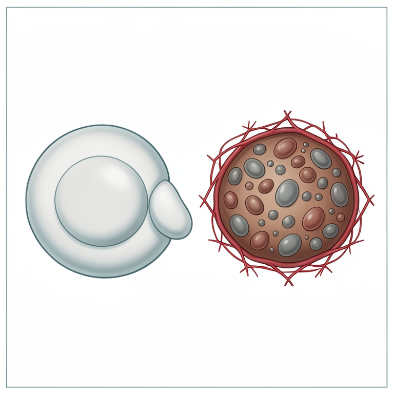
BAT vs WAT Cellular Comparison
AI-generated conceptual illustration (human adipose tissue cells). It explains the idea and does not show study data.
View source evidence ↗Introduction
There are two types of adipose tissue that make up the adipose organ: white adipose tissue, which is often known as white fat, and brown adipose tissue (BAT). [1] BAT may be found almost everywhere in the animal kingdom. Brown fat is composed of two separate cell types, both of which perform roles that are comparable to one another. The first kind will be seen in larger "classic" deposits and will have an embryological origin that is comparable to that of muscle cells. [2] White adipocytes are stimulated by the sympathetic nervous system, which ultimately leads to the impact that is being considered. Adipose tissue that is white in color contains adipocytes that are either beige or brite in color. There is a particularly high concentration of brown adipose tissue (BAT) in newborns and species that are hibernating. [3] The frequency of its occurrence decreases with age in humans; nonetheless, it continues to be present and physiologically active into maturity. This function's principal purpose is to regulate temperature. In addition to the heat that is generated by muscles that are shivering, brown adipose tissue (BAT) also generates heat via a process known as non-shivering thermogenesis. Brown adipocytes, in contrast to white adipocytes, which only contain a single lipid droplet, contain several smaller lipid droplets and a much larger concentration of iron-rich mitochondria. This is one of the factors that contributes to the deeper pigmentation of brown adipose tissue (BAT). [4] When compared to white adipose tissue, brown adipose tissue contains a higher number of capillaries, which makes it easier for the tissue to receive oxygen and nutrients, as well as distribute the heat that is created.
Anatomical location and histology
FDG-PET imaging was used to identify metastatic tumors in adult patients, which led to the identification of BAT in those individuals. These scans, together with the data from human autopsies, have led to the discovery of many BAT depots. Depots of brown adipose tissue may be seen in neonates in a number of different locations, including the interscapular, supraclavicular, suprarenal, pericardial, and para-aortic areas. These depots are located around the pancreas, kidneys, and trachea. [5] These depots gradually take on the appearance of white adipose tissue as the growth process progresses for them.
The mediastinal, supraclavicular, paravertebral, para-aortic, and suprarenal regions are among the depots that are often seen in adult FDG-PET scans from a clinical perspective. There is ongoing debateon whether or not these depots should be classified as "classical" BAT or as beige or brite fat. [6] Two different cell types are referred to as "brown fat" in both academic papers and general media. This designation is based on the cellular morphology and anatomical placement of these two cell types. Both have a large number of mitochondria that are rich in iron and minute lipid droplets, both of which contribute to the dark pigmentation of the cells. Brown adipose tissue, which is recognized by its highly vascularized deposits, may be found in certain anatomical sites, such as between the scapulae, around the kidneys, in the cervical and supraclavicular regions, and along the spinal column. These places are situated in the body. There are a few minute lipid droplets that are included in this kind, which is the less significant of the two. When it comes to adipose tissue, the cell type that may be formed as a result of adrenergic stimulation is referred to as "beige fat." Its lighter brown color is due to the increased variety in lipid droplet dimensions as well as the increased ratio of lipid droplets to mitochondria both of which contribute to the color. [16]
Development
The genesis of brown adipocytes, myocytes, chondrocytes, and adipocytes may be traced back to the mesoderm, which is the intermediate layer of the embryo. Both the traditional muscle and brown adipose tissue populations may trace their roots back to the paraxial mesoderm, which is the stem cell population of the mesoderm. The intrinsic potential to activate the promoter of myogenic factor 5 (Myf5) is a characteristic that is unique to myocytes and this particular population of brown adipose tissue. Both of these cells possess this innate ability. There is no evidence that brown adipose tissue that has been produced by adrenergic stimulation and the progenitors of conventional white adipocytes are able to activate the Myf5 promoter. The cells known as pericytes, which are found inside white adipose tissue and surround the blood vessels, have the potential to give birth to both adipocytes and brown adipocytes. [7] Different from this is the existence of the Myf5 protein, which is involved in the development of a number of different tissues. This is an important distinction. Additionally, brown adipocytes that were not cultured with the transcription factor PRDM16 were able to change into myocytes, but myocytes that were grown with the transcription factor PRDM16 were able to transform into brown adipocytes. [8]
Function
The mitochondria of a eukaryotic cell make use of resources in order to produce adenosine triphosphate (ATP), which is the major source of energy. The proton motive force (PMF) is a term that is often used to describe the accumulation of energy that occurs as a result of a proton gradient across the inner mitochondrial membrane during this process. The energy that is gained from the movement of protons between the ATP synthase enzyme and the membrane (which follows the concentration gradient of the protons) is then used in the production of ATP. The process in question is identified as chemiosmosis. Through a mechanism known as proton leak, endotherms are able to save the body's heat by guiding mitochondria to let protons to flow back down the gradient without facilitating the synthesis of ATP. [9] An uncoupling protein is present in the inner membrane, which provides protons with an alternate path for returning to their orbital. After being actively expelled from the mitochondria by means of the electron transport chain, protons are able to return to the mitochondria with the assistance of uncoupling protein 1, also known as thermogenin. During the process of uncoupling oxidative phosphorylation via this alternate pathway for protons, the energy that is contained within the proton motive force is released as heat. All endothermic cells produce heat to a certain extent, particularly when the temperature of the body falls below a threshold that regulates thermal regulation. Although this is the case, brown adipose tissue is highly specialized for thermogenesis that does not include shivering. When compared to usual cells, each cell initially contains a bigger number of mitochondria than the average cell. These mitochondria have a higher concentration of thermogenin in their inner membrane than other mitochondria they include. [10] Infants Brown adipose tissue is located on the dorsal side, along the superior portion of the vertebral column, and around the shoulders of newborns, constituting about 5% of their total mass. Premature newborns are particularly vulnerable to the deadly cold, thus preventing hypothermia is crucial. Infants are more vulnerable to cold than adults for a variety of reasons: The increased ratio of body volume, which correlates with heat production, to body surface area, which correlates with heat dissipation. A deficiency in thermal insulation, such as subcutaneous adipose tissue and fine body hair (notably in premature infants); The head's relatively larger surface area; The absence of muscular development and the inability or reluctance to shiver. The inability to relocate from cold environments, air currents, or materials that dissipate heat; The incapacity to utilize alternative methods for warmth, such as donning clothing, drying the skin, or engaging in physical activity;
The nervous system's underdevelopment, resulting in inadequate or delayed responses to cold stimuli (e.g., vasoconstriction of blood vessels in and just beneath the skin). Brown fat produces heat, which gives a baby another way to regulate their body temperature.
Adults
It was previously thought that as children become older, the majority of the mitochondria in brown adipose tissue, which are responsible for the brown pigmentation of the tissue, undergo a reduction. This results in the tissue becoming more similar to white fat in terms of both appearance and physiology. In very rare cases, brown fat continues to expand rather than involuting, which may lead to the development of a tumor known as a hibernoma. Recent research has shown that brown fat, as opposed to white fat, is more closely associated with skeletal muscle. [11] Brown adipose tissue (BAT) is almost exclusively found in the upper chest and neck areas, particularly paravertebrally, in the majority of people, according to study that was conducted utilizing positron emission tomography scanning. The residual deposits are less noticeable when an adrenergic beta blocker is administered before to the scan. On the other hand, they are more noticeable when the patient is exposed to cold, which enhances tracer absorption and indicates that metabolic activity is increased. It is possible that these results may lead to the development of innovative weightloss treatments. Brown fat is responsible for burning calories from normal fat. It has been shown that brown fat may be effectively generated in mice by these researchers. [12] According to the findings of a research that was conducted on mice that were defective in APOE, exposure to cold may hasten the formation of atherosclerotic plaque and make it more unstable and unstable. The study mice were kept at a constant low temperature of 4 degrees Celsius for a period of eight weeks, which may have caused stress owing to the abrupt change in temperature rather than a gradual acclimatization to the new environment. For the purpose of determining the effects of ambient temperature reductions of merely 5 to 10 degrees Celsius on adult humans, this might be carried out. Furthermore, a number of recent research have shown that being exposed to cold environment has considerable positive effects on a wide variety of species, including people. Researchers came to the conclusion that "activation of BAT is an effective therapeutic approach to alleviate hyperlipidaemia and protect against atherosclerosis". [26] Additionally, they discovered that activating brown fat may reduce plasma triglyceride and cholesterol levels while also decreasing the progression of diet-induced atherosclerosis. [14] It is before the present increase in health issues connected to excessive accumulation of white fat, it is vital to conduct longitudinal studies that include adults in order to evaluate the amount of benefits and risks associated with the buildup of white fat. The use of β3-adrenoceptor agonists in pharmacological therapies has shown an increase in glucose metabolic activity in brown adipose tissue in animal models and has been documented in reference. [15] Furthermore, studies have revealed: The stimulation of the basal adipose tissue (BAT) improves glucose homeostasis and insulin sensitivity in humans [16] , indicating that it may be beneficial for those who have impaired insulin function. Nonetheless, there exists a broader applicability, as research indicates that even marginally elevated blood glucose levels in healthy, non-diabetic individuals correlate with longterm damage to various organs, including the brain, tendons, eyes, and the endothelial/cardiovascular system, alongside increased concentrations of detrimental advanced glycation end products. BAT activation has the potential to have a major impact on bone density as well as bone health. The activation of brown adipose tissue resulting from exposure to cold causes an increase in adiponectin concentrations; in adult men, even two hours of cold exposure led to a 70% increase in circulating adiponectin levels. [38] It is possible that there is a relationship between longevity and the production of adiponectin, given that studies have shown that both male and female centenarians have greater levels of circulating adiponectin and genetic factors that enhance adiponectin synthesis. [39] In addition, centenarians who had greater levels of plasma adiponectin had improved metabolic indicators, as well as lower levels of Creactive protein and E-selectin. [18] There is a correlation between longer life expectancy and the generation of fibroblast growth factor 21 (FGF-21). [41] The activation of brown adipose tissue (BAT) in humans is triggered by cold exposure, which results in a 37% rise in the amount of circulating fibroblast growth factor 21 (FGF21). Because it improves insulin sensitivity and glucose metabolism, FGF21 may be responsible for some of the aspects that contribute to its longevity-promoting properties.
As a result of exposure to cold, circulation irisin levels increase. [19] Irisin, which is similar to exercise, has a variety of benefits, including improved insulin sensitivity, increased bone density and quality, the promotion of the formation of lean muscle, and a reduction in obesity via the conversion of white fat to brown fat. In comparison to healthy centenarians, who have larger levels of serum irisin, young persons who have had a myocardial infarction have much lower levels of this protein. Based on these results, more study might be conducted to investigate the function that irisin plays in the regulation of lifespan and vascular illnesses. [20] Insulin sensitivity and thermogenesis are both increased when brown adipose tissue and skeletal muscle are exposed to cold. This is because cold exposure causes an increase in SIRT1 phosphorylation and activity. Increased levels of SIRT1 have been shown to have a favorable correlation with human lifespan. In conjunction with other sirtuins, SIRT1 is responsible for a variety of metabolic effects; however, its ability to enhance insulin sensitivity, regulate glucose levels in skeletal muscles, drive white adipose tissue to brown, and boost the activity of brown adipose tissue is of utmost importance for improving health and extending longevity.
Altering the gut flora, reducing obesity and metabolic disorders brought on by a high-fat diet, and encouraging the browning of white adipose tissue are all benefits of intermittent fasting, especially when it is done every other day.
Brown fat solves the problem of obesity:
Since obesity rates are high right now, researchers are searching for methods to help people lose weight, such as reducing food consumption or increasing energy expenditure. Exercise is one way to burn energy, but BAT also plays a role in energy consumption. By dispelling energy as heat, it may be able to counterweight gain. Although scientists are still unsure of how humans could boost their brown fat composition, there is some hope that figuring out how to convert more white fat to brown fat could aid in weight loss or stop further weight gain. It is uncertain, nevertheless, whether this is feasible or beneficial.
Conclusion
To enable such treatments, basic understanding of the synthesis of brown fat and the control of its activity is essential. We incorporate the latest findings on how hormonal and developmental cues control the production and activity of brown fat in connection to its metabolic role.
References
20 in the original paper- Cinti S. The adipose organ. Prostaglandins Leukot Essent Fatty Acids. 2005 Jul;73(1):9-15.
- Enerbäck S. The origins of brown adipose tissue. N Engl J Med. 2009 May 7;360(19):2021-2023.
- Petrovic N, Walden TB, Shabalina IG, Timmons JA, Cannon B, Nedergaard J. Chronic peroxisome proliferator-activated receptor gamma (PPARγ) activation of epididymally derived white adipocyte cultures reveals a population of thermogenically competent, UCP1-containing adipocytes molecularly distinct from classic brown adipocytes. J Biol Chem. 2010 Mar 5;285(10):7153-7164.
- Wu J, Boström P, Sparks LM, Ye L, Choi JH, Giang AH, et al. Beige adipocytes are a distinct type of thermogenic fat cell in mouse and human. Cell. 2012 Jul 20;150(2):366-376.
- Gesta S, Tseng YH, Kahn CR. Developmental origin of fat: tracking obesity to its source. Cell. 2007 Oct 19;131(2):242-256.
- Nedergaard J, Bengtsson T, Cannon B. Unexpected evidence for active brown adipose tissue in adult humans. Am J Physiol Endocrinol Metab. 2007 Aug;293(2):E444-E445.
- Saito M, Okamatsu-Ogura Y, Matsushita M, Watanabe K, Yoneshiro T, Nio-Kobayashi J, et al. High incidence of metabolically active brown adipose tissue in healthy adult humans: effects of cold exposure and adiposity. Diabetes. 2009 Jul;58(7):1526-1531.
- Graja A, Schulz TJ. Mechanisms of aging-related impairment of brown adipocyte development and function. Gerontology. 2015;61(3):211-217.
- Cohade C, Osman M, Pannu HK, Wahl RL. Uptake in supraclavicular fat ("USA-Fat"): description on 18F-FDG PET/CT. J Nucl Med. 2003 Feb;44(2):170176.
- Yeung HW, Grewal RK, Gonen M, Schöder H, Larson SM. Patterns of (18)F-FDG uptake in adipose tissue and muscle: a potential source of false-positives for PET. J Nucl Med. 2003 Nov;44(11):1789-1796.
- Heaton JM. The distribution of brown adipose tissue in the human. J Anat. 1972 Jan;112(Pt 1):35-39.
- van Marken Lichtenbelt WD, Vanhommerig JW, Smulders NM, Drossaerts JM, Kemerink GJ, Bouvy ND, et al. Cold-activated brown adipose tissue in healthy men. N Engl J Med. 2009 Apr 9;360(15):15001508.
- Shinoda K, Luijten IH, Hasegawa Y, Hong H, Sonne SB, Kim M, et al. Genetic and functional characterization of clonally derived adult human brown adipocytes. Nat Med. 2015 Apr;21(4):389-394.
- Lidell ME, Betz MJ, Enerbäck S. Two types of brown adipose tissue in humans. Adipocyte. 2014 Jan 1;3(1):63-66.
- Cedikova M, Kripnerová M, Dvorakova J, Pitule P, Grundmanova M, Babuska V, et al. Mitochondria in white, brown, and beige adipocytes. Stem Cells Int. 2016 Mar 17;2016:1-11.
- Haldar M, Karan G, Tvrdik P, Capecchi MR. Two cell lineages, myf5 and myf5-independent, participate in mouse skeletal myogenesis. Dev Cell. 2008 Mar 11;14(3):437-445.
- Nedergaard J, Bengtsson T, Cannon B. Unexpected evidence for active brown adipose tissue in adult humans. Am J Physiol Endocrinol Metab. 2007 Aug;293(2):E444-E452.
- Celi FS. Brown adipose tissue-when it pays to be inefficient. N Engl J Med. 2009 Apr 9;360(15):15531556.
- Kolata G. Calorie-burning fat? Studies say you have it. The New York Times. 2009 Apr 8;Sect. A:1.
- Kajimura S. Initiation of myoblast/brown fat switch through a PRDM16-C/EBP-β transcriptional complex. Nature. 2009 Aug 20;460:1154-1158.