
Research article · 2024 · Full text
International Journal of Biology SciencesSerum enzyme profiles as predictive biomarkers in acute myocardial infarction: A biochemical perspective
Volume 6(1) · Pages 55–61 · DOI: 10.33545/26649926.2024.v6.i1a.440 ↗
Research areasComputational Drug Discovery MethodsLipoproteins and Cardiovascular Health
Abstract
Author-writtenBackground: Acute myocardial infarction (AMI) remains one of the leading causes of global mortality and morbidity. Timely and accurate diagnosis is critical to initiate effective treatment and improve patient outcomes. While electrocardiography and imaging modalities are standard, biochemical markers—particularly serum enzymes—play a vital role in confirming myocardial injury. Objective: This paper presents a comprehensive biochemical perspective on the use of serum enzyme profiles as predictive biomarkers in AMI. It explores their diagnostic kinetics, clinical significance, and integration into multi-marker strategies. Methods & Content Overview: We review classical and modern biomarkers including creatine kinase-MB (CK-MB), cardiac troponins (cTnI and cTnT), lactate dehydrogenase (LDH), aspartate aminotransferase (AST), and emerging markets such as heart-type fatty acid binding protein (H-FABP), copeptin, ischemia-modified albumin (IMA), myeloperoxidase (MPO), and GPBB. The pathophysiology of enzyme release is discussed in relation to infarct timing and myocardial necrosis. Comparative analysis outlines each biomarker’s rise time, peak, half-life, specificity, and clinical application. The study also highlights the advantages of multi-marker strategies in early detection and risk stratification. Results & Significance: Troponins were reaffirmed as the gold standard due to their high specificity and long diagnostic window, while early-rising markers like H-FABP and copeptin demonstrated value in pre-hospital and emergency triage settings. Prognostic relevance was established for markers such as MPO and persistent troponin elevation, which correlate with infarct size, reinfarction risk, and long-term outcomes. Conclusion: Serum biomarkers, when interpreted in the context of clinical findings and used in a time-sensitive, multi-marker approach, offer a powerful framework for diagnosing, managing, and prognosticating AMI. Understanding their kinetics, limitations, and complementary roles is essential for delivering precise and timely cardiac care.
Summary
Source-checkedPlain-language reading of the full paper, prepared with AI and checked against the text. Each point links to the authors' own words.
Why it matters
- Acute myocardial infarction (AMI) is a critical condition where heart muscle tissue dies irreversibly because of a prolonged lack of blood flow (ischemia). View evidence ↗
- Cardiovascular diseases cause nearly 18 million deaths every year globally, with AMI serving as a major cause of death and disability. View evidence ↗
- Up to one-third of AMI patients, particularly elderly individuals and those with diabetes, may not show classic symptoms, making biochemical confirmation necessary. View evidence ↗
The question
- The paper aims to evaluate serum enzyme profiles as predictive biomarkers for AMI, focusing on their timing (kinetics), clinical importance, and use in combined marker strategies. View evidence ↗
What they did
- The authors reviewed a range of biomarkers, including classical ones (CK-MB, LDH, AST), the current gold standard (troponins), and emerging markers (H-FABP, copeptin, IMA, MPO, and GPBB). View evidence ↗
- The analysis considers how the molecular size and cellular location of these markers influence when they enter the bloodstream after injury. View evidence ↗
What they found
- Myeloperoxidase (MPO) rises in 2-4 hours and can indicate plaque instability and higher risk of adverse events even if troponin levels are normal. View evidence ↗
- Peak troponin concentrations serve as a strong predictor of infarct size, left ventricular dysfunction, and the risk of major adverse cardiovascular events. View evidence ↗
What it means
- The authors conclude that a time-sensitive, multi-marker approach is the most powerful framework for AMI care, and suggest that future diagnostics should integrate AI and point-of-care technologies. View evidence ↗
- The authors note that uniform troponin cut-offs may lead to under-diagnosis in women, who may present with lower absolute levels during an AMI. View evidence ↗
Figures & tables
From the original paper Table 1
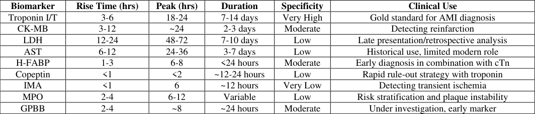 Table 2
Introduction
Acute myocardial infarction (AMI), commonly known as a heart attack, is a life-threatening condition characterized by the irreversible death of myocardial tissue due to prolonged ischemia. Globally, cardiovascular diseases (CVDs) are responsible for nearly 18 million deaths annually, with AMI being a principal contributor to mortality and disability-adjusted life years (DALYs) across all age groups (World Health Organization, 2023). The success of AMI management is time-sensitive. The earlier the condition is diagnosed and treated-particularly with reperfusion therapies such as percutaneous coronary intervention (PCI) or thrombolytics-the better the preservation of myocardial function and survival outcomes. However, clinical presentations of AMI can be highly variable. While classic symptoms include chest pain, dyspnea, and diaphoresis, up to one-third of patients, especially diabetics and the elderly, may present with atypical or silent symptoms (Thygesen et al., 2000) [9] . This diagnostic uncertainty underlines the need for biochemical confirmation of myocardial necrosis. Biochemical markers are integral to the universal definition of AMI, which requires a rise and/or fall of cardiac troponin with at least one value above the 99 th percentile upper International Journal of Biology Sciences https://www.biologyjournal.net reference limit (URL) in the context of ischemia (Thygesen et al., 2018) [1] . These biomarkers not only aid in the diagnosis of AMI but also offer valuable insights into the extent of myocardial damage, prognosis, and risk of future cardiovascular events. Enzymes such as creatine kinase-MB (CK-MB), lactate dehydrogenase (LDH), and aspartate aminotransferase (AST) have historically been used, though their specificity has been superseded by cardiac troponins (cTnI and cTnT) in modern practice. Nevertheless, each marker has a distinct kinetic profile and window of detection, making them valuable under different clinical circumstances-e.g., distinguishing reinfarction, evaluating late presenters, or confirming perioperative myocardial injury.
Objectives of the Paper
This paper adopts a biochemical lens to evaluate the diagnostic and prognostic significance of serum enzyme markers in AMI. Specifically, it: Explores the biochemical basis of enzyme release from injured cardiac tissue. Analyzes the temporal profiles, specificity, and sensitivity of classical and novel biomarkers.
Highlights the advantages and limitations of each biomarker. Examines how multi-marker strategies improve clinical decision-making. Discusses future perspectives in biomarker innovation and point-of-care testing (POCT).
Pathophysiology of Acute Myocardial Infarction
Acute myocardial infarction (AMI) is a clinical event that occurs when there is a prolonged mismatch between myocardial oxygen supply and demand, typically resulting from a coronary artery occlusion. The occlusion, in the majority of cases, is due to a rupture or erosion of an atherosclerotic plaque followed by platelet aggregation and thrombus formation, causing total or subtotal obstruction of coronary blood flow (Libby, 2021) [10] .
When the coronary blood flow is abruptly reduced or halted, oxygen delivery to myocardial tissue ceases, leading to ischemia. Within minutes, aerobic metabolism fails, and cardiac myocytes begin shifting to anaerobic glycolysis, resulting in decreased ATP production, increased lactate, and intracellular acidosis. If perfusion is not restored, irreversible myocardial cell death (necrosis) begins to occur, typically within 20-40 minutes of complete occlusion. [1] . Persistent elevation can reflect ongoing ischemia or microvascular injury. [9] .
Aspartate Aminotransferase (AST)
AST is a mitochondrial enzyme released following cellular injury and was historically used to detect AMI. Rise: 6-12 hours Peak: 24-36 hours Return to Baseline: 3-7 days
Its diagnostic value is diminished by its presence in liver, skeletal muscle, and other tissues.
Heart-type Fatty Acid Binding Protein (H-FABP)
H-FABP is a small cytosolic protein involved in fatty acid transport in cardiac cells. Due to its low molecular weight, it is one of the earliest markers of myocardial injury.
Rise: 1-3 hours Peak: 6-8 hours Return to Baseline: 24 hours Although highly sensitive, its lack of specificity necessitates pairing with troponins for diagnostic accuracy (Chan et al., 2004) [4] .
Emerging and Auxiliary Biomarkers
Although cardiac troponins remain the cornerstone of acute myocardial infarction (AMI) diagnostics, there is growing recognition that a single-marker strategy may not always suffice-especially in the hyperacute window or in atypical clinical presentations. Consequently, several emerging biomarkers have gained attention for their potential to complement troponins and enhance diagnostic precision in specific contexts. Heart-type fatty acid binding protein (H-FABP) is one such marker, released rapidly from damaged cardiomyocytes due to its small molecular size. Its serum concentration rises within one to three hours of myocardial injury, making it particularly useful in early presenters-those arriving within the first two hours of symptom onset. H-FABP's fast kinetics may help bridge the diagnostic gap before troponins become detectable. However, its specificity is modest, as it is also present in skeletal muscle and is renally excreted, making standalone interpretation unreliable.
Copeptin, a stable peptide derived from the precursor of vasopressin, offers a different angle. Rather than reflecting necrosis, copeptin indicates acute endogenous stress, with levels rising sharply during hemodynamic instability. Because copeptin levels increase almost immediately following symptom onset and peak within one hour, it has found utility when used in combination with troponin assays to rapidly rule out AMI in low-risk patients. Studies suggest that a negative copeptin and a normal troponin level can safely exclude infarction at the time of presentation, thus facilitating early discharge decisions in emergency settings. Another promising, albeit less specific, marker is ischemiamodified albumin (IMA). It results from oxidative alterations to albumin during ischemia, which impair its ability to bind transition metals. IMA can rise within minutes of transient ischemia-even in the absence of irreversible damage-rendering it potentially useful for detecting unstable angina or early ischemic changes. Despite this, its clinical application remains limited due to poor specificity and susceptibility to interference from noncardiac conditions such as liver dysfunction or chronic inflammation.
In contrast, myeloperoxidase (MPO) represents a biomarker not of myocardial damage per se but of underlying inflammatory processes. Released by activated neutrophils and monocytes, MPO contributes to oxidative stress and is implicated in atherosclerotic plaque destabilization. Elevated MPO levels in patients with chest pain-even in the absence of troponin elevation-have been associated with a higher risk of adverse cardiac events, positioning it as a candidate for prognostic stratification in troponinnegative, high-risk individuals. Finally, glycogen phosphorylase isoenzyme BB (GPBB) is gaining research interest as an early marker of metabolic stress in myocardial tissue. This enzyme, involved in glycogen breakdown, is released early in ischemia and may reflect hypoxic injury before cell necrosis. While early data on GPBB are encouraging, especially for its detection in the first few hours post-ischemia, its clinical utility is still under evaluation due to assay limitations and limited availability in routine settings. Taken together, these auxiliary biomarkers do not seek to replace troponins but rather enhance the sensitivity, specificity, and timing of AMI diagnostics. In particular, the integration of early-phase markers like copeptin and H-FABP with established troponin assays holds promise for faster and more reliable triage, especially in overcrowded emergency departments. The future of AMI diagnosis may lie in personalized, multi-marker panels capable of distinguishing between ischemia, infarction, inflammation, and long-term risk-all within the critical window when clinical decisions matter most.
Comparative Evaluation: Biomarker Sensitivity, Specificity, and Timing
The diagnostic accuracy of serum biomarkers in acute myocardial infarction (AMI) depends heavily on their kinetic properties, specificity to cardiac tissue, and the temporal pattern of release post-injury. A comprehensive comparison reveals that each marker brings distinct strengths and limitations to clinical practice. Understanding these nuances is crucial for clinicians seeking to optimize diagnostic workflows, particularly in time-sensitive scenarios.
Troponins, both cTnI and cTnT, have established themselves as the most cardiac-specific and sensitive biomarkers. Their high sensitivity allows detection of even minor myocardial injury, which has redefined diagnostic thresholds for non-ST elevation myocardial infarction (NSTEMI). They typically rise within 3 to 6 hours post-symptom onset, peak by 18-24 hours, and may remain elevated for up to 14 days, offering a wide diagnostic window. However, their delayed rise compared to other markers limits early detection, and chronic elevations in renal failure, myocarditis, or sepsis require careful interpretation within a clinical context. CK-MB, once the mainstay of AMI diagnosis, now serves a more focused role. It rises faster than troponin-within 3 to 12 hours-and returns to baseline within 2 to 3 days, making it valuable in detecting reinfarction or infarction recurrence after percutaneous coronary intervention (PCI) or coronary artery bypass grafting (CABG). However, CK-MB lacks high specificity, as it can be elevated in skeletal muscle damage, trauma, or intense physical exertion. LDH and AST are considered historical biomarkers, with slower kinetics and poor specificity. LDH peaks after 48-72 hours and remains elevated for up to 10 days, while AST peaks at 24-36 hours. Their utility has declined significantly due to the availability of more precise and faster markers.
In contrast, emerging markers like H-FABP and copeptin bring added value in ultra-early diagnostics. H-FABP rises within 1-3 hours and may help identify infarction in its earliest stages, although its specificity is relatively low due to expression in non-cardiac tissues. Copeptin, which also rises rapidly (within 30 minutes), serves as a stress response biomarker and has shown promise when paired with troponin in dual-marker rule-out strategies for low-risk patients. Myeloperoxidase (MPO) and IMA, while not direct indicators of necrosis, contribute to a broader understanding of inflammatory status and ischemia, respectively. Elevated MPO levels have been linked to plaque instability and may be prognostically significant even when traditional necrosis markers remain within normal range. IMA, detectable within minutes of ischemia, helps identify transient ischemic episodes and unstable angina-conditions where structural cardiac damage may not have occurred yet.
To consolidate these findings, Table 2 below provides a comparative summary of key biomarkers: This comparative evaluation underscores the value of a multi-marker strategy. While no single biomarker is flawless, combining the strengths of several-early rising markers, cardiac-specific proteins, and inflammatory indicators-can yield a more reliable and timely diagnosis. Such integrative approaches are especially critical in early presenters, complex cases, and resource-limited settings where imaging or serial testing may not be readily available.
| Biomarker | Rise Time (hrs) | Peak (hrs) | Duration | Specificity | Clinical Use |
|---|---|---|---|---|---|
| Troponin I/T | 3-6 | 18-24 | 7-14 days | Very High | Gold standard for AMI diagnosis |
| CK-MB | 3-12 | ~24 | 2-3 days | Moderate | Detecting reinfarction |
| LDH | 12-24 | 48-72 | 7-10 days | Low | Late presentation/retrospective analysis |
| AST | 6-12 | 24-36 | 3-7 days | Low | Historical use, limited modern role |
| H-FABP | 1-3 | 6-8 | <24 hours | Moderate | Early diagnosis in combination with cTn |
| Copeptin | <1 | <2 | ~12-24 hours | Low | Rapid rule-out strategy with troponin |
| IMA | <1 | 6 | ~12 hours | Very Low | Detecting transient ischemia |
| MPO | 2-4 | 6-12 | Variable | Low | Risk stratification and plaque instability |
| GPBB | 2-4 | ~8 | ~24 hours | Moderate | Under investigation, early marker |
Table text extracted from the original PDF; superscript letters are shown inline. The original table image is below.
View original table image
Multi-Marker Approach in AMI Diagnosis
The heterogeneity of clinical presentations in acute myocardial infarction (AMI), coupled with the limitations inherent in relying on a single biomarker, has prompted a paradigm shift towards multi-marker strategies. Rather than depending solely on cardiac troponins, clinicians and researchers increasingly advocate for the integration of complementary biomarkers that reflect different pathophysiological stages of ischemic injury, including necrosis, inflammation, ischemia, and neurohormonal stress. Each biomarker in isolation presents a trade-off between sensitivity, specificity, and timing. For instance, troponins are highly specific and remain elevated for days, but they rise slowly and may miss infarctions if tested too early. In contrast, markers like H-FABP and copeptin rise much earlier-often within the first hour-but lack sufficient specificity when interpreted alone. By combining these complementary temporal and mechanistic profiles, a more accurate and time-sensitive diagnostic picture can be established. The classic example of this is the copeptin-troponin dualmarker strategy. Copeptin rises almost immediately following acute stress and reflects neurohormonal activation, while troponin captures necrosis occurring hours later. Studies have shown that patients presenting early with chest pain and negative results for both copeptin and highsensitivity troponin have a very low probability of AMI, allowing for safe early discharge in low-risk populations (Möckel et al., 2012) [6] . This approach has gained traction in emergency departments globally, improving patient throughput without compromising diagnostic safety. Another successful application is the pairing of H-FABP with troponin, which is particularly useful in the first 3-6 hours post-onset. H-FABP's rapid release helps detect myocardial stress earlier than troponin, while the latter confirms necrosis. Together, they enhance both early detection and subsequent confirmation, reducing the risk of missed diagnoses in early presenters or borderline troponin elevations. Emerging technologies have also enabled the development of multi-analyte point-of-care panels, combining multiple markers into a single assay platform. Such multiplexed diagnostics not only expedite results but also offer quantitative interpretation of risk, helping clinicians stratify patients based on likelihood of infarction and predicted outcomes. For example, a panel including troponin, NT-proBNP, MPO, and H-FABP has been proposed to simultaneously assess myocardial injury, cardiac strain, inflammation, and metabolic stress. In more complex or ambiguous clinical scenarios-such as patients with renal impairment, chronic heart failure, or suspected reinfarction-the multi-marker approach provides additional biochemical clarity. For instance, CK-MB and troponin together may help distinguish between acute reinjury and residual elevation from a prior infarction. Similarly, MPO and IMA can identify patients at higher risk of plaque rupture or transient ischemia, even when necrosis has not yet occurred. From a biochemical standpoint, this approach embraces the complex pathophysiology of AMI, recognizing that myocardial infarction is not a singular event but a continuum involving vascular dysfunction, ischemia, inflammation, cell death, and remodeling. By targeting multiple stages of this cascade, multi-marker diagnostics align more closely with the biological reality of the disease. Nevertheless, the implementation of such strategies must be context-specific. Not all hospitals have access to highsensitivity assays or multiplex platforms, and the added cost of additional markers must be balanced against clinical benefit. Interpretation also requires training to avoid misdiagnosis due to false positives from non-cardiac causes of biomarker elevation. Despite these caveats, the multi-marker model represents a forward-looking framework for AMI diagnostics. It acknowledges that no single biomarker can fully capture the dynamic complexity of myocardial injury, and offers a more robust, nuanced, and patient-centered method to guide early diagnosis, triage, and treatment in one of the most timesensitive emergencies in clinical medicine.
Prognostic Applications and Clinical Significance
Beyond their primary diagnostic utility, serum biomarkers have emerged as indispensable tools for prognostic assessment in acute myocardial infarction (AMI). The levels, patterns, and kinetics of these biomarkers not only confirm myocardial injury but also provide deep insight into infarct size, risk of complications, treatment response, and long-term cardiovascular outcomes. The prognostic role of biomarkers is especially relevant in guiding personalized therapy, resource allocation, and follow-up strategies in both acute and post-discharge settings. One of the strongest predictors of infarct size and patient outcome is the peak concentration of cardiac troponins. Higher troponin levels have been repeatedly correlated with more extensive myocardial necrosis, greater left ventricular dysfunction, and increased risk of major adverse cardiovascular events (MACE), including heart failure and mortality (Omland et al., 2002). Persistent elevation of highsensitivity troponin (hs-Tn) beyond 72 hours has been associated with microvascular obstruction, recurrent ischemia, or ongoing myocardial stress, all of which portend a poor prognosis. Serial troponin measurements also play a role in dynamic risk stratification. A rising trend in serial measurements confirms active myocardial injury, while a falling trend typically indicates stabilization or infarct resolution. This has implications not only for diagnosis but also for determining need for invasive therapy, discharge timing, and rehabilitation planning. In the context of reinfarction, where prior troponin levels may still be elevated from an earlier event, markers like CK-MB regain their value. Due to its shorter half-life, CK-MB levels that re-elevate after an initial decline strongly suggest reinfarction, especially in patients post-percutaneous coronary intervention (PCI) or coronary artery bypass grafting (CABG). Thus, in selected clinical scenarios, CK-MB provides incremental prognostic information where troponins are less informative due to their prolonged elevation. Inflammatory and stress-related markers, such as myeloperoxidase (MPO) and copeptin, extend prognostic insights beyond necrosis. Elevated MPO levels reflect plaque instability and systemic inflammation, which have been linked to increased risk of recurrent ischemic events even in patients without overt myocardial necrosis. Likewise, copeptin, through its association with hypothalamic-pituitary-adrenal axis activation, has been independently associated with short-term mortality and hemodynamic deterioration. These markers help to stratify risk in troponin-negative patients, particularly those with non-specific chest pain, comorbid conditions, or borderline ECG findings. Markers such as NT-proBNP or high-sensitivity CRP, though outside the core panel of myocardial necrosis markers, often accompany biomarker panels in prognostic scoring models. When combined with troponin, they significantly improve risk prediction algorithms like the GRACE and TIMI scores, which influence clinical decisions such as admission, intensive monitoring, and early invasive strategy selection. Another critical prognostic domain is therapeutic response monitoring. Biomarkers aid in evaluating efficacy of reperfusion. A rapid fall in biomarkers, especially in conjunction with symptom relief and ECG normalization, typically reflects successful reperfusion. Conversely, a delayed biomarker peak or persistent elevation may suggest incomplete reperfusion, re-occlusion, or additional myocardial damage. In post-AMI management, biomarkers continue to influence care. Persistently elevated troponins after discharge may suggest subclinical myocardial stress, silent ischemia, or heart failure risk, warranting closer surveillance. Furthermore, emerging studies indicate that integrating biomarker trends with imaging parameters, such as echocardiography or cardiac MRI, enhances the precision of post-infarct risk modelling. In summary, serum biomarkers offer far more than a binary diagnosis of infarction. They act as quantitative surrogates for the biological magnitude of disease, inform the intensity of therapeutic intervention, and help anticipate short-and long-term complications. As biomarker assays become more refined and integrated with digital health platforms, their prognostic applications will continue to expand, bringing us closer to precision cardiology-where every value shapes a tailored treatment plan for each patient.
Limitations and Interferences
While serum biomarkers have dramatically improved the diagnostic and prognostic precision in acute myocardial infarction (AMI), they are not without inherent limitations and are often influenced by non-cardiac factors that can confound clinical interpretation. Understanding these limitations is essential to avoid false positives, unnecessary interventions, or overlooked infarctions. A primary limitation lies in the lack of absolute specificity of many biomarkers, even those regarded as highly cardiacspecific. Cardiac troponins, though unmatched in sensitivity and specificity for myocardial injury, can be elevated in a broad range of non-ischemic conditions. These include chronic kidney disease, congestive heart failure, pulmonary embolism, sepsis, stroke, myocarditis, and even strenuous physical exercise. In such scenarios, elevated troponin may reflect myocardial strain or injury, but not necessarily an infarction due to coronary occlusion. This makes it imperative to interpret biomarker levels in clinical contextconsidering symptoms, ECG findings, imaging, and hemodynamic status. Another limitation is time-dependent release kinetics, particularly in the early phase of AMI. For example, troponin levels typically take 3-6 hours to rise after symptom onset. If a patient presents early and only a single test is performed, results may be falsely negative. This necessitates serial sampling, ideally at 0, 3, and 6 hours, to capture dynamic changes. In contrast, early markers such as H-FABP and copeptin rise quickly but lack cardiac specificity, making them prone to false positives in conditions such as skeletal muscle injury, renal impairment, or systemic inflammation. Laboratory and technical factors also influence biomarker reliability. Hemolysis, which releases intracellular components like LDH and AST from red blood cells, can artificially elevate these markers, potentially mimicking infarction. Similarly, hyperbilirubinemia and lipemia can interfere with colorimetric or immunoassay-based detection methods, particularly in point-of-care testing. In the case of high-sensitivity troponin assays, although their lower detection limits allow for earlier diagnosis, they also detect chronic low-level elevations that may not be related to acute ischemia. Distinguishing between chronic myocardial injury (e.g., from left ventricular hypertrophy or cardiomyopathy) and acute AMI becomes challenging without clear clinical correlation or upward trending levels. Moreover, intra-individual biological variation must be considered, especially in elderly patients or those with systemic illnesses. Another layer of complexity arises with gender-based and age-based variability in reference values. Several studies have shown that females may present with lower absolute troponin levels in the setting of AMI, potentially leading to under diagnosis if uniform cut-offs are applied. Likewise, older adults may exhibit chronically elevated baseline troponin, necessitating the use of age-adjusted reference intervals to avoid misclassification. From a systems perspective, the availability of highsensitivity assays and multiplex panels may be limited in rural or resource-constrained settings. Additionally, cost considerations may deter the widespread adoption of multimarker strategies, particularly when their additive clinical value is still under ongoing evaluation in diverse populations. Finally, false reassurance from negative biomarkers can delay treatment in early presenters if clinicians are unaware of the temporal limitations. No biomarker can substitute for sound clinical judgment, especially in high-risk individuals presenting with ongoing chest discomfort but non-diagnostic test results. This is particularly critical in women, diabetics, and elderly patients, whose AMI presentations may be subtle or atypical.
Conclusion
The diagnostic and prognostic landscape of acute myocardial infarction (AMI) has been fundamentally transformed by the advent and evolution of serum enzyme biomarkers. What began with non-specific markers like AST and LDH has progressed into a sophisticated era led by high-sensitivity troponins, with the continued emergence of novel biochemical indicators that capture distinct dimensions of myocardial pathology. These biomarkers have not only enhanced the speed and accuracy of AMI diagnosis but have also deepened our understanding of the disease continuum-ranging from silent ischemia to extensive necrosis, from transient stress to structural damage. Cardiac troponins remain the gold standard for diagnosing myocardial necrosis due to their superior specificity and prognostic strength. Yet, they are not infallible. Their delayed rise in early presenters and persistent elevation in non-ischemic conditions highlight the need for thoughtful clinical interpretation. In this context, auxiliary biomarkers such as H-FABP, copeptin, IMA, MPO, and GPBB offer valuable support-especially when integrated into multimarker strategies that balance speed with specificity. The utility of biomarkers extends well beyond diagnosis. Their role in prognostic stratification, infarct size estimation, re infarction detection, therapeutic response monitoring, and long-term risk assessment cannot be overstated. High biomarker levels correlate with larger infarcts and worse outcomes, while serial measurements inform clinical trajectory and guide post-discharge care. Importantly, these tools empower clinicians to personalize therapy, escalate care in high-risk patients, or safely expedite discharge in low-risk individuals. However, limitations persist. Biological variability, assay interferences, and non-cardiac elevations require cautious application, particularly in vulnerable populations such as the elderly, women, and patients with chronic comorbidities. Diagnostic decisions based solely on biomarker levelswithout contextual correlation-can lead to both over diagnosis and under treatment. Looking ahead, the future lies in integrated, algorithm-based diagnostics that combine biochemical markers, clinical scoring systems, artificial intelligence, and point-of-care technologies. Such systems promise to deliver real-time, individualized assessments with unprecedented precision. Equally important is the global effort to standardize assays, adjust reference intervals, and ensure equitable access to high-sensitivity testing across healthcare systems.
Figures and tables from the paper
| Time After Occlusion | Cellular Changes |
|---|---|
| 0-30 minutes | Reversible injury, depletion of ATP |
| 30-60 minutes | Onset of irreversible injury, mitochondrial swelling |
| 1-4 hours | Beginning of necrosis, sarcolemmal disruption |
| 4-12 hours | Edema, inflammation, infiltration by neutrophils |
| 12-24 hours | Coagulative necrosis, continued inflammation |
| >24 hours | Tissue remodeling, scar formation begins |
Table text extracted from the original PDF; superscript letters are shown inline. The original table image is below.
View original table image
References
9 in the original paper- Thygesen K, Alpert JS, Jaffe AS, et al. Fourth universal definition of myocardial infarction (2018). Circulation. 2018;138(20):e618-e651. https://doi.org/10.1161/CIR.0000000000000617
- World Health Organization. Cardiovascular diseases (CVDs) -Fact sheet. 2023. https://www.who.int/newsroom/fact-sheets/detail/cardiovascular-diseases-(cvds)
- Apple FS, Jesse RL, Newby LK, Wu AHB, Christenson RH. National Academy of Clinical Biochemistry and IFCC Committee for Standardization of Markers of Cardiac Damage. Clin Chem. 2007;53(4):552-574. https://doi.org/10.1373/clinchem.2006.078063
- Chan CPY, Sanderson JE, Glatz JFC, Cheng W, Li AM, Hempel A, et al. A novel cardiac biomarker, hearttype fatty acid-binding protein (H-FABP): measurement and clinical significance. Clin Chim Acta. 2004;350(12):151-172.
- Omland T, de Lemos JA, Sabatine MS, et al. A sensitive cardiac troponin T assay in stable coronary International Journal of Biology Sciences https://www.biologyjournal.net artery disease. N Engl J Med. 2009;361(26):2538-2547. https://doi.org/10.1056/NEJMoa0805299
- Möckel M, Searle J, Hamm C, et al. Early discharge using single cardiac troponin and copeptin testing in patients with suspected acute coronary syndrome (ACS): a randomized, controlled clinical process study. Eur Heart J. 2012;33(18):2165-2173. https://doi.org/10.1093/eurheartj/ehr203
- Brennan ML, Penn MS, Van Lente F, et al. Prognostic value of myeloperoxidase in patients with chest pain. N Engl J Med. 2003;349(17):1595-1604. https://doi.org/10.1056/NEJMoa035003
- Bar-Or D, Lau E, Winkler JV. A novel assay for cobaltalbumin binding and its potential as a marker for myocardial ischemia-a preliminary report. J Emerg Med. 2001;21(1):1-5.
- Alpert JS, Thygesen K, Antman E, Bassand JP. Myocardial infarction redefined-A consensus document of The Joint European Society of Cardiology/American College of Cardiology Committee. J Am Coll Cardiol. 2000;36(3):959-969.