
Research article · 2025 · Full text
International Journal of Biology SciencesCorrelation between infant mortality in the Europe of nineteenth-century and malnutrition. An overview of selenium and seleno-enzymes: The last novelties about their role in human health and a focus on SelR (MsrB1)
Volume 7(7) · Pages 52–57 · DOI: 10.33545/26649926.2025.v7.i7a.414 ↗
Research areasBirth, Development, and Health
Abstract
Author-writtenChild malnutrition in sub-Saharan Africa is still a very high mortality factor that now is unacceptable in the common thinking of the Western country’s citizens. The situation was not always this way: in fact, in nineteenth-century Europe, infant mortality was two orders of magnitude higher than actual Europe and one higher than present-day Africa. This issue was linked to the poor and incorrect nutrition to which children were subjected from birth and their mothers during pregnancy and breastfeeding, making infant mortality a very frequent e dramatic phenomenon. Medical research did not yet know the good nutritional rules, and chemistry research had not yet identified vitamins and micronutrients that today allow maintaining good health and higher life expectancy. Among the latest micronutrients identified and widely studied, there is selenium, a trace mineral present in the soil, that is essential for humans in small amounts which, through refined processes of cellular biosynthesis, becomes part of some proteins in the form of very reactive seleno-cysteine instead of classical cysteine. These proteins, called indeed seleno-proteins, are essential for the execution of many metabolic pathways relevant for life.
Summary
Source-checkedPlain-language reading of the full paper, prepared with AI and checked against the text. Each point links to the authors' own words.
Why it matters
- Child malnutrition continues to be a significant cause of death in sub-Saharan Africa, a situation that contrasts with modern Western perspectives on child health. View evidence ↗
What they did
- The author analyzes historical mortality data and literature to describe a period where about one-third of infants died in their first year and children accounted for over 50% of total population mortality. View evidence ↗
- The review examines the role of selenium in breast milk, noting concentrations of 25–32 μg/L in colostrum and 10–25 μg/L in mature milk. View evidence ↗
What they found
- Moderate selenium concentrations in Eastern European soil are associated with selenium deficiency in people, which may explain the higher infant mortality rates in that region compared to the rest of Europe. View evidence ↗
- The recommended daily intake of selenium ranges from 15 μg at birth to 55 μg for adults, with a tolerable upper limit of 400 μg/day for adults. View evidence ↗
- The use of selenocysteine (Sec) instead of cysteine (Cys) in the active site of MsrB1 is critical; mutating Sec to Cys reduces activity 85-fold, while adding Sec to other MsrBs increases activity 173-fold. View evidence ↗
- MsrB1 is highly expressed in energetic tissues like the brain, liver, digestive tract, and bone marrow to protect them from the high levels of reactive oxygen species they produce. View evidence ↗
What it means
- The author concludes that implementing informed nutritional strategies to address micronutrient deficiencies is essential for reducing global child mortality. View evidence ↗
Visual summary
1 visualsIllustrations explain the paper; they do not show study data.
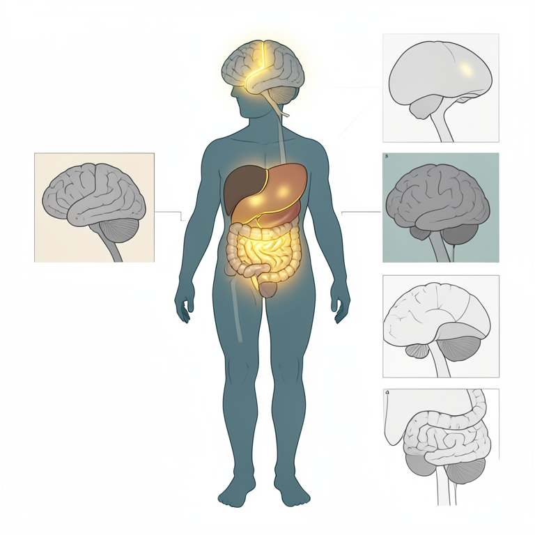
MsrB1 Distribution in the Body
AI-generated conceptual illustration (human anatomy). It explains the idea and does not show study data.
View source evidence ↗Figures & tables
From the original paper Figure 1
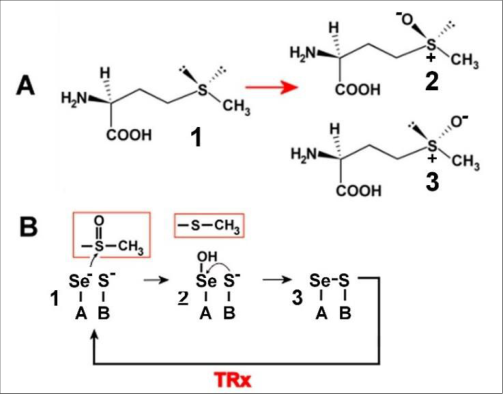 Figure 2
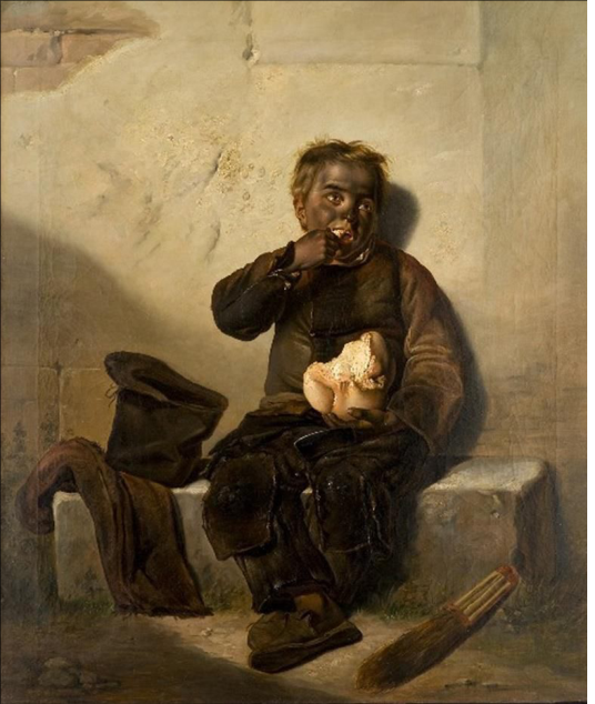 Figure 3
Introduction
"The boys raised the coffin, but as they passed the mother, they stopped for a moment and lowered it that she might say good-by to Ilusha. But on seeing that precious little face, which for the last three days she had only looked at from a distance, she trembled all over and her gray head began twitching spasmodically over the coffin." Fëdor Dostoevskij, "The Brothers Karamazov" [1] .
Nineteenth-century literature offers a privileged point from which to observe the desperation that particularly gripped European society in that historical period, struck by the extremely high mortality rate of the new generations in the prime of their life. However, literature is not the only one that expresses these feelings. The slice of life described by the painter Giuseppe Molteni (figure 1), evoking the most heartbreaking feelings of pain that a mother can experiment, touches the chords of that dramatic reality still in existence in the nineteenthcentury, in a society gripped by the ferocious plague of infant mortality. With her eyes wet with tears and raised to the sky in a pointless search for comfort for her inexhaustible suffering, the young mother is retracted next to the small white coffin that holds the lifeless body of her little child. Ready for his final earthly journey, the small corpse will be restored by the warmth of the mother's tears and the scent of the crown of flowers that she herself has woven with trembling hands, a last gesture of love for the poor little one. Although decreased significantly in the last decades, the current situation in Sub-Saharan Africa regarding infant mortality is well known and reaches about 4% of die in the first year of life, mainly due to endemic malnutrition of these countries and infectious diseases, including HIV, diarrhea and pneumonia, exacerbated from the organic debilitation caused by malnourishment [2] . Number very different from the one found to the Western Word, and particularly to the European Union, when the current percentage of children died before reaching one year of age is 0,3%, although in some Eastern European countries the data is superior (more than 50% of the European average) [3] . This data is, however, far from the European numbers found in the nineteenth-century. In fact, the representative picture reported in Figure 1 is not pure fiction but is a real and widespread fact in that historical period. Death decimated a frightening contingent of children from every social background, since about a third of live births died within the first year of life and only half reached five years of age [4] ; the data of mortality referred to children is superior then 50% of the overall mortality rate of the total population [5] . Getting and exceeding the adolescent age could therefore give the hope of reaching an advanced age.
A turning point would be recorded only in the last decade of the century, in conjunction with the first changes in the hygiene and living conditions of a considerable proportion of the population. The level of infant mortality is undoubtedly the result of a combination of social, economic, cultural, geographical factors, and specific behaviors regarding the care and upbringing of children. Primarily, malnutrition increases morbidity and mortality and affects physical growth and development; some of these effects result from specific micronutrient deficiencies. Nonetheless, the progress of scientific research has contributed, and continues to do so, to the increase in life expectancy. Much attention had to be paid to the environment in which gestation and birth took place, to the type of care provided to the newborn, to the ways and times of breastfeeding (maternal, wet-nursing or artificial), to weaning, and to nutritional deficiencies during pregnancy and breastfeeding. Above all, the nutritional factor helps to explain the reasons for the insufficient life expectancy in children. In fact, if the milk was scarce, of poor quality and unsuitable for the nutritional needs of the child, today we know that it could impact metabolism and resistance to certain infantile, or non-infantile, pathologies transmitted by the mother or wetnursing (primarily syphilis and tuberculosis) or by artificial nutrition with non-pasteurized milk [4] . In an era in which microbiology, biochemistry and analytical chemistry were dawn, the effects of malnutrition were exclusively experimental knowledge. At that time, it was not even possible to establish the chemical composition of breast milk or that by other animal sources. Empirically, it had been established that human mother milk was better than that from other animal sources and that it had to follow the age of the child. Today we know that the reason is attributable to the change in chemical composition over time: the first milk (colostrum) and the mature milk are specifically suitable for the different evolutionary moments of the child. Only today, the chemical composition of
International Journal of Biology Sciences https://www.biologyjournal.net maternal milk is known in detail and the important role of micronutrients, including vitamins and trace elements, has been recognized on children's health. A special mention with respect to the selenium (chemical symbol: Se), that it is very important component of human breast milk, with relevant differences in concentration between the colostrum (25-32 μg/L) and mature milk (10-25 μg/L), which makes it suitable for the various stages of infant development [6] . Se is a fundamental micronutrient for cellular biochemistry, and it exists in the lithosphere regions of the earth, water, soil, and in open environments. Jöns Jacob Berzelius discovered this element in 1817, among the residues of a sulfuric acid plant in Gripsholm, Sweden [7] . Se is one of the major essential trace non-metal element, having atomic number 34, belonging to the same group as sulfur (S) (XVI), and to the period below it (IV versus III). The word "selenium" originates from the Greek word "Selene" (Σελήνη), which means "Moon". The study of Se started with great enthusiasm and continued until modern times, identifying its undeniable biological role. In trace amounts, Se is present in the soil (0.1-2.0 μg/g dry weight of normal soil) under the form of inorganic compounds with different oxidation states, such selenate (SeO 4 2-) and selenite (SeO 3 2-). These compounds are absorbed by plants, that transform inorganic Se into a variant of the common αamino acid cysteine (Cys), named Se-Cys (Sec), Semethionine (Se-Met) or other rare Se-amino acids [8] . Sec and Cys exhibit similar chemical properties and participate in analogous chemical reactions; furthermore, compared with Cys, Sec is more chemically reactive, having higher nucleophilicity, oxidation susceptibility, and acidity [9] . Homo sapiens principally utilize Se-amino acids, and particularly Sec (the "twenty-first" naturally amino acid) as organic Se sources digested from plant and animal foods. These compounds are indispensable for a correct human diet, with a recommended daily dietary allowance of Se at birth of 15 µg, up to 55 µg for adults [10] . Se enters the food chain through plants and from the soil in which they grow. The seleniferous nature of the region determines the amount of Se available in the food. Se levels in the soil vary worldwide. In Europe, for example, moderate concentration is observed in eastern countries, associated with a Se deficiency status in adults and children [11] . This data could explain why in Eastern Europe a significantly higher infant mortality rate, than the remaining part of the continent, is observed [3] . Despite the importance of this micronutrient for human health, exposure to high levels of dietary Se is deleterious and leads to selenosis, a condition who is observed when Se level reaches toxic concentration, whose symptoms includes, in the worst-case scenario, pulmonary oedema [12] . The tolerable upper intake for adults is 400 μg/day [13] .
Main enzymatic property of Se originates from its incorporation, in the form of Sec, in the active site of a limited number of Se-proteins (or rather enzymes, in fact all Se-proteins known, especially from Homo sapiens, have enzymatic activity). In 1973, the presence of Sec was discovered in the antioxidant enzyme glutathione peroxidase (only one isoform of that protein was identified at that time) [14] . Over the decades, the number of known Se-proteins has grown, and they have turned out to be fundamental for human health. The increased reactivity of oxidoreductase enzymes having Sec in place of Cys is usually regarded as the flagship for Se-proteins, despite their costly and inefficient synthesis machinery [15] . Despite many details of the mechanism behind the biosynthesis of Se-proteins remain unclear, it can be described simply: the incorporation of the Sec to a polypeptide happens by means of tRNA specific to amino acid serine, that first bound serine residue, then converted into Sec by adding an activated Se moiety instead of S [16] . Se-enzymes are involved in the redox reactions implicated in several biological roles and they are stored in different organs and tissues: 30% in liver, 30% in muscle, 15% in kidney, 10% in plasma, and 15% in other organs [17] .
In the last twenty years, the list of Se-proteins has grown in every kingdom, from prokaryotes to eukaryotes, and in those times the biological function of some of them was not jet known, since they were so-called "orphan proteins".
Since then, research on Se-proteins, especially those expressed in Homo sapiens, has advanced significantly. The classification of the 25 Se-enzymes encoded in the human genome is as follows [18] : antioxidant enzymes: glutathione peroxidase (GP1, GP2, GP3, GP4 and GP6, while GP5 is not a Se-enzyme), SelK, SelR (also named MsrB1 or, formerly, SelX), SelW; transport and storage of Se: SepP1; redox signaling: thioredoxin reductase (TR1, TR2 and TR3); thyroid hormone metabolism: iodothyronine deiodinase (DIO1, DIO2 and DIO3); protein folding: Sep15, SelN, SelM, SelS; Sec synthesis: SPS2; unknown: SelH, SelI, SelO, SelT, SelV. Se and Se-enzymes are very important for many aspects of human health and are implicated in many physiological processes. A reduced intake of Se leads to Se-protein deficiency. A diet rich in Se appears to protect against a wide range of diseases. The roles of Se/Se-proteins in human health are the following: anti-oxidation, antiinflammation, anti-cancer, anti-bacterial/viral, prevention from brain disorders, immune system health, thyroid function, bone stability, sperm functionality, and embryo growth. Numerous Se-proteins show the capacity to neutralize and remove the oxidative stress, that is a byproduct of cellular metabolism, principally generated during electron transport chain of mitochondria. Overproduction of oxidative stress can damage different cellular structures (nucleic/ribonucleic acids, lipids, proteins) and can prevent signal transduction pathways, depressing the cellular function. As a result, oxidative stress has been linked to a wide range of human disorders, cardiovascular and neurodegenerative/cognitive disease, in addition to the processes related to aging [19,20] . Furthermore, epidemiological studies have suggested an association between low level of Se and inflammatory bowel disease, such as Crohn's disease, and ulcerative colitis, that can potentially progress to colon cancer [21] .
In the metabolic pathways of prokaryotic and eukaryotic cells, some amino acids, both free and inserted in polypeptide chains, can undergo covalent modifications catalyzed, or more frequently not, by enzymes. One of the modifications is the oxidation at the level of the side chain, promoted by various by-products of aerobic metabolism. These processes, generally harmful to the organism, most commonly involve tyrosine, tryptophan, histidine and Met. Met, the only standard α-amino acid with a thioether group, . The oxidation of one or more Met residues can cause the decrease or the complete loss of the biological activity of some proteins or be essential for the activation of others [22] . The oxidative damage of Met is reversible by a family of highly stereospecific enzymes called "Methionine Sulfoxide Reductase" (Msr): MsrAs reduce Met-S-(O) while MsrBs reduce Msr-R-(O) [23] . They are mainly involved in the repair of oxidative damage at the cellular level and in the regulation of cellular metabolism by the cyclic oxidation/reduction of specific Met residues. Depending on the isoforms, they have one, two or three catalytically active Cys, one in the active site and the others used for the transfer of electron by thiol-disulfide exchange (recycling Cys), or Sec instead a Cys in the active site of the Seenzyme MsrB1 (figure 2). When the catalytic Sec of MsrB1 was mutated to Cys, the enzymatic activity decreased up to 85-fold, whereas the replacement of the catalytic Cys of the other MsrBs by a Sec proportionally increased the activity up to 173-fold [24] . (the scenario is essentially the same of the other MsrAs and MsrBs [25] ), equipped with a "catalytic" Sec and only one "recycling" Cys. Mammalians possess one gene for MsrA and three for MsrBs: MsrB1, MsrB2, MsrB3 (which, due to alternative splicing, originates two distinct isoforms: MsrB3A and MsrB3B), localized in different subcellular compartments.
Present in almost all organisms, few Msrs are however Se-enzymes, either MsrAs or MsrBs, with a Sec as catalytic role in place of Cys. While MsrAs containing Sec are usually present to a very few organisms in each family, MsrB1 is the unique MsrB containing Sec in mammalian, including Homo sapiens [24] . MsrB1 possesses, in addition to the recycling Cys, four Cys structured in two domains, involved in the coordination of a Zn 2+ ion, that plays a structural role and is not involved in catalysis. MsrB1 is expressed in nearly all mammalian cell types, particularly at a great level in those of brain, liver, digestive tract, and bone marrow [24] , that are highly energetic tissue and produce a high concentration of reactive oxygen species during their metabolism. Therefore, these organs/tissues require very strong protection from oxidative stress themselves generate. [26, 27] . For these reasons, evolution has ensured that MsrB1 Se-enzyme is strongly expressed in these tissues. Even in humans, neutrophil granulocytes (phagocytic cells involved in the nonspecific immune response through the production of high concentrations of oxidant compounds at bactericidal power) display high concentrations of MsrBs, mainly MsrB1 [28] , evolved for the purpose of self-defense of the cell from the oxidant species itself produces. The role of MsrB1 in humans and other mammals appears to be directed at protecting cellular structures from oxidative damage and, unlike many other Se-enzymes that have been identified as having highly specific biological functions, no definite metabolic role has been observed for this protein in the past. However, recent evidence supports the action of MsrB1 in the cyclic oxidation/reduction of Met residues in the regulation of actin assembly concertedly with Mical protein, that oxidizes two specific Met of actin, resulting in disassembly of actin polymer, whereas MsrB1 reduces Met-R-O to Met and this induces actin polymerization. Because Se in foods is chemically bound to certain proteins principally such as Sec, foods that are high in protein tend to be the best sources of Se. Seafood, beef and pork meat, poultry and eggs are the food with the highest Se content. Grains are not particularly rich in Se, consequentially bread is not nutritionally significant regarding this micronutrient [10] . In the Europe of nineteenth-century, due to the high and unsustainable costs for most of the population, few animalbased foods were eaten, and in many parts of central and northern Europe seafood was not commonly consumed. Grain-based dishes, including bread, were therefore among the main components of the diet of those populations and, due to their chemical composition, it could not contribute significantly to the intake of Se. These harmful eating habits carried nutritional deficiencies in the entire population, including children. It follows that Se (together to other fundamental micronutrients, including vitamins, minerals and iodine) deficiency was most likely endemic in those latitudes at all ages, and maybe it is included in it the child portrayed by Giuseppe Molteni shown in figure 3, intent on calming the pangs of hunger with a piece of bread gained by his job as chimney sweep to which he is ignobly subjected.
In the poor child it is not possible perceive any yearning of hope for a future that, invariably, will reserve for him only suffering and humiliation, and which led from the end of the century to that real mass exodus towards new countries, primary the USA, in search of a better future.
Conclusion
Child malnutrition remains a leading cause of infant mortality in Sub-Saharan Africa, echoing the tragic experiences of nineteenth-century Europe. Scientific advancements, especially the understanding of micronutrients like Se and their biological roles in human development, have significantly improved child survival in the modern world. Se, through its incorporation in vital Seproteins, plays a key role in oxidative stress defense and immune function. Addressing micronutrient deficiencies with informed nutritional strategies is essential to reducing global child mortality and achieving equitable health outcomes across all regions.
References
26 in the original paper- Dostoevskij F. The Brothers Karamazov. Translated from the Russian to the English by Garnett C. New York: The Lowell Press; 2009. Ebook 28054, p. 997.
- World Earth Organization, African Region. Integrated African Health Observatory. Atlas of African Health Statistics 2022, Health situation analysis of the African Region.
- Eurostat, statistics explained. 2022. Available from: https://ec.europa.eu/eurostat/statisticsexplained/index.php?title=Mortality_and_life_expectan cy_statistics.
- Pasi A. Mortalità infantile e cultura medica in Italia nel XIX secolo. Bollettino di Demografia storica. 1995;23:71-96.
- Pozzi L. La tutela della salute materno-infantile in Italia fra Otto e Novecento. In: Popolazione e Storia. 2003;63-90.
- Kim S, Yi DY. Components of human breast milk: from macronutrient to microbiome and microRNA. Clin Exp Pediatr. 2020 Aug;63(8):301-309. DOI: 10.3345/cep.2020.00059.
- Waldemar BM. Selenium Metabolism and Biosynthesis of Selenoproteins in the Human Body. Biochemistry (Mosc). 2022 Jan;87(Suppl 1):S168-S177. DOI: 10.1134/S0006297922140139.
- Liang R, Cheng A, Lu S, Zhang X, Ren M, Lin J, et al. Seleno-amino Acid Metabolism Reshapes the Tumor Microenvironment: from Cytotoxicity to Immunotherapy. Int J Biol Sci. 2024 May;20(7):27792789. DOI: 10.7150/ijbs.95484.
- Tinggi U. Selenium: its role as antioxidant in human health. Environ Health Prev Med. 2008 Feb;13(2):102108. DOI: 10.1007/s12199-007-0019-4.
- Stoffaneller R, Morse NL. A review of dietary selenium intake and selenium status in Europe and the Middle East. Nutrients. 2015 Feb;7(3):1494-1537. DOI: 10.3390/nu7031494.
- Wrobel JK, Power R, Toborek M. Biological activity of selenium: Revisited. IUBMB Life. 2016 Feb;68(2):97105. DOI: 10.1002/iub.1466.
- Harvard, School of Public Heart. The nutrition source: Selenium. 2023. Available from: https://nutritionsource.hsph.harvard.edu/selenium/
- Gromer S, Johansson L, Bauer H, Arscott LD, Rauch S, Ballou DP, et al. Active sites of thioredoxin reductases: why selenoproteins? Proc Natl Acad Sci U S A. 2003 Oct;100(22):12618-12623. DOI: 10.1073/pnas.2134510100.
- Wright DE, O'Donoghue P. Biosynthesis, Engineering, and Delivery of Selenoproteins. Int J Mol Sci. 2023 Dec;25(1):223. DOI: 10.3390/ijms25010223.
- Zhang F, Li X, Wei Y. Selenium and Selenoproteins in Health. Biomolecules. 2023 May;13(5):799. DOI: 10.3390/biom13050799.
- Reszka E, Jablonska E, Gromadzinska J, Wasowicz W. Relevance of selenoprotein transcripts for selenium status in humans. Genes Nutr. 2011 Sep;7(2):127-137. DOI: 10.1007/s12263-011-0246-6.
- Tsuji PA, Santesmasses D, Lee BJ, Gladyshev VN, Hatfield DL. Historical Roles of Selenium and Selenoproteins in Health and Development: The Good, the Bad and the Ugly. Int J Mol Sci. 2021 Dec;23(1):5. DOI: 10.3390/ijms23010005.
- Kieliszek M, Bano I. Selenium as an important factor in various disease states -a review. EXCLI J. 2022 Jul;21:948-966. DOI: 10.17179/excli2022-5137.
- Andoh A, Hirashima M, Maeda H, Hata K, Inatomi O, Tsujikawa T, et al. Serum selenoprotein-P levels in patients with inflammatory bowel disease. Nutrition. 2005 May;21(5):574-579. DOI: 10.1016/j.nut.2004.08.025.
- Levine RL, Moskovitz J, Stadtman ER. Oxidation of Methionine in Proteins: Roles in Antioxidant Defense and Cellular Regulation. IUBMB Life. 2000;50:301307. DOI: 10.1080/713803735.
- Achilli C, Ciana A, Minetti G. The discovery of methionine sulfoxide reductase enzymes: An historical account and future perspectives. Biofactors. 2015 May;41(3):135-152. DOI: 10.1002/biof.1214.
- Tarrago L, Kaya A, Kim H-Y, Manta B, Lee B-C, Gladyshev VN. The selenoprotein methionine sulfoxide reductase B1 (MSRB1). Free Radic Biol Med. 2022 Oct;191:228-240. DOI: 10.1016/j.freeradbiomed.2022.08.043.
- Boschi-Muller S. Molecular Mechanisms of the Methionine Sulfoxide Reductase System from Neisseria meningitidis. Antioxidants (Basel). 2018 Oct;7(10):131. DOI: 10.3390/antiox7100131.
- Pontes Oliveira de Almeida AJ, Pinheiro Lúcio de Oliveira JC, Virgolino da Silva Pontes L, de Souza Júnior JF, Felisberto Gonçalves TA, Dantas SH, et al. ROS: Basic Concepts, Sources, Cellular Signaling, and its Implications in Aging Pathways. Oxid Med Cell Longev. 2022 Oct;2022:1225578. DOI: 10.1155/2022/1225578.
- Wang J, Erlacher M, Fernandez-Orth J. The role of inflammation in hematopoiesis and bone marrow failure: What can we learn from mouse models? Front Immunol. 2022 Aug 11;13:951937. DOI: 10.3389/fimmu.2022.951937.
- Achilli C, Ciana A, Rossi A, Balduini C, Minetti G. Neutrophil granulocytes uniquely express, among human blood cells, high levels of Methionine-sulfoxidereductase enzymes. J Leukoc Biol. 2008 Jan;83(1):181189. DOI: 10.1189/jlb.0707492.