
Research article · 2025 · Full text
International Journal of Biology SciencesIn vitro antagonistic activity of Trichoderma isolates against Rhizoctonia solani by production of lytic enzymes
Volume 7(7) · Pages 14–20 · DOI: 10.33545/26649926.2025.v7.i7a.408 ↗
Research areasPlant Disease Resistance and Genetics
Abstract
Author-writtenTwenty-six Trichoderma isolates were tested for their potentiality in the production of cell wall degrading enzymes and mycoparasitic activity against Rhizoctonia solani, sheath blight of rice. Statistically significant variation was observed in production of lytic enzymes by Trichoderma. In the production of cellulase, eleven Trichoderma isolates showed positive result. Among these, the maximum cellulase production was observed with T. reesei (TR), followed by T. longibrachiatum (M6Ms). Thirteen Trichoderma isolates were positive in amylase enzyme production. In the assay T. longibrachiatum (M9Ms) produced maximum amylase, followed by T. longibrachiatum (M3Ps). In the chitinase assay highest producer was T. longibrachiatum (M1Ps), followed by T. asperellum (M4Ps). Eight isolates of Trichoderma produced protease enzyme viz., T. asperellum (M4Ps), T. aureoviride (TA1), T. reesei (TR), T. longibrachiatum (TL3, M1Ps, M5Ms, M8Ps), T. virens (TV1). Among this, maximum protease producer was T. aureoviride (TA1) and the least producer was T. longibrachiatum (M5Ms). When the total enzyme activity of cellulase and amylases was compared it clearly showed that only seven isolates of Trichoderma produced both enzymes. T. asperellum (M3M) and T. reesei (TR) produced all the three enzymes, whereas T. aureoviride (TA3), T. longibrachaitum (TL1, M3P), T. virens (TV1) and T. viride (TD1) did not show any enzyme activity. Four isolates of Trichoderma, belonging to T. harzianum (M10P), T. koningii (TR), T. longibrachiatum (M6Ms) and T. viride (M1M) showed maximum mycoparasitism on R. solani than other 22 isolates which showed moderate and minimum mycoparasitic activity. A positive correlation was observed between mycoparasitic activity against R. solani and production of cell wall degrading enzymes (CWDEs) by Trichoderma. T. reesei (TR) and T. longibrachaitum (M1Ps) was found to be the best strains to be used as a bio control agent to control rice pathogen R. solani
Summary
Source-checkedPlain-language reading of the full paper, prepared with AI and checked against the text. Each point links to the authors' own words.
Why it matters
- Sheath blight, caused by the soil-borne fungus Rhizoctonia solani, is one of the most significant diseases affecting rice crops. View evidence ↗
- In India, sheath blight is estimated to cause yield losses ranging from 10% to 50%. View evidence ↗
- Due to the undesirable attributes of fungicides, Trichoderma species are preferred as novel biological control agents. View evidence ↗
The question
- The study aimed to evaluate the ability of various Trichoderma isolates to produce hydrolytic enzymes for the control of R. solani. View evidence ↗
What they did
- Mycoparasitic activity was assessed using a modified dual culture method, placing 5 mm discs of both R. solani and Trichoderma on water agar. View evidence ↗
- The study measured the production of four lytic enzymes: cellulase (using CMC), amylase (using soluble starch), chitinase (using 1% chitin), and protease (using 1% casein). View evidence ↗
What they found
- The highest mycoparasitic activity (+++) was observed in T. harzianum (M10P), T. koningii (TR), T. longibrachiatum (M6Ms), and T. viride (M1M). View evidence ↗
- T. reesei (TR) produced the most cellulase (0.433 U/mL/1'), while T. virens (TV2) produced the least (0.029 U/mL/1'). View evidence ↗
- The maximum amylase production was recorded for T. longibrachiatum (M9Ms), followed by T. longibrachiatum (M3Ps). View evidence ↗
- Protease production peaked with T. aureoviride (TA1) at 590µg/ml and was lowest in T. longibrachiatum (M5Ms) at 360µg/ml. View evidence ↗
- The study found a positive correlation between the production of cell wall degrading enzymes and the mycoparasitic activity against R. solani. View evidence ↗
What it means
- The authors conclude that T. reesei (TR) and T. longibrachiatum (M1Ps) are the most promising biocontrol agents for managing rice sheath blight. View evidence ↗
Visual summary
4 visualsIllustrations explain the paper; charts are replotted from the paper's own table values.
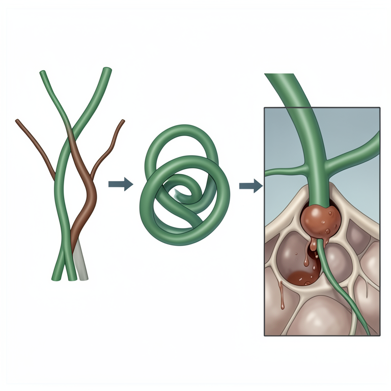
Mycoparasitism Process
AI-generated conceptual illustration (Trichoderma fungus and Rhizoctonia solani fungus hyphae). It explains the idea and does not show study data.
View source evidence ↗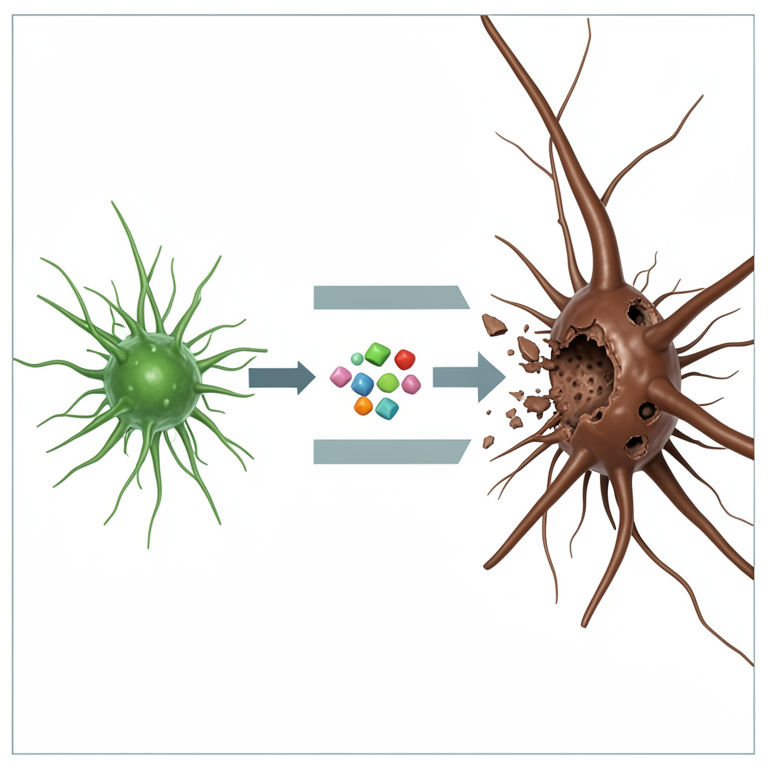
Enzymatic Attack Mechanism
AI-generated conceptual illustration (Trichoderma fungi and Rhizoctonia solani hyphae). It explains the idea and does not show study data.
View source evidence ↗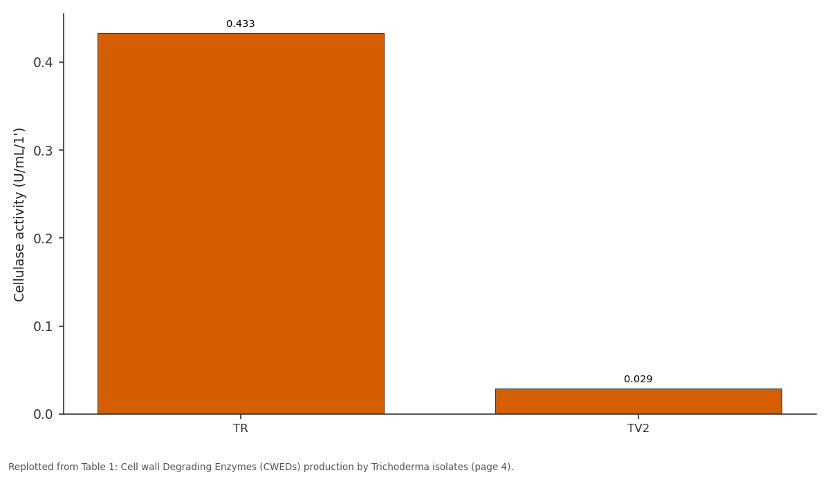
Cellulase activity (U/mL/1'): TR vs TV2
Replotted from Table 1 (page 4) of the original paper. Every bar is the paper's own table value.
View source evidence ↗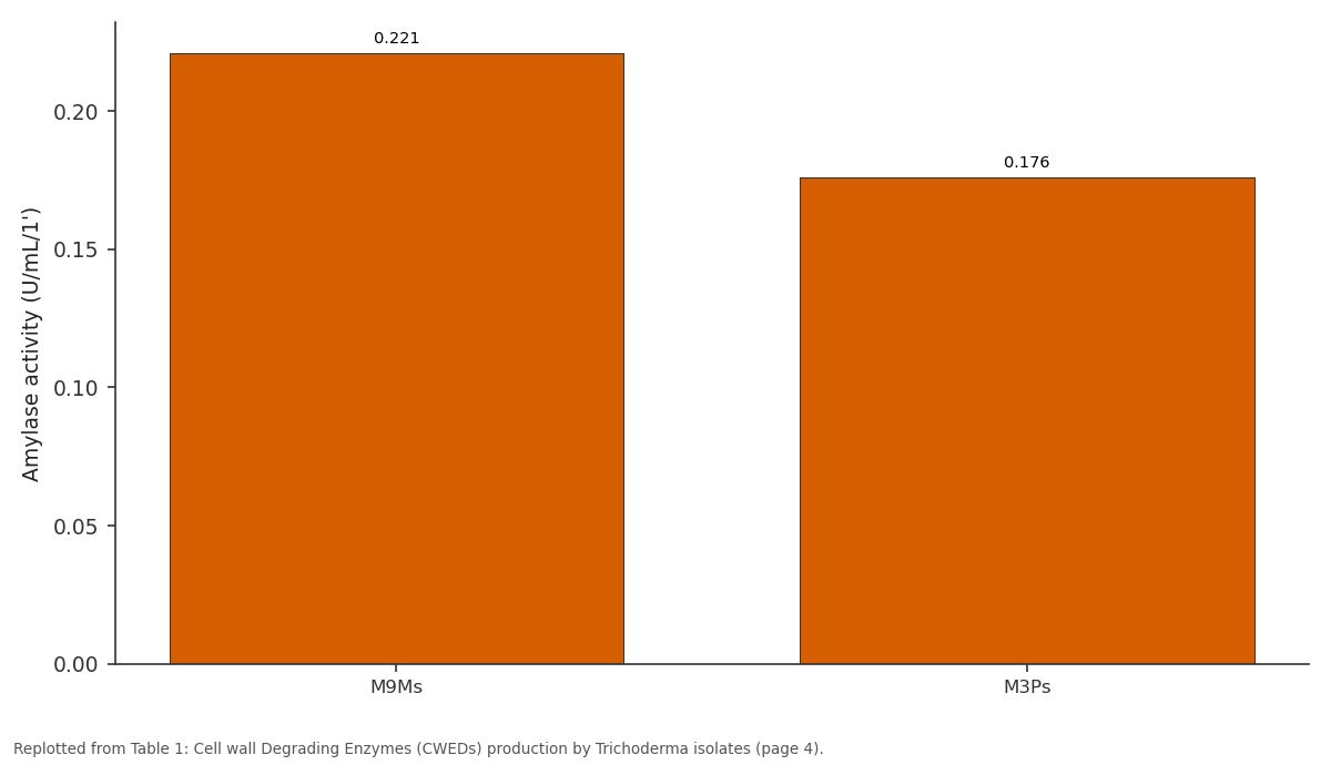
Amylase activity (U/mL/1'): M9Ms vs M3Ps
Replotted from Table 1 (page 4) of the original paper. Every bar is the paper's own table value.
View source evidence ↗Figures & tables
From the original paper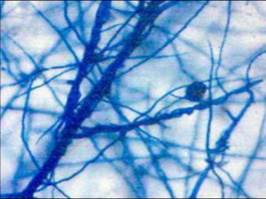 Figure 1
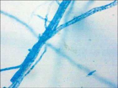 Figure 1
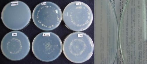 Figure 2
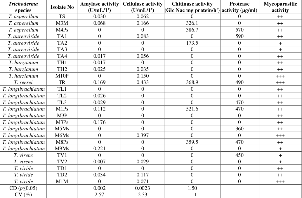 Table 1
Introduction
Sheath blight is considered to be an important disease of rice next to rice blast (DRR 2007). It is caused by a soil borne fungus Rhizoctonia solani (Teleomorph Thanetophorus cucumeris (A B Frank, Donk, Ou 1985) [37] . The loss of yield due to this disease has been estimated to be 10-50% in India. The disease appears both in the nursery and on transplanted rice (Banniza and Holderness 2001) [3] , inflicting heavy losses in many Asian countries and it is most serious fungal diseases worldwide (Manalo et al. 2001) [31] . The sclerotia of R. solani remain viable in the soil for several years and it causes spots or lesions mostly on the leaf sheath (Ou 1985; Damicone et al. 1993) [10] . Its wide occurrence is brought about by intensive methods of rice cultivation involving the use of short culmed, high tillering, high yielding varieties, high inputs of nitrogen fertilizer and application of fungicides. Nevertheless, fungicides have many undesirable attributes, novel bio control agents have therefore been looked for and Trichoderma species have been the preferred choice. Several modes of action have been proposed to explain the bio control of plant pathogens by Trichoderma these include production of antibiotics and cell wall degrading enzymes, mycoparasitism, competition for key nutrients, parasitism, stimulation of plant defense mechanisms and combination of these possibilities (Kubicek et al. 2019) [29] . Trichoderma spp.
International Journal of Biology Sciences https://www.biologyjournal.net are considered to inhibit the growth of phytopathogenic fungi via the production of hydrolytic enzymes which degrade the cell walls of the pathogens (Yao et al. 2023) [48] . Mycoparasitic activity is one of the major mechanisms proposed to explain the antagonistic activity of Trichoderma spp. against many soil borne plant pathogenic fungi (Weindling 1932, 1934; Elad et al. 1982, Lynch 1990, Melo and Faull 2000). This has been long considered as an important means of action of bio control by Trichoderma spp., and is a complex process that involves growth of the biocontrol agent towards the target fungi, lectin mediated coiling of attachment of Trichoderma hyphae to the pathogen, and finally attack the target fungus by the dissolution of cell wall of the pathogen by the activity of enzymes. The active principle responsible for mycoparasitic activity is by the production of either toxins or hydrolytic enzymes or both by Trichoderma (Howell 2003) [23] . The parameter for the successful use of Trichoderma as a potential biocontrol agent is its efficient coiling process followed by a substantial production of hydrolytic enzymes. Synergism between hydrolytic enzymes and antibiotics that affect cell wall synthesis facilitates the penetration of further active compounds suppressing fungal growth (Witkowska and Maj 2002) [45] . Trichoderma spp. was shown to be very efficient producers of extracellular enzymes, and some of these have been implicated in the biological control of plant diseases (Monte, 2001 [35] ; Harman, 2006 [21] ; Jayalakshmi et al. 2009) [24] . The cell wall degrading enzymes such as chitinases, cellulases, β-1,3-glucanases, and proteases have been associated with the ability of Trichoderma spp. to control plant pathogens (De Marco et al. 2003) [13] . As the growth control of the pathogen is mostly by several strains of Trichoderma. However, the quantitative production of these enzymes has an effect directly on lysis of the cell walls of the pathogens. So, the products of lytic enzymes to be identified, to select as an efficient isolate for field application. Hence, a study was undertaken to evaluate the potential of Trichoderma isolates to produce hydrolytic enzymes.
Materials and Methods
Materials and Methods
Mycoparasitic activity of Trichoderma isolates against R. solani
This study was conducted using dual culture method (Dennis and Webster 1971c) with modifications. The 5 mm mycelial disc of Rhizoctonia solani was placed on 2% water agar medium and after one day of incubation a 5 mm disc of Trichoderma inoculum was placed on the inoculum of test pathogen. A block of medium (10 × 20 cm) was cut at the juncture of two colonies and mycelium of the two fungi was mounted in lactophenol, and examined under microscope for hyphal interactions.
Preliminary screening test for production cellulase by Trichoderma isolates
The preliminary screening for cellulase enzyme production by Trichoderma was assayed in Czapek-Mineral salt agar medium and the pH was adjusted to 6.5. Trichoderma isolates were inoculated on the Petri plates and incubated at room temperature (27±2°C) for 2-5 days. Then, the plates were flooded with 1% aqueous solution of hexadecyltrimethyl ammonium bromide. The plates were observed for the formation of a clear zone around the fungal growth (Aneja 1996) [2] .
Cellulase assay: To study the cellulase production of the Trichoderma isolates, Czapek-Dox broth medium with 10g/l Carboxy Methyl Cellulose (1% CMC) was employed. Ten drops of Tween-80 was added. The pH of the medium was adjusted to 5.5, dispensed into 100 ml conical flasks and autoclaved. Later, one ml (2 X10 7 / ml) spore suspension of each Trichoderma isolate were inoculated and incubated in orbital shaker (120 rpm) at room temperature (27±2°C) for seven days. Harvest the culture filtrate and the supernatant was used as enzyme source. CMC activity was determined by 1 ml of enzyme solution with 10 mg of CMC diluted with acetate buffer, pH 6.5, and incubated for 30 min at 60 0 C. To this enzyme solution, DNS reagent was added and the optical density was measured at 520 nm with a UV-visible spectrophotometer (UV 1100, Shimadzu). The reducing sugars were estimated by DNS method as described by Miller (1959) [33] .
Preliminary screening test for production amylase by
Trichoderma isolates: Starch Agar medium was used for the preliminary screening of amylase and the pH was adjusted to 7.0. The Petri plates were inoculated with the Trichoderma species and incubated at room temperature (27±2°C) for 2-5 days. Then, the plates were flooded with Grams iodine solution and the plates were observed for the formation of a clear zone around the fungal growth (Aneja 1996) [2] .
Amylase assay: To study the amylase activity of the Trichoderma isolates, Czapek-Dox broth medium with 10g/l soluble starch (1% soluble starch) was employed. Ten drops of Tween-80 was added. The pH of the Czapek-Dox broth medium was adjusted to 5.5, dispensed into 100 ml conical flasks and autoclaved. Later, one ml (2 X10 7 / ml) spore suspension of each Trichoderma isolate were inoculated and incubated in orbital shaker (120 rpm) at room temperature (27±2°C) for seven days. Harvest the culture filtrate and the supernatant was used as enzyme source. Amylase activity was determined by 1 ml of enzyme solution with 1 ml of 1% soluble starch in acetate buffer, pH 6.5, and incubated for 30 min at 37°C. After incubation period add NaOH and reducing sugars were estimated by DNS method as described by Miller (1959) [33] .
Preliminary screening for chitinases: Trichoderma isolates were grown on synthetic medium where carbon source was substituted by colloidal chitin and incubated at room temperature. Controls were maintained with and without any carbon source. Colloidal chitin for enzymatic studies was prepared according to the procedure of Berger and Reynolds (1958) [5] .
Chitinase assay: Chitinase-β-1,4-Poly N-Acetyl Dglucosaminidase assay was carried out by Reissing et al. (1959) [38] . The 200μl reaction mixture containing 1% chitin, 20 μg of protein (crude enzyme) in 0.1M acetate buffer (pH 5.0) is incubated for the desired time in water bath shaker (100 rpm) at 37°C. The reaction is stopped by boiling at 100°C and then centrifuged at 10,000 rpm for 10 min; the supernatant is used for assay. OD values were taken at 580 nm. Specific activity of chitinase was expressed as μ moles of Glc NAc released/ mg of protein/ h.
Protease assay: Protease assay was done by following the modified procedure of Anson's method (Yang and Huang 1994) [47] . The substrate used (1% casein in 50mM phosphate buffer, pH 7.0) was denatured at 100 0 C for 15min in a water bath and cooled at room temperature. The reaction mixture containing 1ml of substrate and 1ml of enzyme solution were incubated at 37°C for 20 min with shaking and was stopped with 3ml of 10% trichloroacetic acid (TCA). The absorbance of the liberated tyrosine in the filtrate was measured at 280nm. One unit of protease activity was defined as the amount of enzyme that produced an absorbance at 280nm equivalent to 1μmol of tyrosine in one minute under the assay condition. Average value of triplicate data was presented.
Results
Twenty-six isolates of Trichoderma were obtained from the rice soil samples collected from two districts (Nalgonda and Ranga Reddy) of Telangana and forest soil samples of Uttaranchal (Chamoli), India. A preliminary qualitative assay was conducted to screen 26 Trichoderma isolates for mycoparasitic activity against R. solani and cell wall degrading enzymes production (CWDEs) i.e., Celluases, Amylases, Chitinases and Proteases. Based on the results, the isolates were subjected to quantify the enzymes in in vitro. All the twenty-six isolates of Trichoderma showed some degree of mycoparasitic activity against the test pathogen Rhizoctonia solani. The degree of hyphal coiling is varied with different isolates (Table 1). T. harzianum (M10P), T. koningii (TR), T. longibrachiatum (M6Ms) and T. viride (M1M) showed maximum mycoparasitism (+++) on R. solani (Figure 1), the intense coiling of Trichoderma hyphae around the mycelium of the pathogen was observed which led to the distortion of pathogen hyphae, finally the death of the R. solani mycelium was noticed. T. asperellum (TS, M3M, M4Ps), T. aureoviride (TA4), T. harzianum (TH1, TH2), T. longibrachiatum (TL1, TL2, TL3, M1Ps, M3P, M3Ps, M5Ms, M8Ps) and T. viride (TD1, TD2) showed moderate mycoparasitic activity with less hyphal coiling, moderate mycoparasitism (++), mycelial growth of Trichoderma along with the pathogen hyphae was observed, but the intensity of coiling is less and distortion of the pathogen hyphae is moderate. In minimum mycoparasitc activity (+), the hyphal coiling of Trichoderma around the pathogen and distortion of the pathogen was very less. The isolates are T. aureoviride (TA2, TA3), T. longibrachiatum (M9Ms) and T. virens (TV1, TV2) and the occurrence of parallel mycelial growth of Trichoderma isolates against R. solani hyphae was rare in the test plate (Figure 1). andT. viride (TD2, M1M). These are further analyzed by quantitative enzyme assay (Table 1). Among these isolates, the maximum cellulase production was observed with T. reesei (TR) (0.433 U/mL/1'), followed by T. longibrachiatum (M6Ms) (0.397 U/mL/1'). The least activity was recorded with T. virens (TV2) (0.029 U/mL/1'). Thirteen isolates of Trichoderma spp. were positive in amylase enzyme production and these belong to T. asperellum (TS, M3M), T. aureoviride (TA4), T. harzianum (TH1, TH2), T. reesei (TR), T. longibrachiatum (TL2, TL3, M1Ps, M3Ps, M9Ms), T. virens (TV2), and T. viride (TD2). In the assay T. longibrachiatum (M9Ms) produced maximum amylase, followed by T. longibrachiatum (M3Ps) (Table 1). In the preliminary screening test, twelve isolates of Trichoderma showed chitin degrading ability. Among these six isolates T. asperellum (M3M, M4Ps), T. aureoviride (TA2), T. reesei (TR), and T. longibrachiatum (M1Ps, M8Ps) completely degraded the chitin (Fig 2 ).
| Trichoderma species | Isolate No | Amylase activity (U/mL/1') | Cellulase activity (U/mL/1') | Chitinase activity (Glc Nac mg protein/h') | Protease activity (µg/ml) | Mycoparasitic activity |
|---|---|---|---|---|---|---|
| T. asperellum | TS | 0.030 | 0.062 | 0 | 0 | ++ |
| T. asperellum | M3M | 0.068 | 0.166 | 326.1 | 0 | ++ |
| T. asperellum | M4Ps | 0 | 0 | 386.7 | 570 | ++ |
| T. aureoviride | TA1 | 0 | 0.083 | 0 | 590 | ++ |
| T. aureoviride | TA2 | 0 | 0 | 173.5 | 0 | + |
| T. aureoviride | TA3 | 0 | 0 | 0 | 0 | + |
| T. aureoviride | TA4 | 0.017 | 0.056 | 0 | 0 | ++ |
| T. harzianum | TH1 | 0.017 | 0 | 0 | 0 | ++ |
| T. harzianum | TH2 | 0.025 | 0.035 | 0 | 0 | ++ |
| T. harzianum | M10P | 0 | 0.150 | 0 | 0 | +++ |
| T. reesei | TR | 0.169 | 0.433 | 368.9 | 490 | +++ |
| T. longibrachiatum | TL1 | 0 | 0 | 0 | 0 | ++ |
| T. longibrachiatum | TL2 | 0.026 | 0 | 0 | 0 | ++ |
| T. longibrachiatum | TL3 | 0.029 | 0 | 0 | 470 | ++ |
| T. longibrachiatum | M1Ps | 0.112 | 0 | 521.6 | 470 | ++ |
| T. longibrachiatum | M3P | 0 | 0 | 0 | 0 | ++ |
| T. longibrachiatum | M3Ps | 0.176 | 0 | 0 | 0 | ++ |
| T. longibrachiatum | M5Ms | 0 | 0 | 0 | 360 | ++ |
| T. longibrachiatum | M6Ms | 0 | 0.397 | 0 | 0 | +++ |
| T. longibrachiatum | M8Ps | 0 | 0 | 359.5 | 470 | ++ |
| T. longibrachiatum | M9Ms | 0.221 | 0 | 0 | 0 | + |
| T. virens | TV1 | 0 | 0 | 0 | 450 | + |
| T. virens | TV2 | 0.007 | 0.029 | 0 | 0 | + |
| T. viride | TD1 | 0 | 0 | 0 | 0 | ++ |
| T. viride | TD2 | 0.034 | 0.117 | 0 | 0 | ++ |
| T. viride | M1M | 0 | 0.071 | 0 | 0 | +++ |
| CD (p≤0.05) | 0.002 | 0.0023 | 1.50 | |||
| CV (%) | 2.57 | 2.33 | 1.11 |
Table text extracted from the original PDF; superscript letters are shown inline. The original table image is below.
View original table image
Right: T. reesei test pate is transparent because of chitin degradation
Left: Control plate is opaque because of no chitin degradation Chitinase assay was done for these six isolates of Trichoderma. The highest of chitinase produced by T. longibrachiatum (M1Ps) (521.6 Glc Nac mg protein/h'), followed by T. asperellum (M4Ps) (386.7 Glc Nac mg protein/h'). Eight isolates of Trichoderma produced protease enzyme viz., T. asperellum (M4Ps), T. aureoviride (TA1), T. reesei (TR), T. longibrachiatum (TL3, M1Ps, M5Ms M8Ps), T. virens (TV1). Maximum protease production was observed with T. aureoviride (TA1) (590µ g/ml) and the least producer was T. longibrachiatum (M5Ms) (360µ g/ml)
(Table 1). T. asperellum (M3M) and T. koningii (TR) produced all the three enzymes, whereas T. aureoviride (TA3), T. longibrachaitum (TL1, M3P), T. virens (TV1) and T. viride (TD1) did not show any enzyme activity. A positive correlation was found between mycoparasitic activity and Cell Wall Degrading Enzymes production (CWDEs). harzianum strains against R. solani exhibited that coiling around the hyphae of R. solani followed by substantial production of hydrolytic enzymes (Almeida 2007). Risoli and co-workers (2022) [39] reported that Trichoderma is capable of producing putrescence under specific environmental conditions, which contributes to its mycoparasitic activity against Botrytis cinerea. Chet and Baker (1981) suggested the involvement of cellulase production in mycoparasitism on Pythium spp. by T. hamatum. The isolates belonging to T. aureoviride (TA1, TA2), T. longibrachiatum (M9Ms) and T. virens (TV1) are non-producers of cellulase (Table .1) and T. virens (TV2) showed slight production. This may be the reason for least mycoparasitic activity which did not allow the antagonist to dissolute cell walls of R. solani. This study showed correlation between cellulase production and mycoparasitic activity. Melo et al. (1997) reported high levels of cellulases that hydrolyse the cell walls of other fungi. The test pathogen R. solani cell wall consists of high levels of glucans.
Hence, these antagonistic Trichoderma isolates are capable of hydrolyzing the cell walls of the pathogen by the production of lytic enzymes (cellulase) which might have led to the coiling and penetration of the test antagonist. The high mycoparasitic activity with the four isolates of Trichoderma (M10P, TK, M6Ms, M1M) was at later stages resulted in the hyphal collapse of R. solani. In 1983 Elad et al. proved the localized cell wall lysis on the point of interaction between Trichoderma species and R. solani by scanning electron microscopy and fluorescent microscopy studies. Barbosa et al. (2001) [4] reported that T. reesei is a high producer of cellulase when compared with other Trichoderma isolates and T. harzianum, T. reesei, T. polysporum, and T. viride isolates produced amylases and it is one of the enzymes partially necessary for the growth of Trichoderma. In this study also T. reesei (TR) was the highest producer of cellulase. Krishnamurthy et al. (1990) [28] screened 35 strains of T. viride and T. harzianum for antagonism against R. solani and reported that four strains were better for chitinase induction with colloidal chitin. In this study 6 out of 26 isolates showed chitinase activity belonging to the species T. reesei (TR), T. Longibrachaitum, T. asperellum and T. aureoviride. Ulhoa and Peberdy (1991) [43] selected T. harzianum as an effective chitinase producer. Purified chitinolytic enzymes of T. harzianum, inhibited a wide range of fungi (Harman et al. 1993 [20] ; Lorito et al.
1993) [30] . The isolates T. reesei (TR) and T. longibrachiatum (M1Ps) inhibited the growth of R. solani and completely inhibited the sclerotial formation in dual culture than any other selected isolates (Chakravarthy and Nagamani 2007) [9] . T. asperellum (M4Ps), T. longibrachiatum (TL3, M9Ms), T. viride (TD2) isolates inhibited growth of R. solani through the production of volatile antibiotics, whereas T. reesei (TR), T. longibrachiatum (M1Ps, M8Ps) showed least inhibition. But among the three isolates only T. reesei (TR) and T. longibrachiatum (M1Ps) inhibited sclerotial formation completely through the production of volatile metabolites (Chakravarthy and Nagamani 2007) [9] . Several studies have demonstrated that certain Trichoderma species are capable of producing volatile metabolites that can inhibit colony growth to varying extents, with some strains achieving inhibition rates exceeding 80% (Thambugala et al. 2020 [42] ; Kong et al. 2022) [26] . All the three enzymatic activities were observed with the isolate T. reesei (TR). T. longibrachiatum (TL3 and M9Ms) showed only amylase activity, whereas by T. asperellum (M4Ps) and T. longibrachiatum (M8Ps) isolates only chitinases enzyme activity was recorded. With M1Ps enzyme activity of both cellulase and chitinase was observed, but with TD2 isolate cellulase and amylases activity was noticed. T. longibrachiatum (M1Ps) recorded highest chitinase enzyme activity and T. reesei (TR) is the highest cellulase enzyme producer when compared to other isolates. Moreover, Elad et al. (1982) reported that the isolates of T. harzianum, which were found to differ in their ability to attack S. rolfsii, R. solani and P. aphanidermatum, also differed in the levels of mycolytic enzymes produced by them. The enzymes chitinases, b-1, 3 glucanase and protease were released from Trichoderma during antagonism and inhibited the growth of fungal pathogen (Gajera and Vakharia 2012) [16] . Increase of their level and specific activities could be selective advantages in antagonism Trichoderma strains produce a wide variety of CWDEs, among them are chitinases, α-(1, 3), β-(1, 3) and β- (1, 6) glucanases, and proteases, that degrade fungal cell walls (Ait-Lahsen et al. 2001 [1] ; De la Cruz et al. 1995a) [11,12] . Ghasemi et al. 2020 [18] reported that there was a strong link between the diversity of various chitinase proteins and the antagonistic properties of the T. virens mutant M8 against R. solani.
Fungal proteases may also be significantly involved in cell wall degradation, since fungal cell walls contain chitin and glucan polymers embedded in and covalently linked to a protein matrix (Kapteyn et al. 1996) [25] . These observations, together with the fact that chitin and glucans are the main polysaccharides of fungal mycelia, suggest that chitinases and glucanases play an essential role in the lysis of phytopathogenic fungal cell walls during antagonism (Ait-Lahsen et al. 2001 [1] ; Bosah et al. 2010 [8] ; De la Cruz et al. 1995b [11,12] ; Henis and Chet 1975) [22] . The role of Trichoderma proteases in biological control as well as their advantages and disadvantages in biotechnology has been reviewed (Kredics et al. 2005) [27] . These enzymes function by breaking down the polysaccharides, chitin, and glucans that are responsible for the rigidity of fungal cell walls, there by destroying cell wall integrity (Howell 2003) [23] . Involvement of proteases in bio control processes has already been reported (Elad and Kapat 1999) [15] . Besides carbohydrateactive enzymes, proteases are also important for degradation of fungal cell wall and it was evident from the experiments with T. atroviride and T. virens by Gruber and Seidl-Seiboth (2012) [40] . Different types of proteases were reported to be induced during mycoparasitism before and during contact with the prey in different Trichoderma species (Geremia et al. 1993 [17] ; Seidl et al. 2009 [40] ; Suarez et al. 2007) [41] . Although some of the other isolates exhibited superior production of volatile and non-volatile compounds, they were not selected for further investigation, as the primary focus of this study was on cell wall degrading enzyme activity and mycoparasitic potential against Rhizoctonia solani. Hence, in the present investigation out of twenty six isolates of Trichoderma, two strains namely T. reesei (TR) and T. longibrachiatum (M1Ps) were selected because these two isolates showed complete inhibition of sclerotial formation and produced high quantities of chitinase and non-volatiles.
Conclusion
Twenty six Trichoderma isolates screened for their ability to produce cell wall degrading enzymes (CWDEs) and their mycoparasitic activity against Rhizoctonia solani, the causative agent of sheath blight in rice. A significant variability was observed among the isolates in the production of enzymes such as cellulase, amylase, chitinase, and protease. Among the isolates, T. reesei (TR) and T. longibrachiatum (M1Ps) exhibited highest levels of enzyme production and were among the few strains of Trichoderma capable of producing all three key enzymes. Furthermore, these strains exhibited strong mycoparasitic activity against R. solani, showing a clear positive correlation between CWDE production and biocontrol potential. Considering all aspects of our study, T. reesei (TR) and T. longibrachiatum (M1Ps) are identified as the most promising biocontrol agents for sheath blight management in rice.
References
47 in the original paper- Lahsen AH, Soler A, Rey M, Cruz DLJ, Monte E, Llobell A. An antifungal exo-alpha-1,3-glucanase (AGN13.1) from the biocontrol fungus Trichoderma harzianum. Appl Environ Microbiol. 2001;67:58335839.
- Aneja KR. Experiments in microbiology, plant pathology, tissue culture and mushroom cultivation. 2 nd Edition. Wishwa Prakashan (New Age International Pvt. Ltd.), 1996, p. 1-47.
- Banniza S, Holderness. Rice sheath blight-pathogen biology and diversity. In: Major fungal diseases of rice: recent advances. Johnson R, Sreenivasa Prasad S, eds. Kluwer academic publishers, Netherlands, 2001, p. 201-211.
- Berger LR, Reynolds DM. The chitinase system of a strain of Streptomyces griseus. Biochem Biophy Acta. 1958;29:522-534.
- Bernfeld P. Enzymes of starch degradation synthesis. In: Advances in enzymology, XII. Nord F, ed. Interscience Publications, New York, 1951, p. 376.
- Bernfeld P. Amylases (alpha) and beta. In: Methods in enzymology I. Colowick S, Kaplan N, eds. Academic Press, New York. 1955, 149.
- Bosah O, Igeleke CA, Omorusi VI. In vitro microbial control of pathogenic Sclerotium rolfsii. Int J Agric Biol. 2010;12:474-476.
- Chakravarthy SK, Nagamani A. Efficacy of nonvolatile and volatile compounds of Trichoderma species on Rhizoctonia solani. J Mycol Pl Pathol. 2007;37:8286.
- Damicone JP, Patel MV, Moore WF. Density of sclerotia of Rhizoctonia solani and incidence of sheath blight in rice fields in Mississippi. Pl Dis. 1993;77:257260.
- Cruz DLJ, Toro PJA, Benítez T, Llobell A. Purification and characterization of an endo-β-1, 6-Glucanase from Trichoderma harzianum that is related to its mycoparasitism. J Bacteriol. 1995;177:1864-1871.
- Cruz DLJ, Toro PJA, Benítez T, Llobell A, Romero L. A novel endo-β-1,3-glucanase, BGN 13.1, involved in the mycoparasitism of Trichoderma harzianum. J Bacteriol. 1995;177:6937-6945.
- De Marco JL, Valadares-Inglis MC, Felix CR. Production of hydrolytic enzymes by Trichoderma sp. isolate with antagonistic activity against Crinipellis perniciosa, the causal agent of witches' broom of cocoa. Braz J Microbiol. 2003;34:33-38.
- DRR. Production oriented survey, 2006. Directorate of rice research (AICRIP), Hyderabad, India, 2007.
- Elad Y, Kapat A. The role of Trichoderma harzianum protease in the biocontrol of Botrytis cenerea. Eur J Plant Pathol. 1999;105:177-189.
- Gajera HP, Vakharia DN. Production of lytic enzymes by Trichoderma isolates during in vitro antagonism with Aspergillus niger, the causal agent of collar rot of peanut. Brazilian J Microbiol. 2012;43-52.
- Geremia RGH, Goldman D, Jacobs W, Ardiles SB, Vila M, Montagu V, Estrella HA. Molecular characterization of a proteinase-encoding gene, pbr1, related to mycoparasitism by Trichoderma harzianum. Molecular Microbiol. 1993;8:603-613.
- Ghasemi S, Safaie N, Shahbazi S, Shams-Bakhsh M, Askari H. The Role of cell wall degrading enzymes in antagonistic traits of Trichoderma virens Against Rhizoctonia solani. Iranian J Biotechnol. 2020;18(4):e2333.
- Gruber S, Seiboth SV. Self versus non-self: fungal cell wall degradation in Trichoderma. Microbiol. 2012;158:26-34.
- Harman GE, Hayes CK, Lorito M, Broadway RM, Di Pietro A, Peterbauer C, Tronsmo A. Chitinolytic enzymes of Trichoderma harzianum: Purification of Chitobiosidase and endochitinase. Phytopathol. 1993;83:313-318.
- Harman GE. Overview of Mechanisms and Uses of Trichoderma spp. Phytopathol. 2006;96:190-194.
- Henis Y, Chet I. Microbiological control of plant pathogens. Adv Appl Microbiol. 1975;19:85-111.
- Howell CR. Mechanisms employed by Trichoderma species in the biological control of plant diseases: the history and evolution of current concepts. Plant Dis. 2003;87:4-10.
- Jayalakshmi SK, Raju S, Usha Rani S, Benagi VI, Sreeramulu K. Trichoderma harzianum L1 as a potential source for lytic enzymes and elicitor of defense responses in chickpea (Cicer arietinum L.) against wilt disease caused by Fusarium oxysporum f. sp. ciceri. Australian J Crop Sci. 2009;3:44-52.
- Kapteyn JC, Montijn RC, Vink E, Cruz DLJ, Llobell A, et al. Retention of Saccharomyces cerevisiae cell wall proteins through a phosphodiesterlinked β-1,3, β-1,6glucan heteropolymer. Glycobiol. 1996;6:337-345.
- Kong WL, Ni H, Wang WY, Wu XQ. Antifungal effects of volatile organic compounds produced by Trichoderma koningiopsis T2 against Verticillium dahliae. Front Microbiol. 2022;13:1013468.
- Kredics L, Antal Z, Szekeres A, Hatvani L, Manczinger L, Vagvolgyi C, Nagy E. Extracellular proteases of Trichoderma species. A review. Acta Microbiol Immunol Hung. 2005;52:169-184.
- Krishnamurthy J, Samiyappan R, Vidhyasekaran P, Nakkeeran S, Rajeswari E. Efficacy of Trichoderma chitinases against Rhizoctonia solani, the rice sheath blight pathogen. J Biosci. 1999;24:207-213.
- Kubicek CP, Steindorff AS, Chenthamara K, Manganiello G, Henrissat B, Zhang J, et al. Evolution and comparative genomics of the most common Trichoderma species. BMC Genomics. 2019;20:485.
- Lorito M, Harman GE, Hayes CK, Broadway RM, Tronsmo A, Woo SL, et al. Chitinolytic enzymes produced by Trichoderma harzianum: Antifungal activity of purified endochitinase and chitobiosidase. Phytopathol. 1993;83:302-307.
- Manalo JO, Mew TW, Teng PS, Velilla AR, Borromeo ES. Populations of rice sheath blight in the IIocos Norte rice-based ecosystem. In: Exploring biodiversity for sustainable pest management. Mew TW, Borromeo E, Hardy B, eds. IRRI, Philippines, 2001, p. 101.
- Mendels M, Sternberg D. Recent advances in cellulose technology. J Ferment Tech. 1976;54:267-286.
- Miller GL. Use of dinitrosalicylic acid reagent for determination of reducing sugar. Analytical Chem. 1959;31:426-428.
- Miller HN, Sauve RJ. Etiology and control of Pythium stem rot of geranium. Plant Dis Rep. 1975;59:122-126.
- Monte E. Understanding Trichoderma: between biotechnology and microbial ecology. Intl Microbiol. 2001;4:1-4.
- Naidu VD. Influence of sheath blight of rice on grain and straw yield in some popular local varieties J Res Publ. 1992;10:78-80.
- Ou SH. Rice Diseases. 2nd edition. Commonwealth Mycological Institute, 1985, p. 272.
- Reissing JG, Strominger JL, Lelior LF. A modified colorometric method for the estimation of N-Acetyl sugars. J Biological Chem. 1959;217:959-962.
- Risoli S, Cotrozzi L, Sarrocco S, Nuzzaci M, Pellegrini E, Vitti A. Trichoderma induced resistance to Botrytis cinerea in Solanum species: a meta-analysis. Plan Theory. 2022;11:180.
- Seidl V, Song L, Lindquist E, Gruber S, Koptchinskiy A. Transcriptomic response of the mycoparasitic fungus Trichoderma atroviride to the presence of a fungal prey. BMC Genomics. 2009;10:567.
- Suarez MB, Vizcaino JA, Llobell A, Monte E. Characterization of genes encoding novel peptidases in the biocontrol fungus Trichoderma harzianum CECT 2413 using the TrichoEST functional genomics approach. Curr Genet. 2007;51:331-342.
- Thambugala KM, Daranagama DA, Phillips A, Kannangara SD, Promputtha I. Fungi vs. fungi in biocontrol: an overview of fungal antagonists applied against fungal plant pathogens. Front Cell Infect Microbiol. 2020;10:604923.
- Ulhoa CJ, Peberdy JF. Regulation of chitinase synthesis in Trichoderma harzianum. J Gen Micro. 1991;137:2163-2169.
- Vishnu C, Sudha Rani K, Gopal Reddy, Seenayya G. Amylolytic bacteria producing lactic acid. J Sci Ind Res. 1998;57:600-603.
- Witkowska D, Maj A. Production of lytic enzymes by Trichoderma sp. and their effect on the growth of phytopathogenic fungi. Folio Microbiologica. 2002;47:279-282.
- Xie G, Xuefang Y, Xiaoping R. Biological control agents against major pathogens of rice in the Yangtze Delta area of China. In: Exploring biodiversity for sustainable pest management. Mew TW, Borromeo E, Hardy B, eds. IRRI, Philippines, 2001.
- Yang SS, Huang CI. Protease production by amylolytic fungi in solid state fermentation. J Chin Agric Chem Soc. 1994;32:589-601.
- Yao X, Guo H, Zhang K, Zhao M, Ruan J, Chen J. Trichoderma and its role in biological control of plant fungal and nematode disease. Frontiers in Microbiol. 2023;14:1160551.