
Research article · 2025 · Full text
International Journal of Biology SciencesMinerals profiles in the epididymal luminal milieu with respect to maturation of buck (Capra hircus) spermatozoa
Volume 7(6) · Pages 381–384 · DOI: 10.33545/26649926.2025.v7.i6f.402 ↗
Research areasAgriculture and Biological Studies
Abstract
Author-writtenThe aim of the present study was to investigate about micro environment of epididymis towards maturation of buck spermatozoa. Normal testes attached with intact epididymis collected from adult and healthy bucks (n=60) from local abattoir immediately after slaughter were used for assessment of minerals profiles in the epididymal luminal fluid. It was revealed that in caput, corpus and caudal regions, the level of Zn and Cu found to be increased gradually, however, the level of Fe, Na and K diminished steadily. Concentration of Zn and Cu was highest in caudal segment, whereas concentration of Fe, Na and K was highest in caput segment. The overall relationship of spermatozoan motility was found to be positive and highly significant (p
Summary
Source-checkedPlain-language reading of the full paper, prepared with AI and checked against the text. Each point links to the authors' own words.
Why it matters
- Mammalian sperm cannot fertilize an egg immediately after leaving the testes; they must undergo changes while passing through the epididymis to become capable of fertilization. View evidence ↗
- The specific environment within the epididymal lumen is critical for the successful maturation of sperm. View evidence ↗
- Trace elements are essential for male reproduction as they often serve as enzyme cofactors during testicular development and spermatogenesis. View evidence ↗
The question
- The study aimed to measure the levels of zinc, copper, iron, sodium, and potassium in the epididymal luminal fluid (ELF) across the caput, corpus, and cauda segments of bucks. View evidence ↗
What they did
- The researchers used testes and intact epididymides from 60 healthy adult bucks collected from a local abattoir. View evidence ↗
- To collect the luminal fluid, 3.5 g of tissue from each epididymal segment was minced and flushed with 2 ml of chilled phosphate-buffered saline (PBS). View evidence ↗
- Sodium and potassium were measured using flame photometry, while zinc, copper, and iron were analyzed via spectrophotometry after microwave digestion. View evidence ↗
- Spermatozoa motility was evaluated from the right epididymis using a standard subjective ranking method. View evidence ↗
What they found
- Zinc levels increased gradually from the caput (124.25 ± 0.76 µg/dl) to the corpus (130.45 ± 0.75 µg/dl) and reached the highest concentration in the cauda (138.40 ± 1.28 µg/dl). View evidence ↗
- Copper concentrations also increased along the epididymis, from 36.54 ± 0.73 µg/dl in the caput to 51.25 ± 1.21 µg/dl in the cauda. View evidence ↗
- Iron levels decreased steadily, with the highest concentration in the caput (181.62 ± 1.62 µg/dl) and the lowest in the cauda (136.27 ± 1.12 µg/dl). View evidence ↗
- Sodium concentrations diminished from the caput (152.34 ± 1.29 mEq/l) to the cauda (132.69 ± 1.18 mEq/l). View evidence ↗
- Potassium levels also decreased along the transit, from 15.82 ± 0.64 mEq/l in the caput to 8.96 ± 0.81 mEq/l in the cauda. View evidence ↗
- Sperm motility showed a highly significant positive correlation with copper (r=0.626, p<0.01) and a significant positive correlation with sodium (r=0.384) and zinc (r=0.743) at the 5% level. View evidence ↗
- A negative but non-significant relationship was observed between sperm motility and the levels of iron (r=0.301) and potassium (r=0.296). View evidence ↗
What it means
- The authors conclude that the changing concentrations of zinc, copper, iron, sodium, and potassium in the luminal fluid are key factors in regulating the environment necessary for sperm maturation in bucks. View evidence ↗
- The study suggests that these mineral profiles could serve as simple analytical indicators to monitor the sperm maturation process. View evidence ↗
- The authors state that further research is needed to understand the molecular, biochemical, and physiological mechanisms by which these minerals influence male fertility. View evidence ↗
Visual summary
2 visualsIllustrations explain the paper; charts are replotted from the paper's own table values.
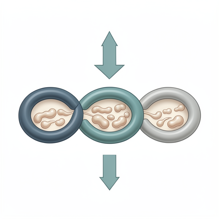
Mineral Gradient in the Epididymis
AI-generated conceptual illustration (buck (Capra hircus) epididymis and spermatozoa). It explains the idea and does not show study data.
View source evidence ↗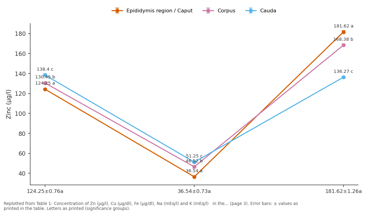
Epididymis region / Caput and Corpus and more: 124.25±0.76a vs 36.54±0.73a vs 181.62±1.26a
Replotted from Table 1 (page 3) of the original paper. Every bar is the paper's own table value.
View source evidence ↗Figures & tables
From the original paper Table 1
 Table 2
 Table 3
Introduction
In mammals, testicular sperm are not able to fertilize an oocyte and require additional post testicular changes during their transit through the epididymis to achieve fertilization competence. Proper luminal microenvironment of the epididymis is essential and plays pivotal role for successful sperm maturation [15] . Maturation of sperm occurs by the interaction of sperm cells with the unique epididymal luminal environment during epididymal transit [19] . Numerous reports have demonstrated that during sperm maturation process, many epididymal secretory proteins including hormone, enzymes, vitamins, ions, cations, trace elements are associated with the sperm surface and play important roles in male reproduction [9] . Several studies have postulated that trace elements are also present in epididymal luminal fluid (ELF) assuming their probable roles in sperm maturation process [16, 20, 21] . Trace elements which often act as the cofactors of many enzymes, play important roles in the process of normal spermatogenesis and testicular development and are indispensable elements in semen of male animals [7] . Depending on the concentration, trace/inorganic elements influence the epididymal spermatozoan characters during maturational phases and play key roles on the metabolism of carbohydrates, proteins, lipids and with other biomolecules. However, the influence and / or mode of action by which these elements act are not completely understood due to complexity in mode of action of the metallo-biomolecule's and the neurohormonal relationship. Hence, the aims of present study on buck was to estimate the physiological levels of zinc (Zn), copper (Cu), Iron (Fe), Sodium (Na) and Potassium (K), in the ELF collected from different segments of epididymis viz. caput, corpus and cauda from the testis of the slaughtered bucks.
Materials and Methods
Materials and Methods
Source of the material and retrieval of epididymal spermatozoa
Testes with intact epididymis from healthy bucks (n=60) immediately after slaughter were collected for present study.
Tunica albuginea, fat-pad, blood vessels, adipose and connective tissues were removed and washed thoroughly with NSS. Ligatures were placed unilaterally at the proximal end of the vasdeferenses, ampulla and cauda epididymis separately and distal to caput epididymis and vasefferentia. After ligations, epididymis along with vasdeferenses were dissected out from each testis and washed thoroughly by NSS. Each ligated portion of the right epididymis was cut gently into three parts, viz caput, corpus and cauda. The individual portions were minced carefully and luminal content from each segment was collected by giving gentle pressure on the excised tissues into the medium with separate clean glass rods. The resultant suspensions were aspirated and filtered through individual nitex membrane (150 mm pore size) into glass test tubes, centrifuged (Remi, India) at 500 g for 10 min. and the supernatants were discarded. Finally, each sperm pellet was re-suspended with 2 ml of PBS separately, vortexed for 3s and kept at 37°C into an incubator provided with 5% CO₂ in air for 20 min.
allowing sperm cells to swim up into the medium.
Assessment of spermatozoan motility
Right epididymis was considered for evaluation of spermatozoa motility by standard subjective ranking method [28] .
Collection of epididymal luminal fluid
Left epididymis of each animal was considered for mineral estimation. Each portion of the ligated caput, corpus and cauda epididymis was cut gently and placed individually into the sterile dry watch glasses and weighted. A total 3.5 g of each caput, corpus and cauda from each epididymis was weighted followed by carefully mincing of each portion. 2 ml of chilled PBS was then poured into each watch glass. Individual glass roads were used to give gentle pressure on the excised tissues, and tissues were thoroughly flushed several times with same PBS solution remaining into each watch glass with separate syringes. Whole content of each segment including the PBS solution were transferred to coded sterile test tubes and cold centrifuged at 15,000 g for 30 min. After centrifugation supernatant from each test tube was collected very carefully and alliqoted and kept in 1.5 ml Eppendort tubes. All the tubes were then properly labelled, and kept at -20°C till experimentation.
Mineral estimation
All the samples were thawed first. Each sample was digested in a microwave (MLS-1200 MEGA, Milestone, USA) using a 5 ml of nitric oxide and 1ml of HCl/g, then assessed by spectrophotometer (Unicam Solar 939 USA). The flame conditions were those recommended by the instrument manufacturer for Zn Cu, and Fe (wavelength 324.8 nm, 213.0 nm, 248.3 nm, respectively). Analyzing reference material (MBH Anal.Ltd.UK) were employed to test the reproducibility of the method. The graphite furnaces were optimized for maximum absorbency and liner response while aspirating as per known strands. The concentration of Na and K from the ELF were estimated by Flame photometry [2] .
Statistical Analysis
All the data were subjected to analysed by SPSS 21.0 (Statistical Package for Social Science) and Pearson's correlation coefficient. The mean were compared using Duncan Multiple Range tests [14] .
Results
Results
Zinc:
The levels of Zn (µg/dl) recorded were 124.25 ± 0.76, 130.45 ± 0.75 and 138.40 ± 1.28 in caput, corpus and cauda epididymis respectively (Table 1). Concentration of Zn was highest caudal segment. Highly significant differences (p< 0.01) were found among the three different segments. The correlation of coefficients (Table 2) showed significant differences (p<0.05) between caput and corpus (r = 0.468), caput and cauda (r = 0.489) and between corpus and cauda (r = 0.725) the differences varied highly significantly (p<0.01).
| Parameter Variable | Zinc | Copper | Iron | Sodium | Potassium |
|---|---|---|---|---|---|
| Caput and Corpus | .468* | .526** | .427* | .463** | .314 |
| Caput and Cauda | .489* | .578** | .498* | .537** | .524** |
| Corpus and Cauda | .725** | .639** | .567** | .613** | .305 |
Table text extracted from the original PDF; superscript letters are shown inline. The original table image is below.
View original table image
| Parameter | Epididymis region | — | — |
|---|---|---|---|
| Caput | Corpus | Cauda | |
| Zinc (µg/l) | 124.25±0.76a | 130.45±0.75b | 138.40±1.28c |
| Copper (µg/l) | 36.54±0.73a | 46.52±1.31b | 51.25±1.21c |
| Iron (µg/l) | 181.62±1.26a | 168.38±1.21b | 136.27±1.12c |
| Sodium (mEq/l) | 152.34 ±1.29a | 147.68±1.25b | 132.69±1.18c |
| Potassium(mEq/l) | 15.82±0.64a | 12.42±0.72b | 8.96±0.81c |
Table text extracted from the original PDF; superscript letters are shown inline. The original table image is below.
View original table image
Copper: The mean Cu (µg/dl) concentration were 36.54 ± 0.73, 46.52 ± 1.31 and 51.25 ± 1.21 in caput, corpus and cauda epididymis respectively with highly significant difference (p<0.01) among three different segments (Table 1). Concentration of Cu was found highest in caudal segment. The correlation of coefficients (Table 2) between caput and corpus (r = 0.526), caput and cauda (r = 0.578), and between corpus and cauda (r = 0.639) varied highly significantly (p<0.01).
Iron:
The levels of Fe (µg/dl) recorded were 181.62 ± 1.62, 168.38 ± 1.21 and 136.27 ± 1.12 in caput, corpus and cauda epididymis respectively with highly significant difference (P<0.01) among the three segments (Table 1). Concentration of Fe was highest in the caput segment. The correlation of coefficients (Table 2) between caput and corpus (r = 0.427), caput and cauda (r = 0.498) were found significant (p<0.05) and between corpus and cauda (r = 0.567) was highly significantly different (p<0.01).
Sodium:
The mean Na (mEq/l) concentration were 152.34 ± 1.29, 147.68 ± 1.25 and 132.69 ± 1.18 in caput, corpus and cauda epididymis respectively with highly significant difference (p<0.01) among the three segments (Table 1). Concentration of Na was found highest in the caput. The correlation of coefficients (Table 2) between caput and corpus (r = 0.463), caput and cauda (r = 0.537) and corpus and cauda (r = 0.613) varied highly significantly (p<0.01).
Potassium:
The levels of K (mEq/l) were 15.82 ± 0.64, 12.42 ± 0.72, 8.96 ± 0.81 in caput, corpus and cauda epididymis respectively with highly significant difference (P<0.01) among the three segments (Table 1). Concentration of K was found highest in the caput segment. The correlation of coefficients (Table 2) between caput and cauda (r = 0.524) was found highly significant (p<0.01), whereas between caput and corpus (r = 0.314) and between corpus and cauda (r = 0.305) were non significant. In the epididymis overall relationship of spermatozoan motility (Table 3) was found to be positive and highly significant (p<0.01) with the total concentration of Cu (r= 0.626) and at 5% level with Na (r=0.384) and Zn (0.743), however, negative but nul significant (p< 0.01) relationship was observed in case of Fe (r= 0.301) and K (r= 0.296).
Discussion
The present investigation confirms that Zn levels and sperm motility, which exhibited a good correlationship as the concentration of Zn increases gradually from caput to cauda and hence might be used as a simple analytical tool to monitor as one of the indicators for sperm maturation process. An auto metallographic analysis of different sections of the epididymis revealed that Zn²⁺ levels within the lumen increased from caput to cauda [25] which simulate with the present findings.
The role of Zn in spermatogenesis and sperm maturation is of great importance. Zn deficiency is related to alterations in sperm parameters that lead to male infertility [22] . Gonadal dysfunction, decreases testicular weigh, and shrinkage of seminiferous tubules may occur due to deficiency of Zn [6] . A Zn deficiency could seriously affect spermatogenic process, as well, as primary and secondary sex organs development [24] . Zn is a vital component of enzymes involved in steroiodgenesis. Additionally, it has been shown that Zn may act indirectly through the pituitary to regulate the gonadotropics hormones [18] . Present data indicates exhibited a positive correlationship between spermatozoan motility and the level of Cu. The level of Cu found to be increased gradually. This may be due to that during journey through the epididymal regions, spermatozoan motility that gradually enhanced requires more ATP for energy metabolism for which more cupro enzymes is required in mitochondria. Moreover, it is also essential for suppression of excessive ROS production to maintain the integrity of sperm cell membrane. Cu is an important trace and essential element for all organisms [11] . Moreover, it is a component of a member of metallo enzmes and metallo proteins [1] which are involved in energy and antioxidant metabolism [17,4] . The present observation revealed that concentration Fe, Na and K were highest in the caput region and diminished gradually, which may indicate that immature sperm cells probably require more Fe, Na and K for their metabolism than the sperm of other regions. Semen contains certain amount of Fe, as its physiological level is required for normal spermatozoa production. Studies on the Fe transport into the testies via the transferrin receptors have focused primarily on the Sertoli cell function [26] , but the Leydig cell through its storage of Fe via ferritin may also play a role in the Fe homeostasis and may be a primary source of Fe for the Sertoli transport to developing sperm. Storage of Fe in Leydig cells also provides an extra layer of protection to germinal cells and still maintains easy availability of Fe to the Sertoli and germ cells. The demands for Fe are the` greatest during sperm production [27] . Levels of Na and K delicately control the sperm motility, and quiescence and viability [8] . The Na concentration of the spermatozoa decrease concomitantly with the level of sodium in the epididymal plasma but an extracellular / intracellular gradient is maintained in the ejaculate [10,23] . Intracellular potassium has been shown to be related to sperm motility in ram [13] . K plays vital roles in the regulations of sperm motility, chemotaxis and the acrosome reaction [12,5] , an indication of sperm plasma membrane integrity [3] and in situations when spermatozoa encounter a hypo-osmotic challenge upon ejaculation into the female tract [29] .
Conclusion
Present study concluded that alteration in the physiological concentration of Zn, Cu, Fe, Na, K, in the ELF are obvious along with other factors in regulating the intraluminal milieu that are responsible for sperm maturation process in the epididymis of bucks. Present investigation also confirms that all the parameters studied might be used as a simple analytical tool to monitor as one of the indicator for sperm maturation process in comparison with other conventional evaluations. Molecular as well as biochemical and physiological mechanism of minerals involved in the male fertility are however require more investigations.
References
29 in the original paper- Agarwal K, Sharma A, Talukder G. Clastogenic effects of copper sulphate on the bone marrow chromosomes of mice in vivo. Mutat Res. 1990;243:1-6.
- Amrutkar RD, Thube AE, Kulkarni SC. Determination of sodium and potassium content present in water sample collected from Girna and Godavari River by flame photometry. J Pharm Sci Biosci Res. 2013;3(3):105-7.
- Asadpour R. Relationship between mineral composition of seminal plasma and semen quality in various ram breeds. Acta Sci Vet. 2012;40(2):1027-34.
- Aydemir B, Kiziler AR, Onaran I, Alici B, Ozkara H, Akyolcu MC. Impact of Cu and Fe concentrations on oxidative damage in male infertility. Biol Trace Elem Res. 2006;112(3):93-113.
- Barfield JP, Yeung CH, Cooper TG. Characterization of potassium channels involved in volume regulation of human spermatozoa. Mol Hum Reprod. 2005;11(12):891-7.
- Bedwal RS, Bahugana A. Zinc, copper and selenium in reproduction. Experientia. 1994;5:626-40.
- Cigankova V, Mesaros P, Bires J, Ledecky V, Ciganek J, Tomajakova E. Morphological structure of the testis in stallions at zinc deficiency. Slov Vet J. 1998;23:97100.
- Cooper TG. The epididymis, sperm maturation and fertilization. Berlin: Springer-Verlag; 1986.
- Cornwall GA. New insights into epididymal biology and function. Hum Reprod Update. 2009;15:213-27.
- Crabo BG. Studies on the composition of epididymal content in bulls and boars. Acta Vet Scand. 1965;6(Suppl.5):1-94.
- Craig PM, Galus M, Wood CM, McClelland GB. Dietary iron alters waterborne copper-induced gene expression in soft water-acclimated zebrafish (Danio rerio). Am J Physiol Regul Integr Comp Physiol. 2009;296(2):R362-73.
- Darszon A, Labarca P, Nishigaki T, Espinosa F. Ion channels in sperm physiology. Physiol Rev. 1999;79:481-510.
- Dott HM, White IG. Effect of potassium on ram spermatozoa studied by a flow dialysis technique. J Reprod Fertil. 1964;7:127-38.
- Duncan DB. Multiple range and multiple range test. Biometrics. 1955;11:1-42.
- Foley GL. Overview of male reproductive pathology. Toxicol Pathol. 2001;29:49-63.
- Gaur M, Pruthi V, Prasad R, Pereira BM. Inductively coupled plasma emission spectroscopic and flame photometric analysis of goat epididymal fluid. Asian J Androl. 2000;2:288-92.
- Halliwell B, Gutteridge JMC. Free radicals in biology and medicine. 3rd ed. London: Oxford University Press; 2000.
- Hurley WL, Doane RM. Recent developments in the roles of vitamins and minerals in reproduction. J Dairy Sci. 1989;72:784-804.
- James ER, Carrell DT, Aston KI, Jenkins TG, Yeste M, Huetos AS. The role of the epididymis and the contribution of epididymosomes to mammalian reproduction. Int J Mol Sci. 2020;21:5377. https://doi.org/10.3390/ijms21155377
- Mann T. Secretory function of the prostate, seminal vesicle and other male accessory organs of reproduction. J Reprod Fertil. 1974;37:179-88.
- Mann T, Lutwak-Mann C. Male reproductive function and semen. J R Soc Med. 1982;75:289-98.
- Marin de Jesus S, Vigueras-Villasenor RM, CortesBarberena E, Hernandez-Rodriguez J, Montes S, Arrieta-Cruz I, et al. Zinc and its impact on the function of the testicle and epididymis. Int J Mol Sci. 2024;25:8991. https://doi.org/10.3390/ijms25168991
- Quinn PJ, White IG. The transport of cations by ram and bull spermatozoa. Aust J Biol Sci. 1968;21:781-7.
- Smith OB, Akinbamijo OO. Micronutrients and reproduction in farm animals. Anim Reprod Sci. 2000;60-61.
- Stoltenberg BM, Ernst E, Andreasen A, Danscher G. Histochemical localization of zinc ions in the epididymis of the rat. Histochem J. 1996;28(3):173-85.
- Toebosch AM, Kroos M, Grootegoed JA. Transport of transferrin-bound iron into rat Sertoli cells and spermatids. Int J Androl. 1987;10:753-64.
- Wise T, Lanstra DD, Rohrer GA, Ford JJ. Relationships of testicular iron and ferritin concentrations with testicular weight and sperm production in boars. J Anim Sci. 2003;81:503-11.
- World Health Organization. WHO laboratory manual for the examination of human semen and semencervical mucus interaction. 3rd ed. Cambridge: Cambridge University Press; 1997.
- Yeung CH, Cooper TG. Potassium channels involved in human sperm volume regulation: quantitative studies at the protein and mRNA levels. Mol Reprod Dev. 2008;75:659-68.