
Research article · 2025 · Full text
International Journal of Agriculture and Food ScienceEstimation of Sterols content in different parts of Moringa oleifera
Volume 7(6) · Pages 321–323 · DOI: 10.33545/2664844x.2025.v7.i6e.474 ↗
Research areasMoringa oleifera research and applications
Abstract
Author-writtenA class of steroid alcohols found naturally in plants are called phytosterols. Many plant steroids that are utilized in industries and pharmaceuticals are known to be derived from sterols. Because plant sterols compete with cholesterol for absorption sites, phytosterols have been proven to be useful in treating excessive cholesterol. The two main phytosterols found in the majority of plants are stigmasterol and β-sitosterol. By regulating the synthesis of inflammatory cytokines, sitosterol and its related molecule sitosterol in reduce the absorption of cholesterol and aid in the modulation of immunological function, inflammation, and pain levels. Prostate enlargement may be lessened and allergies may be managed with this regulation of cytokine production and activity. The sterols of the medicinally valuable plant Moringa oleifera, which belongs to the Moringaceae family, have been investigated both in vitro and in vivo. Unorganized tissue was created on MS medium treated with different growth hormone combinations and doses for in vitro research. Using germinated seed as an explant, Moringa oleifera callus was produced on MS medium supplemented with 1.5 mg/L BAP + 1.5 mg/L 2, 4 D. The chosen plant's unorganized tissues and plant sections (separately) were examined independently for the qualitative and quantitative measurement of sterols. β-sitosterol and stigmasterol were detected and quantified in both unstructured tissue and plant sections of Moringa oleifera. Compared to plant sections, the amount of sterols was higher in disorganized tissues.
Summary
Source-checkedPlain-language reading of the full paper, prepared with AI and checked against the text. Each point links to the authors' own words.
Why it matters
- Phytosterols are useful for treating high cholesterol because they compete with cholesterol for absorption sites in the body. View evidence ↗
- Sitosterol and related molecules help modulate pain, inflammation, and immune function by regulating the synthesis of inflammatory cytokines. View evidence ↗
- Stigmasterol and β-sitosterol are the two primary phytosterols found in most plants. View evidence ↗
The question
- The study aims to support the production of sterols from commercially available parts of the Moringa oleifera plant. View evidence ↗
What they did
- In vitro callus (unorganized tissue) was produced using seeds on MS medium supplemented with 1.5 mg/L BAP and 1.5 mg/L 2,4-D. View evidence ↗
- Fresh plant parts were collected from Bikaner, separated, and shade-dried for phytosterol analysis. View evidence ↗
- The researchers used the Kaul and Staba (1968) method to examine the phytosterol content of both plant parts and tissue samples. View evidence ↗
- Sterol concentrations were determined in mg/g.d.w. by comparing the optical density of samples against regression curves for β-sitosterol and stigmasterol. View evidence ↗
What they found
- Both β-sitosterol and stigmasterol were detected across all tested plant parts and disorganized tissues of M. oleifera. View evidence ↗
- Leaves contained the highest levels of β-sitosterol (9.53 mg/100gdw) and stigmasterol (5.40 mg/100gdw), while flowers had the lowest (7.40 and 3.85 mg/100gdw, respectively). View evidence ↗
- The total sterol content peaked in the leaves at 15.40 mg/100g.d.w. and was lowest in the flowers at 11.31 mg/100g.d.w. View evidence ↗
- The concentration of sterols was found to be higher in disorganized tissues compared to the natural plant sections. View evidence ↗
What it means
- The authors conclude that all plant sections and tissue cultures of M. oleifera contain sufficient phytosterols to serve as precursors for reproductive and growth hormones. View evidence ↗
- The authors suggest that sterol levels in tissue cultures could be further increased by adding specific sterol precursors to the MS medium. View evidence ↗
Visual summary
2 visualsIllustrations explain the paper; charts are replotted from the paper's own table values.
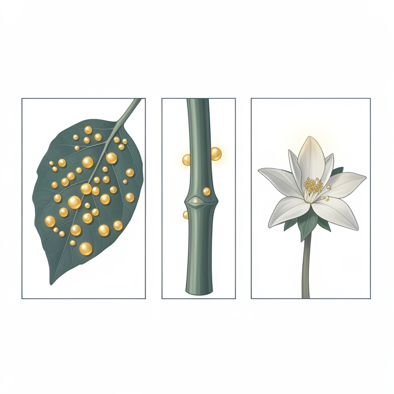
Sterol Distribution in Moringa oleifera
AI-generated conceptual illustration (Moringa oleifera plant). It explains the idea and does not show study data.
View source evidence ↗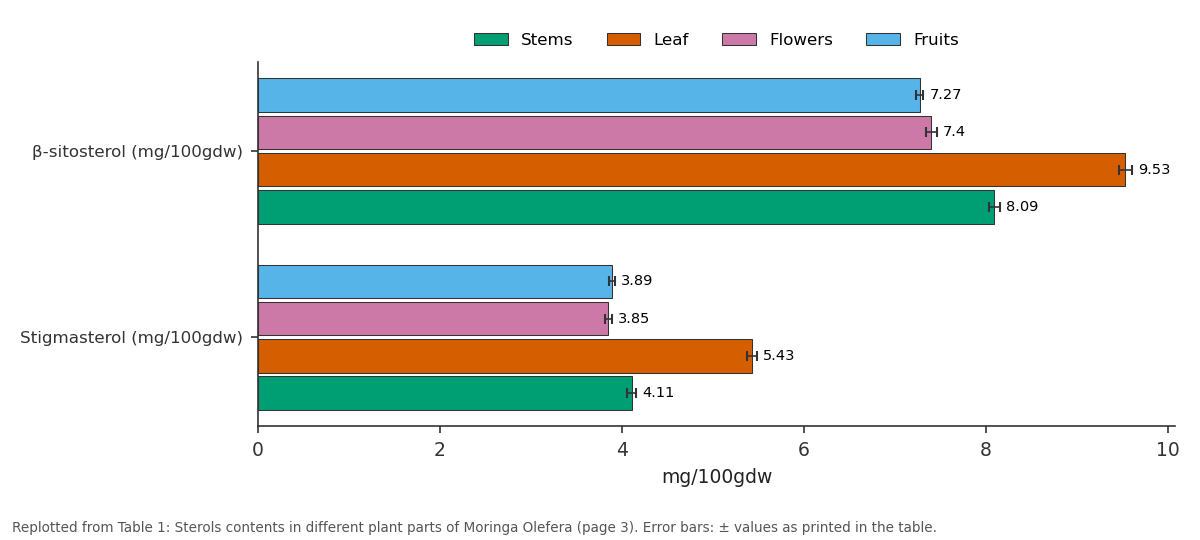
Stems and Leaf and more: β-sitosterol (mg/100gdw) vs Stigmasterol (mg/100gdw)
Replotted from Table 1 (page 3) of the original paper. Every bar is the paper's own table value.
View source evidence ↗Figures & tables
From the original paper Table 1
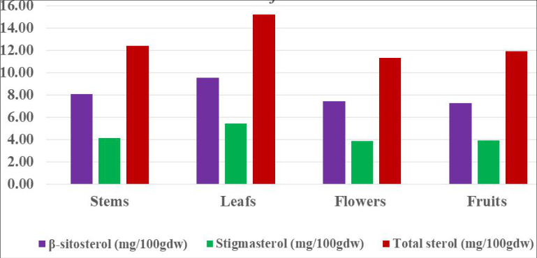 Figure 1
Introduction
A wide range of low molecular weight organic molecules, known as secondary metabolites, with typically complex and distinctive structures can be synthesized by plants. Secondary metabolites, which may be limited to a specific taxonomic group, are substances that are biosynthetically generated from primary metabolites. Previously, these were believed to be first-waste materials, but the function of secondary products has been rather unclear. Secondary metabolites from plants are now thought to be an enormous source of pharmacological value for both clinical and scientific research. Their biological actions are very therapeutic and useful in medication development, health care, and the manufacture of advantageous chemicals. One family of secondary metabolites with practical economic applications is steroids. The texture of steroids is greasy, soapy, or waxy, and they dissolve better in oil than in water. Sterols and sapogenins are among them. The human is the most well-known animal sterol cholesterol. β-sitosterol, camp sterol, and stigmasterol are the most prevalent phytosterols.
Studies have demonstrated the potential of phytosterols to treat a range of illnesses and that they are vital nutrients that guarantee the body's defence systems operate at their best (Ebner MJ et.al. 2006 and Heftmann E. (Ed.), 1973). Because plant sterols compete with cholesterol for absorption sites, they lower the amount of cholesterol absorbed, which is why phytosterols have been proven to be useful in treating high cholesterol. Additionally, phytosterols have an anti-inflammatory impact and are used to treat prostatitis and enlarged prostates. The plant Moringa oleifera, which belongs to the Moringaceae family, has been chosen to produce sterols. The primary goal of this research is to support efforts to produce sterols from commercially available plant parts (Singh D, Nag TN., 1981 andSinghvi S. 1980).
Methods of estimation
Fresh M. oleifera plant pieces were gathered from various Bikaner localities. Plant components were separated, allowed to dry in the shade, and their phytosterol content was examined. For every plant part, five duplicates were obtained, and the mean value was determined. Using seeds as explants, unorganized cultures with abundant callusing were created in M. oleifera on Murashige and Skoogs medium supplemented with 1.5 mg/L BAP+1.5 mg/L 2,4-D. These cultures were kept alive for six months by subculturing them frequently every six to eight weeks at 26 +/-1 °C, 55% relative humidity, and diffused light (3000 lux). After 15 minutes of drying at 100 °C to deactivate enzymes, tissues were next dried at 60 °C until their weight remained constant. To get the mean value, five of these replicates were examined. Using the method of (Kaul and Staba, 1968), the phytosterol content of plant parts and tissue samples was examined independently.
Processes for Sterols production
Using a water bath, tissue samples were hydrolysed for four hours using 30% (v/v) hydrochloric acid (2 gm/20 ml). Before the filtrate reached pH 7.0, the hydrolyzed test samples were individually cleaned with distilled water. The resulting test samples were successively soxhlet extracted in 200 ml of benzene for 24 hours and dried at 60 °C for 8 hours. Different test samples' benzoene extracts were independently dried in a vacuum and then collected in chloroform for additional examination.
Qualitative processes
Thin Layer Chromatography (TLC): The standard reference samples of sterols (β-sitosterol, campesterol, lanosterol, and stigmasterol) were applied separately to thin (0.2-0.3 mm) glass plates coated with silica gel "G" and activated. Following air drying and a 50% sulfuric acid spray, the plates were developed in an organic solvent mixture consisting of hexane and acetone (80:20 v/v), and then heated for ten minutes at 100 °C. There were two purple spots in every sample that matched those of the standard βsitosterol (Rf 0.60) and stigmasterol (Rf 0.64). Thin-layer preparation chromatography (PTLC): The standard reference sterols and each extract were applied independently to silica gel "G" activated glass plates that were thickly covered (0.3-0.4 mm). Hexane and acetone (80:20) were used as an organic solvent mixture to develop the plates. After being allowed to air dry, the produced plates were examined under a UV lamp. Together with silica gel from unsprayed plates, the two fluorescent spots (Rf 0.60 and Rf 0.64), which corresponded to those of the standard reference samples of β-sitosterol and stigmasterol, respectively, were marked and collected separately. After that, each combination was eluted using chloroform, dried in a vacuum, and crystallized independently using methanol and acetone. Together with their corresponding standard reference sterols, the crystallized isolates from every sample tested were put through colorimetry (for quantitative estimation), melting point (using a Toshniwal, India melting point apparatus), and infra-red spectrophotometer (using a Perkin-Elmer, 337, Grating, Infra-red spectrophotometer, employing nujol or potassium bromide pellets) analyses.
Quantitative processes β-sitosterol and stigmasterol test samples were quantitatively estimated using (Das and Banerjee's, 1980) approach. A stock solution containing 500 µg/ml of βsitosterol and stigmasterol in chloroform was made independently. On silica gel "G" coated and activated plates, six concentrations (0.1, 0.2, 0.3, 0.4, 0.5, and 0.6 ml) were made from this solution and spotted. Hexane:acetone (80:20) was the solvent system used to develop the plates. After air drying, these generated chromatograms were subjected to iodine fumes. After marking the iodine-positive spots, the surplus iodine was evaporated by heating them. Following the scraping of the spots and the addition of silica gel, each was eluted in test tubes using five milliliters of chloroform. Every tube was centrifuged, the supernatants were gathered, dried by evaporation, and then the process continued. Five of these repetitions were run for each concentration, and the average optical density was plotted against each concentration to create a regression curve that complied with Beer's law. On silica gel "G" coated and activated glass plates, the crude extracts from different plant sections and culture samples were separately diluted in chloroform, spotted alongside reference β-sitosterol and stigmasterol, and developed in an 80:20 hexane: acetone solvent system. All of the corresponding samples had two spots that matched those of the real β-sitosterol and stigmasterol samples. As previously mentioned, each of these locations was eluted and extracted. Elutes underwent the previously described processing after being dried and dissolved in five millilitres of chloroform. By comparing the optical density of the experimental sample with the regression curves of β-sitosterol and stigmasterol individually, the concentration of each sterol contained in different tissue samples of M. oleifera was determined (in mg/g.d.w.).
Results
β-sitosterol and stigmasterol were found in all plant parts and disorganized tissues of M. oleifera. Regarding the quantitative estimation of sterol content in various M. oleifera plant sections. The leaf had the highest levels of βsitosterol and stigmasterol i.e. 9.53 and 5.40 (mg/100gdw) respectively, whereas flowers had the lowest level i.e. 7.40 and 3.85 (mg/100gdw) respectively (Table .1 and Fig. 1). The total sterol content was lowest in flowers i.e. 11. 31 mg/100g.d.w. and highest in leaf i.e. 15.40 mg/100g.d.w. (Table .1 and Fig. 1).
Discussion
Sterols content in moringa plant parts: Moringa oleifera is a highly prized plant with a remarkable array of therapeutic applications and a high nutritional content. Numerous references to traditional medicine attest to its therapeutic efficacy, and scientific evidence of these widely accepted applications is beginning to substantiate at least some of the claims made by the scientific literature regarding the antibiotic, anti-trypanosomally, hypotensive, antispasmodic, antiulcer, anti-inflammatory, hypercholesterolaemic, and hypoglycaemic properties of moringa preparations. Compounds can undergo additional purification and perhaps be used in animal research. The current investigation demonstrated that all plant sections and M. oleifera tissue cultures have enough amounts of phytosterols, which are thought to be precursors of growth and reproductive hormones. By adding different sterol precursors to MS medium, the amount of sterols in tissue culture can be further increased based on the requirements.
Figures and tables from the paper
| Sterols | Stems | Leaf | Flowers | Fruits |
|---|---|---|---|---|
| β-sitosterol (mg/100gdw) | 8.09±0.06 | 9.53±0.07 | 7.40±0.06 | 7.27±0.04 |
| Stigmasterol (mg/100gdw) | 4.11±0.05 | 5.43±0.05 | 3.85±0.04 | 3.89±0.03 |
| Total sterol (mg/100gdw) | 12.40±0.05 | 15.40±0.05 | 11.31±0.05 | 11.90±0.04 |
Table text extracted from the original PDF; superscript letters are shown inline. The original table image is below.
View original table image
References
6 in the original paper- Das SK, Banerjee AB. A rapid method for quantification of sterols after thin layer chromatography. Indian J Exp Biol. 1980;18:969-971.
- Ebner MJ, Corol DI, Havlikava H, Honour JW, Fry JP. Identification of neuroactive steroids and their precursors and metabolites in adult male rat brain. Endocrinology. 2006;147(1):179-190.
- Heftmann E, editor. Modern methods of steroids analysis. New York: Academic Press; 1973.
- Kaul B, Staba EJ. Dioscorea tissue cultures -I: Biosynthesis and isolation of diosgenin from Dioscorea deltoidea callus and suspension cells. Lloydia. 1968;31:171-179.
- Singh D, Nag TN. Steroidal components of seeds of Peganum harmala growing in Rajasthan. Comp Phys Ecol. 1981;6(3):163-164.
- Singhvi S. Production of sapogenins, steroidal alkaloids and some other products from in vivo and in vitro tissue culture of some Solanaceous and Compositae plants [PhD thesis]. Jaipur: University of Rajasthan; 1980.
Cited by 2
From OpenAlex, OpenCitations and reference lists read by Scinovex
- Estimation of vitamins content in different parts of Moringa oleifera Lam. in Chhatarpur district (MP) ↗
Nidhi, PK Khare · International Journal of Pharmacology and Clinical Research · 2025
The "miracle tree" or "tree of life," Moringa oleifera, is prized for its many medicinal uses and amazing nutritional composition. Traditionally uses have been found for almost every part of the plant, including the leaves, pods, seeds, flowers, roots, and bark. In this research work also critically analysed Moringa oleifera plant parts for medicinal and nutritional properties to cure the
- The major woman diseases in Chhatarpur District and there management by use of Moringa oleifera plant parts ↗
Nidhi, PK Khare · International Journal of Pharmaceutical Sciences and Drug Analysis · 2025
Based on my survey, state-level data, and some other general health studies that cover Chhatarpur, these are some of the main illnesses and health problems impacting women in the Chhatarpur district (Madhya Pradesh). Anaemia (low hemoglobin/iron deficiency); According to NFHS-5 data, more than half of the state's women between the ages of 15 and 49 were anemic. Reproductive Health Challenges: