
Research article · 2025 · Full text
International Journal of Biology SciencesAnalytical techniques for assessing microplastic-induced physiological damage in tissues
Volume 7(6) · Pages 218–222 · DOI: 10.33545/26649926.2025.v7.i6d.378 ↗
Research areasMicroplastics and Plastic Pollution
Abstract
Author-writtenMicroplastic contamination is a growing environmental concern with potential health risks. This study aims to investigate the physiological effects of microplastics and develop strategies to mitigate their impact. The research utilizes advanced analytical techniques to identify, visualize, and characterize microplastics in tissues. Methods such as FTIR and Raman spectroscopy, X-ray Micro-CT, CLSM, LM, TEM, SEM, Fluorescence Microscopy, IHC, and biochemical assays are employed. The findings reveal that microplastics can induce oxidative stress, inflammation, and cellular damage, which may lead to various pathologies. The implications of this research lie in understanding the complex mechanisms underlying microplastic toxicity and developing effective environmental and public health policies. Standardizing protocols and establishing dose-response relationships are suggested for future research. Overall, this study contributes to the comprehension of analytical techniques to analyse microplastic-induced physiological damage and its implications for human health.
Summary
Source-checkedPlain-language reading of the full paper, prepared with AI and checked against the text. Each point links to the authors' own words.
Why it matters
- Microplastic contamination involves plastic fragments smaller than 5 mm and has become a significant environmental problem. View evidence ↗
- These particles are found in a wide range of organisms, from plankton to humans, where they can disrupt biological processes and cause physiological damage. View evidence ↗
- Interaction between microplastics and tissues can lead to inflammation, oxidative stress, and cell damage, which are linked to pathologies like cardiovascular diseases, cancer, and neurodegenerative diseases. View evidence ↗
The question
- The study aims to investigate how microplastics affect physiology and to develop strategies for mitigating these impacts. View evidence ↗
What they did
- The authors review a suite of analytical techniques, including various forms of microscopy (LM, TEM, SEM, CLSM, Fluorescence), spectroscopy (FTIR, Raman), X-ray Micro-CT, IHC, and biochemical assays. View evidence ↗
- FTIR and Raman spectroscopy are used specifically to identify the chemical composition and characterize microplastics in tissue samples. View evidence ↗
- X-ray Micro-CT is utilized for non-destructive 3D visualization to examine the morphology and spatial distribution of microplastics. View evidence ↗
- Light microscopy combined with H&E staining is used as an initial step to assess tissue morphology and identify damage. View evidence ↗
What they found
- Micro-FTIR and FTIR imaging can analyze microplastics in tissue sections as small as 10-20 μm. View evidence ↗
- Raman spectroscopy can detect smaller microplastics, analyzing particles as small as 1 μm with minimal sample preparation. View evidence ↗
- The review finds that microplastics can cause cellular damage, inflammation, and oxidative stress, potentially leading to various diseases. View evidence ↗
- Lipid peroxidation, a sign of oxidative stress, is quantitatively assessed using the TBARS assay by measuring MDA-TBA adducts at 532 nm. View evidence ↗
- Elevations in the cytokines TNF-α and IL-1β are indicators of acute inflammatory responses to microplastic exposure. View evidence ↗
- An increase in IL-6 levels may indicate the presence of chronic inflammation. View evidence ↗
What it means
- The authors conclude that integrating multiple analytical techniques is essential to fully understand microplastic-tissue interactions and the resulting molecular and cellular responses. View evidence ↗
- The authors state that future research needs to standardize protocols, create more sensitive detection methods, and establish dose-response relationships. View evidence ↗
Visual summary
1 visualsIllustrations explain the paper; they do not show study data.
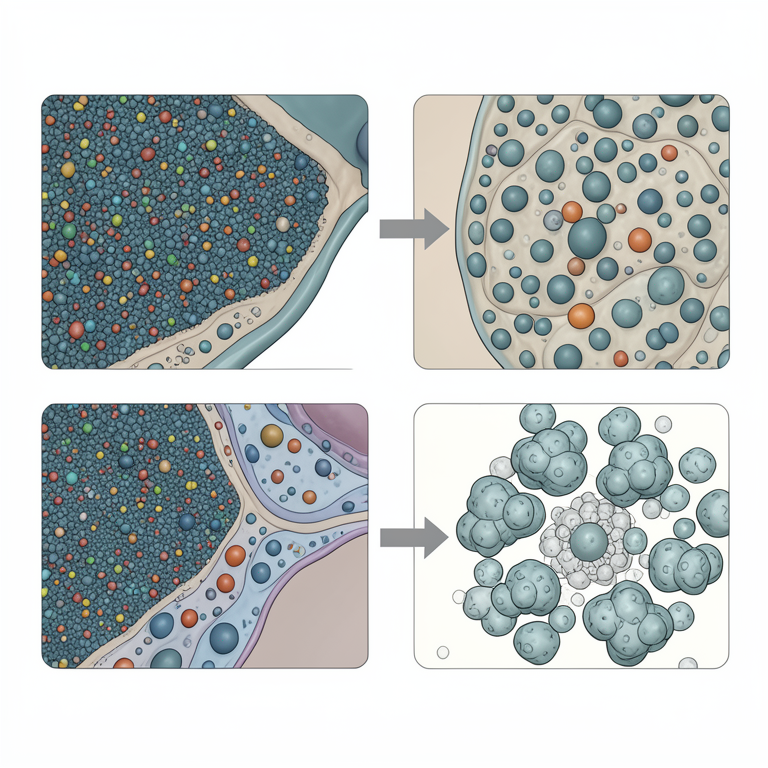
Integrative Analytical Workflow
AI-generated conceptual illustration (fish liver tissue containing microplastic particles). It explains the idea and does not show study data.
View source evidence ↗Introduction
Microplastic contamination, as represented by fragments of plastic of less than 5 mm in size, has been an emergent environmental issue. These particles are of diverse origin such as the breakdown of larger plastic material, personal cosmetics, and chemical fibers, and have become ubiquitous in marine as well as terrestrial environments (Ahmed, M. B., et al., 2021; Andrady, A. L. 2017) [2,6] . Microplastics have been reported to be present in an extremely diverse array of organisms ranging from plankton to the human being and present serious issues regarding their capacity to interfere with biological processes and induce physiologic damage. The persistent presence of such particles in the environment, coupled with their tendency to adsorb toxic chemicals, further contributes to their potential toxicities. (Al-Thawadi, S. 2020; Anbumani, S., et al., 2018) [3,5] . The widespread occurrence of microplastics in biological systems emphasizes the necessity of thoroughly investigating their physiological effects. Microplastics can interact with tissues and organs, causing oxidative stress, inflammation, and cell damage. These effects can interfere with normal cellular processes and are associated with different pathologies, such as cancer, neurodegenerative diseases, and cardiovascular diseases. Moreover, the small size of microplastics facilitates their passage through biological barriers, thereby posing the risk of accumulation in vital organs and impairing their function. Thus, the investigation of physiological effects of microplastics is essential for the development of effective strategies to counteract their adverse effects on health. (Geyer, R., et al., [15,47,36,7] . Analytical techniques are required for the assessment of the nature and degree of tissue injury caused by microplastics. These methods enable investigators to visualize and measure structural changes, identify markers of inflammation and oxidative stress, and characterize the localization and composition of microplastics in tissues. By providing critical information regarding cellular and molecular damage mechanisms of microplastic toxicity, these methodologies are crucial for further progress in comprehending health hazards of microplastic exposure. The following sections outline various analysis techniques that make International Journal of Biology Sciences https://www.biologyjournal.net up an integrative toolkit to investigate microplastic-induced physiological damage.
Microplastic Localization, Visualization, Identification, and Characterization in Tissues
Advanced analytical methods enable Identification of microplastics within biological samples and precise characterization of microplastics in tissues, providing information about their chemical composition, size, shape, and distribution. Fourier Transform Infrared Spectroscopy (FTIR) and Raman Spectroscopy are analytical techniques used to identify and characterize the chemical composition of microplastics within tissues. These methods rely on the interaction of infrared or Raman light with the material to produce characteristic spectra, which can be used to identify specific polymers (Xu, J. L., et al., 2019) [48] . FTIR Spectroscopy is technique to measures the absorption of infrared radiation by the sample, producing a spectrum that serves as a molecular fingerprint. FTIR can identify different types of plastics (e.g., polyethylene, polystyrene, polyvinyl chloride) based on their characteristic absorption bands (Rathore, C., et al., 2023) [38] . Micro-FTIR and FTIR imaging enable the analysis of microplastics as small as 10-20 μm within tissue sections (Mikulec, V., et al., 2023) [33] .
Complementary to FTIR, Raman spectroscopy measures the inelastic scattering of monochromatic light by the sample. It provides information about molecular vibrations and can identify plastic polymers with high specificity. Raman has the advantage of minimal sample preparation and can analyze particles as small as 1 μm, making it suitable for detecting smaller microplastics (Käppler, A., et al., 2016) [23] . X-ray Microtomography (Micro-CT) is an imaging technique that provides 3D visualization of microplastics within tissues and allows researchers to examine the spatial distribution and morphology of microplastics without damaging the sample. Micro-CT generates high-resolution 3D images by taking multiple X-ray projections of the sample from different angles and reconstructing them computationally. This technique can reveal the threedimensional distribution of microplastics within organs and tissues, providing insights into their accumulation patterns and potential migration pathways. The non-destructive nature of Micro-CT allows for subsequent analyses of the same sample using other techniques, enabling multi-modal characterization of microplastic-tissue interactions (Parobková, V., et al., 2025; de Paiva Teles, Á., et al., 2024) [34, 11] . Confocal Laser Scanning Microscopy (CLSM) is a powerful imaging technique that provides high-resolution, 3D images of microplastics within tissues (Li, L., Luo, Y., et al., 2020) [26] . CLSM is particularly useful for studying the interactions between microplastics and cellular structures, as well as for tracking the movement of microplastics within living organisms allowing for the real-time observation of microplastic uptake, intracellular trafficking, and potential exocytosis. By using optical sectioning, CLSM eliminates out-of-focus light, resulting in sharper images with improved contrast compared to conventional fluorescence microscopy. This technique can be combined with fluorescent labelling of microplastics and cellular structures to visualize their spatial relationships with subcellular resolution (Zhu, Y., et al., 2024) [49] .
Examining tissue morphology and assessing microplastic-induced damage
Light Microscopy (LM) and Haematoxylin & Eosin (H&E) staining are fundamental techniques for examining tissue morphology and serve as the first step in assessing microplastic-induced damage. LM provides an overall view of tissue architecture, while H&E staining differentiates between cellular components, highlighting structural changes, inflammation, and necrosis. Haematoxylin stains nucleus blue, while eosin stains cytoplasm pink, allowing for clear visualization of tissue organization and cellular abnormalities (Abdel-Zaher, S., et al., 2023) [1] . In tissues exposed to microplastics, H&E staining can reveal inflammatory cell infiltration, tissue fibrosis, necrotic areas, and alterations in cell morphology. These histopathological changes provide valuable insights into the severity and nature of tissue damage, establishing a foundation for more specialized analyses (Hamed, M., et al., 2021) [17] . Building upon the observations from light microscopy, Transmission Electron Microscopy (TEM) and Scanning Electron Microscopy (SEM) provide high-resolution images of tissue ultrastructure. TEM is particularly useful for detecting intracellular localization of microplastics and examining subcellular damage, including alterations in organelles, membrane integrity, and cytoskeletal organization (Trianni, A., et al., 2024) [42] . SEM offers detailed surface morphology of tissues, revealing physical interactions between microplastics and cellular surfaces (Wang, Z. M., et al., 2017) [46] . These advanced imaging techniques can identify subtle changes that are not visible with conventional light microscopy, such as disruption of cell junctions, alterations in mitochondrial structure, and changes in endoplasmic reticulum morphology. By providing nanometer-scale resolution, TEM and SEM are invaluable for elucidating the mechanisms of microplastic-induced cellular damage (Jeong, C. B., et al., 2016) [22] . Fluorescence Microscopy complements electron microscopy by enabling the visualization of microplastics within tissues through fluorescent labelling. This technique is particularly valuable for tracking the distribution and movement of microplastics in samples. Many plastic polymers exhibit autofluorescence, but specific fluorescent dyes like Nile Red can enhance detection sensitivity and specificity (Maes, T., et al., 2017; Karakolis, E. G., et al., 2019) [31,24] . By using different fluorescent markers, researchers can simultaneously visualize microplastics and cellular structures, allowing for the assessment of spatial relationships and potential interactions. Time-lapse fluorescence microscopy can also be employed to study the dynamic processes involved in microplastic uptake, translocation, and elimination in living tissues (Karakolis, E. G., et al., 2019) [24] . Moving from structural to functional analyses, Immunohistochemistry (IHC) and Immunofluorescence (IF) are techniques used to identify specific protein markers in tissues. These methods involve the use of antibodies to detect proteins associated with inflammation, oxidative stress, and apoptosis. By visualizing these markers, researchers can assess the biological responses to microplastic exposure and understand the underlying mechanisms of tissue damage (Gaspar, L., et al., 2023) [14] . In microplastic toxicology studies, Inflammatory markers (CD68 for macrophages, myeloperoxidase for neutrophils) (Pechiappan, H. 2021; Shao, W., et al., 2024) [35,39] , and apoptotic markers (cleaved caspase-3, TUNEL staining) (Wan, S., et al., 2024; Lu, T., et al., 2024) [44,30] can also be used.
Measuring Oxidative Stress in Microplastic-Exposed Tissues
Once cellular and tissue damage is identified through microscopic techniques, biochemical assays provide quantitative measurements of the molecular changes induced by microplastics. Oxidative stress is a primary mechanism through which microplastics exert their toxic effects, making its assessment crucial for understanding microplastic-induced damage. [22,19,18,9] . Oxidative stress can cause protein carbonylation, which is measured using 2,4-dinitrophenylhydrazine (DNPH) derivatization followed by spectrophotometric analysis. Increased protein carbonyl content indicates protein oxidation and potential functional impairment (Dalle-Donne, I., et al., 2003; Michailidou, K., et al., 2024) [10,32] . Comet Test (Single Cell Gel Electrophoresis) is assay to visualize DNA damage in individual cells. Cells are embedded in agarose, lysed, and subjected to electrophoresis. Damaged DNA migrates further, creating a "comet tail" whose length and intensity correlate with the degree of DNA damage. The Comet Test can detect single and double-strand breaks, providing a sensitive measure of genotoxicity induced by microplastics (Tang, K. H. D. 2025; Berber, A. A. 2019; Farag, A. A., et al., 2023) [41,8,13] . TUNEL Assay is a technique that identifies apoptotic cells by labeling the terminal end of nucleic acids. The enzyme terminal deoxynucleotidyl transferase (TdT) adds dUTPs to the 3'-OH ends of fragmented DNA, which can then be visualized using various detection methods. The TUNEL Assay detects apoptotic cell death by labeling fragmented DNA allowing for the assessment of tissue-specific vulnerability to microplastic-induced damage (Lu, C., et al., 2023; Li, T., et al., 2024) [29,27] . 8-OHdG Assay is a technique in which 8-OHdG is a product of DNA oxidation and a biomarker of oxidative DNA damage. It can be measured using ELISA or HPLC techniques. Elevated 8-OHdG levels suggest potential mutagenic and carcinogenic effects of microplastic exposure (Sincihu, Y., et al., 2023; Ding, R., et al., 2024) [40,12] .
Antioxidant Enzymes: superoxide dismutase, catalase, glutathione s-transferase, and glutathione peroxidase (GPx) and glutathione (GSH) assays assess the activity of enzymes involved in the antioxidant defense mechanisms. SOD catalyzes the dismutation of superoxide radicals, CAT decomposes hydrogen peroxide, and glutathione (GSH) acts as a cellular redox buffer. Changes in the activity of these enzymes reflect the cellular response to oxidative stress induced by microplastics. For instance, initial increases in SOD and CAT activities may indicate an adaptive response, while decreased activities after prolonged exposure suggest antioxidant system depletion (Jaikumar, I.M., et al., 2021; Köktürk, M., et al., 2024) [21,25] .
Assessment of Inflammatory response due to microplastic exposure
The Enzyme-Linked Immunosorbent Assay (ELISA) is used to measure the levels of inflammatory cytokines such as IL-6, TNF-α, and IL-1β. These cytokines are key mediators of inflammation and their elevated levels indicate an inflammatory response to microplastic exposure. ELISA provides quantitative data on cytokine concentrations, allowing for the assessment of inflammation severity and progression (Grellner, W., et al., 2000; Liu, C., et al., 2021) [16, 28] . In microplastic toxicology, specific cytokine profiles can indicate different pathological processes:
• IL-1β and TNF-α elevations suggest acute inflammatory responses (Pechiappan, H. 2021) [35] . • IL-6 increases may indicate chronic inflammation (Pulvirenti, E., et al., 2022) [37] • TGF-β upregulation can signal fibrotic changes (Wang, S., et al., 2023) • IL-10 alterations reflect attempts to regulate inflammatory processes (Pechiappan, H. 2021) [35] .
The temporal patterns of cytokine expression can provide insights into the evolution of the inflammatory response to microplastic exposure (Hwang, J., et al., 2020) [20] .
Conclusion
The Metabolic changes caused by microplastics. The integration of these techniques allows assessment of microplastic-tissue interactions, from the identification, visualization, localization and characterization of microplastics to the evaluation of cellular and molecular responses. This multifaceted approach is essential for understanding the complex mechanisms underlying microplastic toxicity. Future research directions should focus on standardizing analytical protocols, developing more sensitive detection methods, and establishing dose-response relationships between microplastic exposure and physiological effects. Additionally, studies examining the long-term consequences of chronic microplastic exposure are needed to fully understand the health implications of this pervasive environmental pollutant. By employing these analytical techniques, researchers can better understand the health risks associated with microplastic pollution and develop strategies to mitigate their impact on biological systems, ultimately contributing to more effective environmental and public health policies.
References
48 in the original paper- Abdel-Zaher S, Mohamed MS, Sayed AEDH. Hemotoxic effects of polyethylene microplastics on mice. Front Physiol. 2023;14:1072797.
- Ahmed MB, Rahman MS, Alom J, Hasan MS, Johir MAH, Mondal MIH, et al. Microplastic particles in the aquatic environment: a systematic review. Sci Total Environ. 2021;775:145793.
- Al-Thawadi S. Microplastics and nanoplastics in aquatic environments: challenges and threats to aquatic organisms. Arab J Sci Eng. 2020;45(6):4419-40.
- Ammann R, Tanner C, Schulz G, Osmani B, Nalabothu P, Töpper T, et al. Three-dimensional analysis of aligner gaps and thickness distributions, using hard xray tomography with micrometer resolution. J Med Imaging. 2022;9(3):031509.
- Anbumani S, Kakkar P. Ecotoxicological effects of microplastics on biota: a review. Environ Sci Pollut Res. 2018;25:14373-96.
- Andrady AL. The plastic in microplastics: A review. Mar Pollut Bull. 2017;119(1):12-22.
- Avio CG, Gorbi S, Regoli F. Plastics and microplastics in the oceans: From emerging pollutants to emerged threat. Mar Environ Res. 2017;128:2-11.
- Berber AA. Genotoxic evaluation of polystyrene microplastic. Sakarya Univ J Sci. 2019;23(3):358-67.
- Dalle-Donne I, Rossi R, Giustarini D, Milzani A, Colombo R. Protein carbonyl groups as biomarkers of oxidative stress. Clin Chim Acta. 2003;329(1-2):23-38.
- de Paiva Teles Á, da Silva MVS, de Oliveira DF, Lopes RT. High resolution X-ray microtomography as a tool for observation and classification of individual microplastics. Braz J Radiat Sci. 2024;12(4A Suppl.):e2631.
- Ding R, Chen Y, Shi X, Li Y, Yu Y, Sun Z, et al. Sizedependent toxicity of polystyrene microplastics on the gastrointestinal tract: oxidative stress-related DNA damage and potential carcinogenicity. Sci Total Environ. 2024;912:169514.
- Farag AA, Youssef HS, Sliem RE, El Gazzar WB, Nabil N, Mokhtar MM, et al. Hematological consequences of polyethylene microplastics toxicity in male rats: oxidative stress, genetic, and epigenetic links. Toxicology. 2023;492:153545.
- Gaspar L, Bartman S, Coppotelli G, Ross JM. Acute exposure to microplastics induced changes in behavior and inflammation in young and old mice. Int J Mol Sci. 2023;24(15):12308.
- Geyer R, Jambeck JR, Law KL. Production, use, and fate of all plastics ever made. Sci Adv. 2017;3(7):e1700782.
- Grellner W, Georg T, Wilske J. Quantitative analysis of proinflammatory cytokines (IL-1β, IL-6, TNF-α) in human skin wounds. Forensic Sci Int. 2000;113(13):251-64.
- Hamed M, Soliman HA, Badrey AE, Osman AG. Microplastics induced histopathological lesions in some tissues of tilapia (Oreochromis niloticus) early juveniles. Tissue Cell. 2021;71:101512.
- Hamed M, Soliman HA, Osman AG, Sayed AEDH. Antioxidants and molecular damage in Nile Tilapia (Oreochromis niloticus) after exposure to microplastics. Environ Sci Pollut Res. 2020;27:14581-8.
- Hsieh SL, Wu YC, Xu RQ, Chen YT, Chen CW, Singhania RR, et al. Effect of polyethylene microplastics on oxidative stress and histopathology damages in Litopenaeus vannamei. Environ Pollut. 2021;288:117800.
- Hwang J, Choi D, Han S, Jung SY, Choi J, Hong J. Potential toxicity of polystyrene microplastic particles. Sci Rep. 2020;10(1):7391.
- Jaikumar IM, Periyakali SB, Rajendran U, Joen-Rong S, Thanasekaran J, Tsorng-Harn F. Effects of microplastics, polystyrene, and polyethylene on antioxidants, metabolic enzymes, HSP-70, and myostatin expressions in the giant river prawn Macrobrachium rosenbergii: impact on survival and growth. Arch Environ Contam Toxicol. 2021;80:64558.
- Jeong CB, Won EJ, Kang HM, Lee MC, Hwang DS, Hwang UK, et al. Microplastic size-dependent toxicity, oxidative stress induction, and p-JNK and p-p38 activation in the monogonont rotifer (Brachionus koreanus). Environ Sci Technol. 2016;50(16):8849-57.
- Käppler A, Fischer D, Oberbeckmann S, Schernewski G, Labrenz M, Eichhorn KJ, et al. Analysis of environmental microplastics by vibrational microspectroscopy: FTIR, Raman or both?. Anal Bioanal Chem. 2016;408:8377-8391.
- Karakolis EG, Nguyen B, You JB, Rochman CM, Sinton D. Fluorescent dyes for visualizing microplastic particles and fibers in laboratory-based studies. Environ Sci Technol Lett. 2019;6(6):334-40.
- Köktürk M, Özgeriş FB, Atamanalp M, Ucar A, Özdemir S, Parlak V, et al. Microplastic-induced oxidative stress response in turbot and potential intake by humans. Drug Chem Toxicol. 2024;47(3):296-305.
- Li L, Luo Y, Peijnenburg WJ, Li R, Yang J, Zhou Q. Confocal measurement of microplastics uptake by plants. MethodsX. 2020;7:100750.
- Li T, Bian B, Ji R, Zhu X, Wo X, Song Q, et al. Polyethylene terephthalate microplastic exposure induced reproductive toxicity through oxidative stress and p38 signaling pathway activation in male mice. Toxics. 2024;12(11):779.
- Liu C, Chu D, Kalantar-Zadeh K, George J, Young HA, Liu G. Cytokines: from clinical significance to quantification. Adv Sci. 2021;8(15):2004433.
- Lu C, Liang Y, Cheng Y, Peng C, Sun Y, Liu K, et al. Microplastics cause reproductive toxicity in male mice through inducing apoptosis of spermatogenic cells via p53 signaling. Food Chem Toxicol. 2023;179:113970.
- Lu T, Yuan X, Sui C, Yang C, Li D, Liu H, et al. Exposure to polypropylene microplastics causes cardiomyocyte apoptosis through oxidative stress and activation of the MAPK-Nrf2 signaling pathway. Environ Toxicol. 2024;39(12):5371-82.
- Maes T, Jessop R, Wellner N, Haupt K, Mayes AG. A rapid-screening approach to detect and quantify microplastics based on fluorescent tagging with Nile Red. Sci Rep. 2017;7(1):44501.
- Michailidou K, Palisidou C, Feidantsis K, Ainali NM, Kastrinaki G, Lambropoulou DA, et al. Impact of aged and virgin polyethylene microplastics on multi endpoints effects of freshwater fish tissues. Sci Total Environ. 2024;948:174704.
- Mikulec V, Adamović P, Cvetković Ž, Ivešić M, Gajdoš Kljusurić J. Green techniques for detecting microplastics in marine with emphasis on FTIR and NIR spectroscopy-short review. Processes. 2023;11(8):2360.
- Parobková V, Maleček L, Zemek M, Kalčíková G, Vykypělová M, Buchtová M, et al. Advancing microplastic detection in zebrafish with micro computed tomography: a novel approach to revealing microplastic distribution in organisms. J Hazard Mater. 2025;488:137442.
- Pechiappan H. Effects of nano-and microplastics on inflammatory responses in macrophages in vitro [master's thesis]. Trondheim: NTNU; 2021.
- Prata JC, Da Costa JP, Lopes I, Duarte AC, RochaSantos T. Environmental exposure to microplastics: an overview on possible human health effects. Sci Total Environ. 2020;702:134455.
- Pulvirenti E, Ferrante M, Barbera N, Favara C, Aquilia E, Palella M, et al. Effects of nano and microplastics on the inflammatory process: in vitro and in vivo studies systematic review. Front Biosci (Landmark Ed). 2022;27(10):287.
- Rathore C, Saha M, Gupta P, Kumar M, Naik A, de Boer J. Standardization of micro-FTIR methods and applicability for the detection and identification of microplastics in environmental matrices. Sci Total Environ. 2023;888:164157.
- Shao W, Weng Z, Liang J, Liu Q, Zhang H, Xu J, et al. Enterohepatic circulation of nanoplastics induced hyperplasia, epithelial-mesenchymal transition, and neutrophil extracellular traps in gallbladder. Nano Today. 2024;57:102353.
- Sincihu Y, Lusno MFD, Mulyasari TM, Elias SM, Sudiana IK, Kusumastuti K, et al. Wistar rats hippocampal neurons response to blood low-density polyethylene microplastics: a pathway analysis of SOD, CAT, MDA, 8-OHdG expression in hippocampal neurons and blood serum Aβ42 levels. Neuropsychiatr Dis Treat. 2023;73-83.
- Tang KHD. Genotoxicity of microplastics on living organisms: effects on chromosomes, DNA and gene expression. Environments. 2025;12(1):10.
- Trianni A, Brossa A, Catalano F, Saraceni A, Bongiovanni S, Merlo GR, et al. Unraveling the molecular mechanisms of microplastics internalization and their intracellular impact. Eur J Histochem. 2024;68(s1):47-8.
- Vethaak AD, Leslie HA. Plastic debris is a human health issue. ACS Publ. 2016;6825-6.
- Wan S, Wang X, Chen W, Wang M, Zhao J, Xu Z, et al. Exposure to high dose of polystyrene nanoplastics causes trophoblast cell apoptosis and induces miscarriage. Part Fibre Toxicol. 2024;21(1):13.
- Wang S, Chen L, Shi X, Wang Y, Xu S. Polystyrene microplastics-induced macrophage extracellular traps contributes to liver fibrotic injury by activating ROS/TGF-β/Smad2/3 signaling axis. Environ Pollut. 2023;324:121388.
- Wang ZM, Wagner J, Ghosal S, Bedi G, Wall S. SEM/EDS and optical microscopy analyses of microplastics in ocean trawl and fish guts. Sci Total Environ. 2017;603:616-26.
- Wright SL, Kelly FJ. Plastic and human health: a micro issue?. Environ Sci Technol. 2017;51(12):6634-47.
- Xu JL, Thomas KV, Luo Z, Gowen AA. FTIR and Raman imaging for microplastics analysis: state of the art, challenges and prospects. TrAC Trends Anal Chem. 2019;119:115629.
- Zhu Y, Li Y, Huang J, Zhang Y, Ho YW, Fang JK, et al. Advanced optical imaging technologies for microplastics identification: progress and challenges. Adv Photonics Res. 2024;5(11):2400038.