
Research article · 2025 · Full text
International Journal of Biology SciencesHistopathological study of the oviduct in female mice treated with levetiracetam
Volume 7(4) · Pages 105–110 · DOI: 10.33545/26649926.2025.v7.i4b.335 ↗
Research areasSperm and Testicular FunctionReproductive Biology and FertilityReproductive System and Pregnancy
Abstract
Author-writtenThe present study was conducted to investigate the effects of levetiracetam (LEV) at a concentration of 350 mg/kg/day of body weight on oviduct tissue in female albino mice (Mus musculus). The experimental animals were divided into three groups and administered orally once daily for 14, 30, and 45 days. Each treatment group was matched to its own control group. The results of the present study showed that ciliated columnar cells transformed into cuboidal cells, with reduced cilia density, shortened secretory cells, hemorrhage, necrosis, and cell death of the mucosal folds, decreased tissue hyperplasia with the appearance of vacuoles, the presence of inflammatory cells in the lumen, and hyperplasia and hypoplasia of the tunica muscularis when compared to the control group.
Summary
Source-checkedPlain-language reading of the full paper, prepared with AI and checked against the text. Each point links to the authors' own words.
Why it matters
- Levetiracetam (LEV) is a second-generation antiepileptic medication used as an alternative to valproic acid (VPA). View evidence ↗
- Some evidence suggests LEV may affect endocrine function and hormonal balance, such as altering progesterone or follicle-stimulating hormone (FSH) levels. View evidence ↗
The question
- The study aimed to identify and compare histopathological changes in the oviductal tissue of mice treated with LEV versus a control group. View evidence ↗
What they did
- The experiment used 40 female Balb/C Swiss mice, aged 8-12 weeks with an average weight of 24-25 grams. View evidence ↗
- Mice were split into four groups of 10: three treatment groups receiving LEV for 14, 30, and 44 days, and one control group receiving distilled water. View evidence ↗
- The LEV drug was administered orally at a daily dose of 350 mg/kg. View evidence ↗
- Oviduct samples were fixed in Bouin's solution, cut into 5-micrometer sections, and stained with hematoxylin-eosin for microscopic analysis of the annulus and isthmus. View evidence ↗
What they found
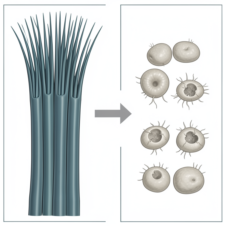
- LEV treatment caused a decrease in the height of ciliated epithelial cells, which shifted from columnar to cuboidal shapes. View evidence ↗
- The study observed hemorrhage and the presence of inflammatory and mononuclear cells within the oviduct's amniotic cavity and connective tissue. View evidence ↗
- Treatment led to the degeneration and disruption of mucosal folds, cell scattering, vacuolation, and areas of necrosis and cell death. View evidence ↗
- The smooth muscle tunica thinned as treatment duration increased, reaching a single row of cells in mice treated for 45 days. View evidence ↗
- The epithelial lining of mucosal folds showed significant damage, including hyperplasia, necrosis, karyolysis of nuclei, and a widespread loss of cilia. View evidence ↗
What it means
- The authors suggest that LEV may reduce estrogen levels, which are critical for maintaining the integrity of the ovarian wall. View evidence ↗
- The authors conclude that LEV causes extensive cell death, degradation, and loss of cilia in the oviductal tissue. View evidence ↗
Visual summary
1 visualsIllustrations explain the paper; they do not show study data.
Cellular Transformation in Oviduct
AI-generated conceptual illustration (female mouse oviduct epithelial cells). It explains the idea and does not show study data.
View source evidence ↗Figures & tables
From the original paper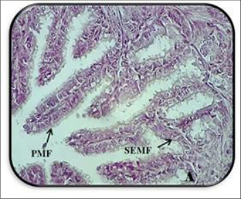 Figure 1
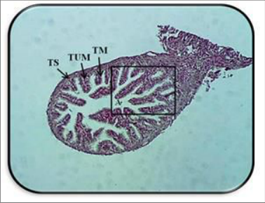 Figure 1
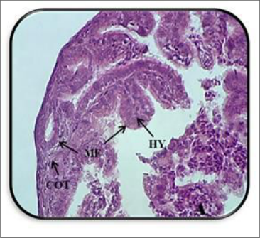 Figure 2
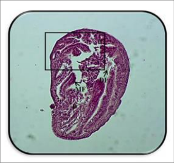 Figure 2
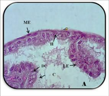 Figure 3
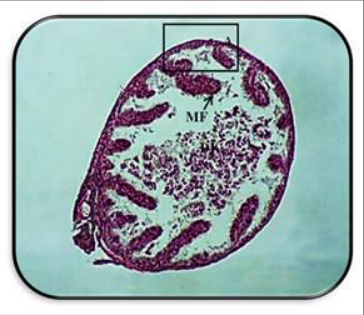 Figure 3
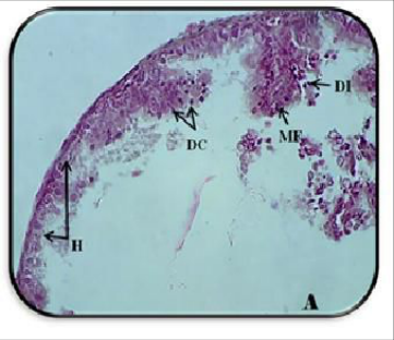 Figure 4
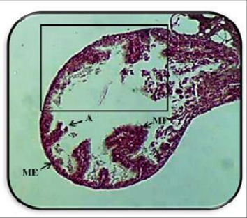 Figure 4
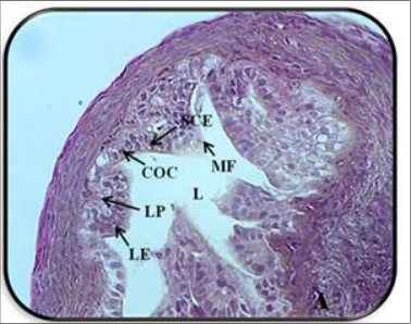 Figure 5
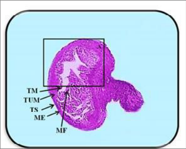 Figure 5
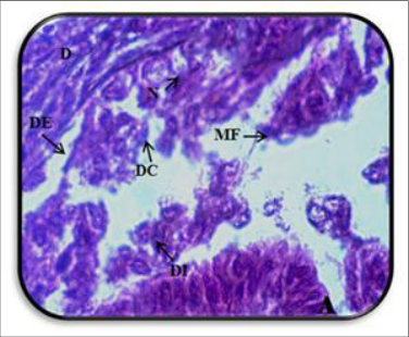 Figure 6
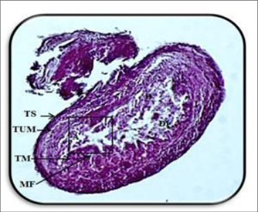 Figure 6
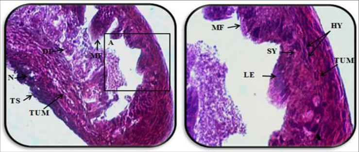 Figure 7
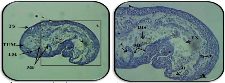 Figure 8
Introduction
Levetiracetam (LEV) is a new antiepileptic drug, widely used for its antiepileptic efficacy, offering an alternative to valproic acid (VPA) for many patients which discovered in the early 1980s. Safety data indicate that LEV is well tolerated by the vast majority of patients and has no endocrine side effects (Harden, 2001) [6] . In 1991, studies were reported clinical trials in patients with epilepsy, and it was considered an adjunctive treatment for the onset of partial seizures (Rossetti and Bromfiled, 2005; Garcia and Rubio, 2009) [11,3] . LEV was first approved by the US Food and Drug Administration (FDA) in 1991 as a new second-generation antiepileptic drug, and was approved for use as tablets in India in 2005 (Kavita et al., 2011) [8] . LEV which knows (Salpha-ethyl-2-oxo-1-pyrrolidine acetamide) also used to treat non-epileptic conditions including bipolar disorder and to prevent hemiplegia, myoclonus, and myoclonus. LEV as a single enantiomer, was reported by pharmacological studies "the therapeutic effects of antiepileptic drugs on sexual function since 1850, when bromides were introduced as a treatment for epilepsy". Later research showed the development of impotence and decreased libido in 11-22% of patients after initiating treatment with carbamazepine (CBZ), phenytoin (PHT), phenobarbital, or primidone. These drugs are known as older or first-generation drugs, as they were widely used. Second-generation drugs have gained favor due to their fewer side effects. However, several studies have demonstrated that the endocrine effects of valerian (VPA) in females occur in the ovary, where it acts as an enzyme inhibitor that prevents the conversion of testicular fat to estradiol (Perucca et al., 2004) [10] . This hypothesis has been supported by numerous studies on porcine ovarian follicle cells, where it exerts a significant sterologenic effect in both gonadotropin-stimulated and non-gonadotropinstimulated genital glands (Gregoraszczuk et al., 2000; Taubøll, 2003) [4,14] . High levels of testicular fat in the ovary inhibit the maturation of ovarian follicles and promote polycystic ovarian syndrome (PCOS) (Nandedkar and Munshi, 1981) [9] . (LEV) has been widely used for its antiepileptic efficacy, representing an alternative to VPA for many patients. Safety data indicate that LEV is well-tolerated and safe in many patients, and it has no endocrine side effects (Harden, 2001) [6] . However, it has an effect on basal steroid hormone secretion in ovarian follicular cells in prepubertal pigs, suggesting its effect on endocrine function and hormonal balance by increasing progesterone levels or decreasing follicle-stimulating hormone (FSH) levels (Gustavsen, 2008; Svalheim, 2010) [5,13] .
Thus, the aim of this study was to identify the histopathological changes in oviductal tissue resulting from the use of the drug and compare them with the control group.
Materials and Methods
Levetiracetam: In the current study, the drug Levetiracetam (LEV) was used. The drug was administered to mice orally at a concentration of 350 mg/kg/day. The dose was multiplied by the average mouse weight (28) g and the drug concentration in the package (100) mg/ml. The dose used in the experiment was (0.1) ml/day (FDA, 2013).
The study samples: consisted of 40 Mus musculus (Swiss mice) of the Balb/C strain, obtained from the Drug Control Center. The mice were 8-12 weeks old and weighed an average of 24-25 grams. The animals were divided into four groups, each containing 10 mice. The female animals were dosed as follows: the first group received a dose for 14 days, the second group for 30 days, and the third group for 44 days, while the control group was treated with distilled water. Vaginal swabs were taken from the females used in this study for two consecutive estrous cycles to ensure the integrity of the estrous cycle.
Animal house: which present in Department of Biology/College of Education for Pure Sciences (Ibn Al-Haitham) at the University of Baghdad plastic and that consist of cages covered with metal mesh lids, measuring 48 x 15 x 7 cm. The house containing wooden shore sawdust and tap water. They were supplied with standard food and were kept under standard laboratory conditions, with uniform temperature, lighting, and ventilation.
Samples of swab:
A swab was taken from the vaginal lining using a metal loop, which was sterilized over a flame until red, and then immersed in normal saline (NaCl). The swabs were placed on clean glass slides and left to dry. They were then stained with methylene blue for three minutes. The slides were then washed with top water and allowed to dry (Husman, 1967) [7] . They were then examined under a light compound microscope.
Anatomy process: After completing the treatment, each group was prepared to anatomy process, by fixing the animal's head and pulling the tail to make a longitudinal incision in the middle region of the lower abdomen up to the sternum of the female mic. The viscera were then removed, the oviduct was liberated and transferred to normal saline, and the surrounding fatty material was removed. The oviduct was also cleaned of the surrounding tissue, and its convolutions were opened so that they extended along its length. Then, the ampulla, isthmus, and uterus regions were separated. They were dried with filter paper and transferred to the fixative for the purpose of preparing microscopic slides (Butler and Jurrlink, 1987) [1] .
Microscopic studies: Microscope slides were prepared according to the method of Bancroft and Stevens (1982) [21] , where the oviduct was fixed in Bouin's solution for 24 hours, after which it was washed several times in a 70% ethanol solution. After that, the samples were passed through a series of ascending concentrations of ethyl alcohol starting from (70%, 100%) for half an hour, then the samples were incubated in xylene for 15 minutes, then the samples were placed in a mixture of xylene and molten paraffin wax at a temperature of (56-58) °C in a ratio of (1:1) for 15 minutes in a convection oven at a temperature of 60°C. After that, the samples were impregnated by placing them in the molten wax twice for an hour each time. Then the samples were stained in the center of the wax and left until the wax hardened. After that, the samples were cut with a thickness of 5 micrometers, and the transverse sections were carried in the oviduct for the regions (the annulus and the isthmus). The sections were stained with hematoxylin-eosin (Eosin-(hematoxylin) Histological and histopathological changes were studied using a compound imaging microscope of type M.
Results and Discussion
Regarding the histopathological changes in the amniotic cavity, the present results indicate that LEV causes histopathological changes in the amniotic cavity wall of animals treated with the drug for periods of 14, 30, and 45 days, respectively. These changes were characterized by a decrease in the height of the folds and a lack of branching into secondary and tertiary folds. According to the figure 1 and 2, the height of the ciliated epithelial cells decreased, as they appeared low columnar to cuboidal, with the nucleus at the apex of the cells, with an eccentric location. All sections were reported in the two figures 1 and 2, which was tunica mucosa TM, tunica muscularis TUM, tunica serosa TS, media mesothelium ME, vasa pellucida BV, columnar epithelium LE, lamina propria LP, columnar cells COC, secretory cells SCE, cilia C, mucosal fold MF, degeneration D, tear DI,and stained (H & E). The non-ciliated secretory cells appeared thin and columnar. Some of these folds extended into the connective tissue and sometimes into the tunica muscularis, especially in groups dosed for a long period. Furthermore, hemorrhage was found, along with the presence of mononuclear cells and inflammatory cells in the connective tissue and in the amniotic cavity of the oviduct. Degeneration and disruption of some mucosal folds were also observed, as well as the scattering of cells. The mucous membrane in the middle of the lumen of the duct, and the shape of the ciliated cells changed to resemble secretory cells, and the appearance of vacuolation within the epithelial tissue and clusters of epithelial cell nuclei in a syncytium with necrosis and cell death. These results are consistent with what Wordinger and Morrill (1985) [20] reached regarding the negative effect of the oviduct upon treatment with the hormone diethylstilbestrol (DES), which is used as a treatment for abortion, as it causes an increase in the number of epithelial cells lining the mucosal folds, and the nuclei of the cells become central and change to a location close to their apical edge. Newbold et al. (1983) [12] demonstrated that the epithelium lining the mucosal folds in the wall of the ampulla region of the oviduct of animals treated with diethylstilbestrol (DES) suffered from hyperplasia of the muscularis and serosa tunica. This is consistent with the present finding, as the mucosal folds extended into the muscularis layer in some ampulla sections.as shown in figure 5 and 6, the lamina propria, which consists of loose connective tissue and blood vessels, was affected and had less tissue in the groups treated with LEV for a prolonged period compared to the control group. This may be due to the death of cells affected by the drug under study. The Magnified part 40x refers to epithelial tissue LE, lamina propria LP, columnar cell COC, secretory cell SCE, lumen L,. figure 6 This is consistent with what Wordinger and Morrill (1985) [20] reported, who indicated that the oviduct wall was affected in rats treated with drugs. The current result also showed a decrease in the smooth muscle tunica with increasing duration of treatment with LEV, as it thinned in animals treated with the drug for 45 days to a single row of smooth muscle cells. This is inconsistent with what Treuting et al. (2012) [18] indicated, which is that the tunica muscularis is formed through the oviduct in the amniotic region. As for the serous tunica, the current result showed that it consists of a layer of loose connective tissue containing blood vessels and covered by simple squamous epithelium. This is consistent with what was shown by Codon and Casanave (2009) [17] , who explained that this tunica consists of a thin layer of loose connective tissue containing blood vessels. Figure 7 and 8 refers to mucosal folds and the absence of the typical arrangement (atypia) of the isthmus wall, especially the mucosal folds that suffered atrophy, degeneration, and sloughing of their mucosal sheath, in addition to their disruption. The epithelial lining of the mucosal folds suffered a lot of damage represented by hyperplasia with degeneration and necrosis and the scattering of its cells in the middle of the cavity. However, the death of a large portion of its cells and their degradation and sloughing, and their presence in a compact cell form (synaptic), swelling, and a lack of cilia in a large portion of them, with changes occurring in their nuclei represented by their karyolysis. The effect of the number of columnar and secretory epithelial cells lining the mucosal folds in the current result may be related to the effect of hormone concentration levels resulting from the use of the drug (LEV) used in this study. This was shown by Gustavsen (2008) [5] and Taubøll et al.
(2009) [15] . LEV has an important role in reducing estrogen, and this hormone plays a very important role in the integrity of the ovarian wall. The epithelial cells lining the mucosal folds are also affected by diethylstilbestrol when adult mice are exposed to it, as an increase in the number of secretory cells was observed, and the nucleus was close to the cell surface (Newbold et al., 1983; Wordinger and Morril, 1985) [12, 20] . This is also consistent with the current result, as indicated by Chit et al. (2013) indicated that the epithelial cells lining the mucosal folds in the oviduct are affected by varying levels of ovarian hormone concentrations, estrogen and progesterone. As for the presence of gaps between epithelial cells in the isthmus region in the current result, this was also indicated by Wordinger et al. (1980) [19] through their study of the oviduct at the cellular level. The presence of a number of white blood cells (lymphocytes), macrophages, and inflammatory cells in this result may indicate an immune response by the animal treated with the drug. This was also indicated by Newbold et al. (1983) [12] , who noted the accumulation of lymphocytes and plasma cells in the oviduct wall of drug-treated animals. Wordinger and Morril (1985) [20] and King (1987) also explained that inflammation in the oviduct is represented by the presence of inflammatory cells, which are macrophages, which are multinucleated giant cells, with the occurrence of hemorrhage, degeneration, and necrosis.
Conclusion
According to the work the number of columnar was influence in addition to secretory epithelial cells lining the mucosal folds in the current result which may be related to concentration levels of hormone when effect on the results when used of the drug (LEV). The studies also agree with some research which include effect of an accumulation of neutrophil white blood cells in the lumen of the oviduct, as well as glomerular and lymphatic macrophages. A lot of damage represented by hyperplasia with degeneration and necrosis and the scattering of its cells in the middle of the cavity due to effect of the epithelial lining of the mucosal folds suffered. According to this studies the death of a large portion of its cells and their degradation and sloughing, and presence in a compact cell form (synaptic), swelling, and a lack of cilia in a large portion of them, causing reduce in the activity of LEV .
Figures and tables from the paper
References
21 in the original paper- Butler H, Jurrlink BHJ. An atlas for staging mammalian and chick embryos. Boca Raton FL: CRC Press; 1987. p. 110-138.
- Farooq MU, Bhatt A, Majid A, Gupta R, Khasnis A. Two years of experience in the treatment of status epilepticus with intravenous levetiracetam. Epilepsy and Behavior. 2009;15:467-469.
- Garcia C, Rubio G. Efficacy and safety of levetiracetam in the treatment of Panayiotopoulos syndrome. Journal of Epilepsia. 2009;24(3):935-940.
- Gregoraszczuk E, Wójtowicz AK, Taubøll E, Ropstad E. Valproate-induced alterations in testosterone, estradiol and progesterone secretion from porcine follicular cells isolated from small and medium-sized ovarian follicles. Seizure. 2000;9:480-485.
- Gustavsen MW. Reproductive endocrine side effects of antiepileptic drugs [thesis]. Oslo: Department of Neurology, University of Oslo; 2008. p. 37.
- Harden C. Safety profile of levetiracetam. Epilepsia. 2001;42(Suppl. 4):36-39.
- Husman GL. Animal tissue techniques. 2nd ed. San Francisco and London: W.H. Freeman Company; 1967. p. 560-569.
- Kavita K, Raut AL, Gohel KH, Dove P. Levetiracetam. Journal of the Association of Physicians of India. 2011;59:652-654.
- Nandedkar TD, Munshi SR. Effect of dihydrotestosterone on follicular development, ovulation and reproductive capacity of mice. Journal of Reproduction and Fertility. 1981;62:21-24.
- Perucca E, Hedges A, Makki KA, Ruprah M, Wilson JF, Richens A. A comparative study of the relative enzyme-inducing properties of anticonvulsant drugs in epileptic patients. Journal of Clinical Pharmacology. 2004;58(58):54-63.
- Rossetti AO, Bromfield EB. Levetiracetam in the treatment of status epilepticus in adults: A study of 13 episodes. European Neurology. 2005;54:34-38.
- Newbold RR, Bullock R, McLachlan JA. Exposure to diethylstilbestrol during pregnancy permanently alters the ovary and oviduct. Biology of Reproduction. 1983;28:735-744.
- Svalheim S. Epilepsy, hormones and antiepileptic drugs [doctoral thesis]. Oslo: Faculty of Medicine, University of Oslo; 2010. p. 55.
- Taubøll E, Gregoraszczuk EL, Kołodziej A. Valproate inhibits the conversion of testosterone to estradiol and acts as an apoptotic agent in growing porcine ovarian follicular cells. Epilepsia. 2003;44:1014-1021.
- Taubøll E, Gregoraszczuk EL, Wójtowicz AK, Milewicz T. Effects of levetiracetam and valproate on reproductive endocrine function studied in human ovarian follicular cells. Epilepsia. 2009;50(8):18681874. DOI:10.1111/j.1528-1167.2009.02131.
- Chen S, Einspanier R, Schoen J. In vitro mimicking of estrous cycle stages in porcine oviduct epithelium cells: Estradiol and progesterone regulate differentiation gene expression and cellular function. Biology of Reproduction. 2013;89(3):54.1-54.12.
- Codon SM, Casanave EB. Morphology and histological analysis of the oviduct of Chaetophractus villosus (Mammalia: Xenarthra, Dasypodidae). International Journal of Morphology. 2009;27(2):355-360.
- Treuting PM, Dintzis SM, Montine KS. Comparative anatomy and histology: A mouse and human atlas. London: Academic Press; 2012. p. 474.
- Wordinger RJ, Highman B, Townsend JW, Greenman DL. The ultrastructure of some histopathological changes seen in oviducts of mice continuously fed diets containing diethylstilbestrol. Journal of Environmental Pathology and Toxicology. 1980;4(1):169-179.
- Wordinger RJ, Morrill A. Histology of the adult mouse oviduct and endometrium following a single prenatal exposure to diethylstilbestrol. Virchows Archiv B Cell Pathology. 1985;50(1):71-79.
- Bancroft J, Stevens A. Theory and practice of histological techniques. 2 nd ed. London: Churchill Livingstone; 1982. p. 662.