
Research article · 2025 · Full text
International Journal of Biology SciencesMolecular and pathological insights into recent outbreaks of Avian Infectious Laryngotracheitis in commercial and native layers of Tamil Nadu
Volume 7(4) · Pages 85–90 · DOI: 10.33545/26649926.2025.v7.i4b.331 ↗
Research areasLivestock and Poultry ManagementMicrobial infections and disease researchVector-Borne Animal Diseases
Abstract
Author-writtenInfectious laryngotracheitis (ILT) is a re-emerging, acute respiratory disease in chicken in various parts of the country including Tamil Nadu, leading to production losses. The present study was conducted to detect and confirm the pathogen associated with mortality among commercial and native chickens in five different flocks in and around Salem district of Tamil Nadu. On necropsy examination, haemorrhagic tracheitis and caseous plugs were noticed along the trachea and no other gross abnormalities were observed. Tracheal samples from the dead birds were collected for laboratory investigation. The suspected field samples were screened positive by PCR targeting the ILTV-ICP4 gene. Virus isolation was performed in 9-11 days old embryonated chicken eggs via chorioallantoic membrane (CAM) route and thickened, haemorrhagic membranes were observed after five days post-infection, with pock lesions. The infected CAM during the serial egg-passages were also found positive for ILTV by PCR. Histopathological examination revealed the presence of pathognomonic syncytia with eosinophilic inclusion bodies in the respiratory tract epithelium and profuse infiltration of heterophils and mononuclear cells along the submucosa, followed by necrosis and deciliation. This study confirmed the presence of ILT in both commercial and native chicken flocks by molecular, histopathological analyses and virus isolation studies, warranting the need for continuous surveillance and improved vaccines for disease control.
Summary
Source-checkedPlain-language reading of the full paper, prepared with AI and checked against the text. Each point links to the authors' own words.
Why it matters
- Avian Infectious Laryngotracheitis (ILT) is a highly contagious and economically damaging respiratory disease in chickens globally, leading to death and reduced productivity. View evidence ↗
- ILT is currently re-emerging in various parts of India, including Tamil Nadu, causing significant production losses. View evidence ↗
- While live attenuated vaccines (CEO and TCO) are the primary global prevention strategy, they are not widely used in India. View evidence ↗
The question
- The study aimed to detect and confirm re-emerging ILT infections in commercial and native layer chickens in the Salem district of Tamil Nadu using molecular and pathological methods. View evidence ↗
What they did
- The study examined five flocks: two commercial Bovans layer farms (approx. 10,000 birds, 12-32 weeks old) and three backyard Aseel chicken flocks (50-100 birds, 16-25 weeks old). View evidence ↗
- Molecular screening was performed using PCR to amplify a 635 bp fragment of the ILTV-ICP4 gene. View evidence ↗
- Virus isolation was attempted by inoculating 9-11 day-old embryonated chicken eggs via the chorioallantoic membrane (CAM) route and incubating them at 37°C for 5-7 days. View evidence ↗
- Histopathological analysis was conducted on tissues including the trachea, lungs, air-sacs, liver, and spleen fixed in 10% neutral buffered formalin. View evidence ↗
What they found
- PCR testing confirmed the presence of ILTV in all five samples collected from the suspected poultry flocks. View evidence ↗
- Virus isolation resulted in stunted growth and thickened CAM with hemorrhagic blood vessels in all five infected embryos 5 days post-infection. View evidence ↗
- Typical pock lesions were observed by the third passage in two isolates derived from native chickens. View evidence ↗
- Gross pathology showed acute haemorrhagic tracheitis and blood-mixed mucous casts in the larynx and trachea of affected birds. View evidence ↗
- Histopathology revealed pathognomonic eosinophilic intranuclear inclusion bodies appearing as 'grape-like clusters' in the respiratory epithelium. View evidence ↗
- Microscopic examination showed syncytia formation of denuded epithelial cells, deciliation, and multifocal haemorrhages. View evidence ↗
What it means
- The authors conclude that ILTV is present in both commercial layers and native Aseel chickens in the studied region. View evidence ↗
- The authors state that developing new and effective vaccines is urgently needed to combat re-emerging ILT infections. View evidence ↗
- The authors suggest that future studies should use real-time PCR to improve diagnostic accuracy and speed. View evidence ↗
Figures & tables
From the original paper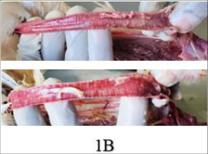 Figure 1
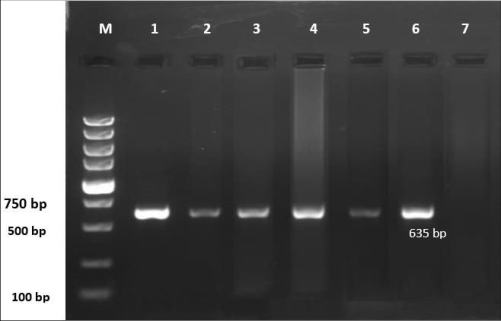 Figure 3
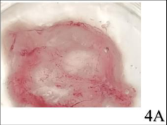 Figure 4A
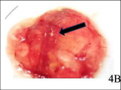 Figure 4A
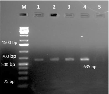 Figure 5
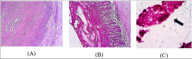 Figure 6
Introduction
Avian Infectious Laryngotracheitis (ILT) is an economically significant viral disease of chickens that affects the poultry industry worldwide. It is an acute, highly contagious respiratory infectious disease that causes mortality, decreased growth rates, and reduced egg production [1] . Infectious Laryngotracheitis Virus (ILTV) belongs to the genus Iltovirus within the family Orthoherpesviridae, classified as Iltovirus gallidalpha1 (previously classified as Gallid alphaherpesvirus 1) [2] . The virion exhibits an icosahedral nucleocapsid surrounded by a protein tegument layer encapsulated by an external envelope containing glycoproteins [1] . The ILTV genome is a linearized double-stranded DNA of approximately 153 kilobases. The genome comprises unique long (UL) and unique short (US) regions flanked by inverted repeat sequences (IRS) and terminal repeat sequences (TRS) [3] . Since ILTV remains conserved antigenically, the complete genome characterization allows for its classification into nine genotypes [4] . Very often, genotyping of ILTV strains are mainly based on molecular characterization by PCR of the infected cell protein (ICP4) gene. This also stands out as the only immediate-early gene of ILTV, whose encoded product regulates the transcription of the other early and late genes of the virus [4][5][6][7][8] . The disease manifestations are characterized by severe dyspnea, gasping, ocular and nasal discharge, and in severe cases, bloody expectoration mixed with mucus [9][10][11] . Gross and histopathological lesions typically include fibrinous, necrotizing and haemorrhagic sinusitis, laryngitis, tracheitis, and conjunctivitis. Bronchitis, bronchopneumonia, eosinophilic intranuclear inclusion bodies with or without syncytia are present [10,12] depending on the severity of the disease including occasional esophagitis and ulcerative pharyngitis [13] .
Birds recovering from ILTV infections act as potential source of infection for the other susceptible birds in the flock, with the virus assuming a latent form along the nerve ganglia. Reactivation of virus may happen under stressed conditions, enabling reinfection by horizontal transmission through virus shedding in the nasal secretions and faecal matter [11] . The virus primarily affects chickens as natural hosts, across all ages and susceptible breeds, although birds of more than 3 weeks are most affected [14][15][16][17][18][19][20][21] . However, ducks and pigeons remain unaffected by ILTV [21] . Occasionally in broilers, lesions are confined to the conjunctiva, nasal passages and trachea with mild mucoid or catarrhal exudate. This can be termed the "Silent form of ILT" due to the absence of characteristic respiratory signs [10] . Although clinical manifestations and post-mortem lesions may be suggestive of ILT, viral etiology can be confirmed using virus isolation in embryonated chicken eggs by the chorio-allantoic membrane (CAM) route or primary chicken liver or kidney cells. These laborious methods have now been simplified by rapid and precise molecular diagnostics like PCR [20] , realtime PCR for detecting ILT infections and RFLP for differentiating vaccine and field strains [16,[20][21][22][23][24][25][26][27][28][29] . Although serology is not practised widely, histopathology has been a preferred screening test for a prompt diagnosis of suspected infections of ILT, by demonstrating the intranuclear inclusion bodies in infected tissues [19][20][21]23] . An effective strategy for preventing ILTV infection is only by vaccination using live attenuated ILT vaccines [chickenembryo-origin (CEO) and tissue-culture-origin (TCO)] in commercial poultry flocks globally, although not widely practised in India. These live vaccines have been studied to be responsible for the emergence of virulent variants [15,17,18,[29][30][31][32] during passage in chicken, synonymously described as "vaccinal ILT" [15,31] .
Therefore, the objective of this study was to detect and confirm the re-emerging infections of ILT in both commercial and native layers of Salem district in Tamil Nadu, as a part of disease surveillance, based on pathological and molecular methods.
Materials and Methods
Materials and Methods
Case history and gross pathology
Poultry farms rearing commercial layer birds and Aseel chicken in adjoining places in the Salem district of Tamil Nadu had reported sudden mortality of 4-5 birds daily, since two days, with respiratory signs, swollen head, nasal discharge, and severe dyspnoea. The flocks in the two commercial poultry farms included layers of the Bovans strain, with a flock size of approximately 10,000 birds of 12-32 weeks of age. The other three reports were from backyard flocks of Aseel breed of chicken, 16-25 weeks, reared in small holdings of 50-100 in close neighbourhood. Necropsy of the dead poultry carcasses were conducted at the Avian Disease Laboratory, Thalaivasal, Salem. Severe haemorrhagic tracheitis filled with blood-mixed mucus was observed in the affected birds from all five farms, suspected for ILT. Observations of any other gross pathologic lesions were also recorded.
Sampling and processing for virological studies
Tracheal samples were aseptically collected in sterile containers and transported to the laboratory under cold chain for virology studies. The samples were triturated to prepare 10% (w/v) tissue suspension with sterile phosphate-buffered saline (PBS) containing 100U/ml penicillin, 100 μg/ml streptomycin and 0.25 μg of amphotericin B and clarified at 3000 rpm for 15 minutes at 4 o C. The supernatant was harvested and filtered through a 0.45µm syringe filter and stored at -80 o C for nucleic acid extraction and virus isolation.
Sampling and processing for pathological studies
Tissue samples of trachea, lungs, air-sacs, liver, spleen were collected and fixed in 10% neutral buffered formalin for histopathological studies. Transverse sections of tracheal samples with haemorrhagic or caseous deposits and other fixed tissues, measuring approximately 0.5 cm in thickness, were taken for further processing. The formalin-fixed tissues including trachea were trimmed to a thickness of about 3 mm, dehydrated, cleared and embedded in paraffin as per standard procedures. Tissue sections of 3 to 5µm thickness were sliced, mounted on glass slides and stained with Haematoxylin -Eosin (H&E) for histopathological examination [34] .
Screening for ILT by diagnostic PCR
Viral DNA was extracted from tracheal tissue suspensions suspected for ILT, using a commercial DNA extraction kit (M/s. Transcience Innovative Technologies, Chennai) as per the manufacturer's instructions. All the samples were screened by Polymerase chain reaction (PCR) amplifying 635 bp fragment of the ICP4 gene [5,33] of ILTV using a pair of forward (5'-CTT-CAG-ACT-CCA-GCT-CAT-CTG-3') and reverse (5' AGT-CAT-GCG-TCT-ATG-GCG-TTG-AC-3') primers. PCR was performed in a total reaction volume of 20 µl containing 10µl of master mix (2x), 1 µM of each forward and reverse primer, 7µl of nuclease-free water, and 2µl of extracted DNA with the following conditions: 94 o C/3min for initial denaturation, followed by 35 cycles of 94 o C/1min, 62 o C/1min, and 72 o C/1.5min and a final extension at 72 o C/10min. About 5 µl of the PCR products were separated by agarose gel (1.5%) electrophoresis, stained with ethidium bromide solution (0.5mg/ml) and visualized in a gel documentation system (Gel Doc™, Bio-Rad, USA) for the target-specific product.
Virus isolation and confirmation by PCR
About 0.1-0.2 ml of the processed tissue suspension was inoculated into 9-11 days-old embryonated chicken eggs through the chorioallantoic membrane (CAM) route. The eggs were incubated at 37 o C for 5-7 days. Any embryonic deaths within 24 hours of inoculation, were considered as non-specific. The infected CAMs were harvested, examined for cytopathic effect and processed for detection of ILTV by PCR. Three to four serial passages of all the ILTV-positive CAMs were carried out in embryonated chicken eggs by CAM route. The infected CAMs of each passage were harvested and processed further for confirmation of ILTV by ICP4 gene PCR.
Results and Discussion
Infectious laryngotracheitis is a highly contagious, upper respiratory disease in poultry continuing as a threat to the poultry industry, due to high mortality rates and substantial economic losses. In the present study, ILTV was detected from five poultry farmstwo of them from commercial layer flocks and three from Aseel chicken reared in the backyards in and around Salem district of Tamil Nadu. While ILT can affect chickens across all age groups, those older than three weeks were reported to exhibit the highest susceptibility to ILTV [35] . The affected birds in this study were in the age group of 12-32 weeks age, indicating a diverse range of age groups affected by ILTV, commonly reported by earlier researchers [36] . Although native chicken are known to be tolerant to diseases in general, compared to commercial chicken, poor management and biosecurity are major predisposing factors for infectious diseases like ILT and many other respiratory diseases of poultry.
Clinical signs and gross pathology
In this report, the ailing birds in the flocks suspected of ILT, exhibited clinical signs of dullness, nasal discharge and dyspnoea presented as 'pump-handle type of respiration' -a characteristic manifestation of ILT. Occlusion of the laryngeal and tracheal lumen by the catarrhal or haemorrhagic exudates have resulted in the birds extending their neck, gasping for air. Similar clinical manifestations have been the classical signs reported in the past [9, 10, 21, 3037] . The post-mortem lesions were predominantly in the larynx and trachea, characterised by haemorrhagic tracheitis and blood-mixed mucous casts along the tracheal lumen of the affected birds (Fig. 1A) indicative of an acute condition. Discrete congestion and generalised catarrhal changes with caseous exudates (Fig. 1B) as deposits were also observed in some birds as reported earlier [19-21, 30-32, 35-37] , while no significant gross lesions were recorded in conjunctiva or any other visceral organs, except for the upper respiratory tract. Although the tracheal casts were formed over a period of time, the infected birds early in an outbreak, died from an acute infection, with no gross pathologic signs. Sometimes, the major pathologic findings have been limited to swelling of the conjunctiva and infraorbital sinuses, in mild cases of ILT reported earlier [33] . The virus seemed to possess a distinct tissue tropism, being limited in its invasion of the mucous membrane of the upper respiratory tract. Although the pathogen might have entered the circulation, no characteristic lesions were visible in any other tissues.
Screening results by diagnostic PCR
The present study detected ILTV in all the five samples collected from different poultry flocks suspected for ILT by PCR targeting the fragment 2 of the infected cell protein 4 (ICP4 gene). The ILTV-ICP4 PCR amplified a 635bp fragment using gene-specific primers (Fig. 3). The ICP4 gene is noteworthy of its vital role in regulating viral gene expression during the initial phase of infection as an immediate-early transcribing gene [38] . This biological property and conserved nature of the ICP4 gene and henceforth its utility in the molecular diagnosis of ILT has been well-documented by WOAH and other researchers [8,9,21,27,33] .
Virus isolation
Virus isolation studies was done using 9-11 days old embryonated chicken eggs inoculated by the CAM route. Thickening of CAM, with hemorrhagic blood vessels were observed after 5 days post infection (5DPI) as described earlier [33] . All the five infected embryos exhibited a stunted growth. The appearance of typical pock lesions were distinct in the third passage (Fig. 4A,4B) of two of the isolates derived from the native birds. The virus isolates from the other three samples produced only thickened, haemorrhagic CAM without any pock lesions in the fourth passage in this study, similar to those documented in the previous studies [9,35] . Further serial passages in embryonated chicken eggs or cell culture may enable the demonstration of the characteristic pock lesions of ILTV. However, to confirm the presence of ILTV, the DNA extracted from the infected CAM harvested from each passage, was subjected to ILTV-ICP4 -PCR. A positive amplification of the target gene could be demonstrated in every passage, confirming the presence of the virus (Fig. 5).
Histopathology
Diagnosis relying solely on clinical signs and lesions is not conclusive and hence requires laboratory confirmation. Histopathology is the gold standard diagnostic method and is also a rapid diagnostic tool, because intranuclear inclusion bodies found in the tracheal and conjunctival epithelial cells are pathognomonic of ILT. Histopathological examination of all the tracheal tissue samples from the five different sources in this study, revealed microscopical lesions pertaining to acute tracheitis. Major portions of trachea in all the samples could be demonstrated for haemorrhagic and necrotic lesions, with multifocal erosion of the mucosal epithelium. The mucosa and submucosa layers were infiltrated by numerous heterophils, lymphocytes and plasma cells. Denuded epithelial cells forming syncytia were present in the lumen. Deciliation with multifocal haemorrhages and cellular infiltrations were observed as characteristic microscopical findings (Fig. 6 A& B).
The desquamated epithelial cells formed syncytia in the respiratory mucosa mixed with the exudates. Intranuclear, eosinophilic inclusion bodies with margination of chromatin, that appeared as "grape-like clusters" were observed in the respiratory epithelium along with haemorrhages (Fig. 6C) suggestive of ILT. The haemorrhagic exudate with cellular debris might have occluded the tracheal lumen, resulting in pump-handle type of respiration in the affected birds. However, these inclusion bodies have been reported to typically disappear by the fifth-day post-infection, following necrosis and desquamation of the superficial epithelial cells [10,18,23] . Previous reports have indicated the limitation in the diagnosis of chronic ILT infections based on histopathology, because the viral inclusion bodies disappear during chronic stages of the disease where desquamation of epithelial cells and necrosis are common [19,33] .
Molecular confirmation of ILTV
Though the gross lesions and histopathological evidences strongly suggest ILTV infection, molecular analysis by PCR targeting the Infected Cell Protein-4 (ICP4) has been extensively used by researchers for the detection of ILTV in the suspected field samples, owing to its rapid, precise, and highly sensitive detection of viral particles. However, the conventional methods of demonstration of the virus by isolation using embryonated eggs or tissue culture [35] remain the gold standard methods. Further, detection by fluorescent antibodies or immunohistochemistry enable to confirm the diagnosis.
The diagnostic PCR results obtained in this study were consistent with the earlier reports including the gross and microscopic pathological findings, except for the minimal involvement of conjunctival mucosa. Although conventional PCR is quite less sensitive than real-time PCR, its efficacy in diagnosing ILT has been well described in previous reports [12,14,22,39,40] . Future studies should incorporate such real-time monitoring to enhance diagnostic accuracy for the rapid identification of ILTV in disease outbreaks [28,41] . However, the demonstration of ILTV-specific PCR product in mild cases of infection may indicate either the presence of low virulent viruses or reactivation of latent virus in the trachea producing a low-level persistent infection. Further exploration by other PCR-based methods like LAMP and real-time PCR will enable the sensitive and reliable detection of latent ILTV infections [21,33,40] .
Control of ILT
Globally, the control of Infectious Laryngotracheitis Virus (ILTV) is achieved through the use of live, attenuated vaccines either of tissue culture origin or chicken embryo origin. Although there are no approved or licensed vaccines against ILT in India, the recent disease outbreaks reported in poultry may be attributed to the re-emerging ILTV, which might be an outcome of the vaccine virus regaining its virulence during passage in birds and causing chronic infections [42] . While ILTV vaccines are effective in preventing mortality, they do not inhibit infection or excretion of the virulent virus. Spontaneous recombination events between vaccine strains and virulent viruses have led to the emergence of new variants, resulting in widespread ILTV outbreaks globally, especially in regions where live attenuated vaccines are employed [16,25,38,45] . Several studies have reported a close genetic relationship between vaccines and virulent viruses, with CEO-related virulent strains circulating and causing disease [36,[43][44][45] . Further studies using PCR-RFLP analysis and sequencing would throw more light on the differentiation of the vaccine virus from circulating field strains [28,35,43] . Stringent biosecurity is an effective preventive measure against ILT. Maintenance of hygiene of farm workers, equipment, and vehicles in the farm premises should be practised to reduce the risk of carrying infected material into contact with the flock. Movement of feed, birds and other farm materials from flocks with a history of ILT, or those using ILT vaccine, or sites containing backyard flocks to the healthy flocks must be avoided.
In conclusion, the present study confirms the presence of ILTV infection in commercial layers and native Aseel chicken through molecular detection, virus isolation and histopathological analyses. A continuous surveillance of the early detection of circulating ILTV strains in such geographical locations, will enable the execution of appropriate biosecurity measures to better control the spread of the disease. Development of novel and effective vaccines remain the need of the hour, to combat the reemerging infections of ILT in poultry.
Acknowledgement
AcknowledgmentsThe authors are thankful to Tamil Nadu Veterinary and Animal Sciences University, Chennai for the necessary facilities and funds for carrying out the research work.
Figures and tables from the paper
References
44 in the original paper- García M, Spatz S. Infectious Laryngotracheitis. In Diseases of Poultry: John Wiley & Sons, Ltd. Hoboken, NJ, USA, 2020. p. 189-209. ISBN 978-1-119-37119-9.
- Current ICTV Taxonomy Release ICTV. Available from: https://ictv.global/taxonomy (accessed on 28 March 2025).
- Spatz SJ, Volkening JD, Keeler CL, Kutish GF, Riblet SM, Boettger CM, et al. Comparative full genome analysis of four Infectious Laryngotracheitis Virus (Gallid Herpesvirus-1) virulent isolates from the United States. Virus Genes. 2012;44:273-285.
- Spatz SJ, Garcia M, Riblet S, Ross TA, Volkening JD, Taylor TL, et al. MinION Sequencing to genotype US strains of Infectious Laryngotracheitis Virus. Avian Pathol. 2019;48:255-269.
- Chacón JL, Ferreira AJP. Differentiation of field isolates and vaccine strains of Infectious Laryngotracheitis Virus by DNA sequencing. Vaccine. 2009;27:6731-6738.
- Choi EJ, La TM, Choi IS, Song CS, Park SY, Lee JB, Lee SW. Genotyping of Infectious Laryngotracheitis Virus using allelic variations from multiple genomic regions. Avian Pathol. 2016;45:443-449.
- Hermann S, Stevens MJA, Sigrist B, Bilic I, Albini S, Wolfrum N. Unveiling the genetic landscape of Infectious Laryngotracheitis Virus in Switzerland: Evidence for vaccine-like and wild-type strains. Virol. J. 2024;600:110217.
- Kardoğan Ö and Sarıçam İnce S. Molecular characterization and phylogenetic analysis of infectious laryngotracheitis virus isolates from commercial chicken flocks in Turkey. Arch. Virol. 2024;169(11):231.
- Mishra A, Thangavelu A, Roy P, Tirumurugaan KG, Hemalatha S, Gopalakrishnamurthy TR, Gowthaman V, Raja A, Shoba K and Kirubaharan JJ. Infectious Laryngotracheitis in layer Birds from Tamil Nadu, India. Indian J. Anim. Res. 2020;54(11):1408-1414.
- García M, Spatz SJ, Guy JS. 2013. Infectious laryngotracheitis. In: Swayne DE, Glisson JR, McDougald LR, Nolan LK, Suarez DL, Nair V. Editors. Diseases of Poultry. Ames, Iowa. Blackwell Publishing; pp. 161-179.
- Oldoni I, Rodriguez-Avila A, Riblet SM, et al. Pathogenicity and growth characteristics of selected infectious laryngotracheitis virus strains from the United States. Avian Pathol. 2009;38(1):47-53.
- Goodwin MA, Smeltzer MA, Brown J, et al. Comparison of histopathology to the direct immunofluorescent antibody test for the diagnosis of infectious laryngotracheitis in chickens. Avian Dis. 1991;35(2):389-391.
- Sary K, Chenier S, Gagnon CA, et al. Esophagitis and pharyngitis associated with avian infectious laryngotracheitis in backyard chickens: two cases. Avian Dis. 2017;61(2):255-260.
- Crespo R, Woolcock PR, Chin RP, et al. Comparison of diagnostics techniques in an outbreak of infectious laryngotracheitis from meat chickens. Avian Dis. 2007;51(4):858-862.
- Guy JS, Barnes HJ, Smith L. Increased virulence of modified-live infectious laryngotracheitis vaccine virus following bird-to-bird passage. Avian Dis. 1991;35(2):348-355.
- Moreno A, Piccirillo A, Mondin A, et al. Epidemic of infectious laryngotracheitis in Italy: characterization of virus isolates by PCR-restriction fragment length polymorphism and sequence analysis. Avian Dis. 2010;54(4):1172-1177.
- Pitesky M, Chin RP, Carnaccini S, et al. Spatial and temporal epidemiology of infectious laryngotracheitis in central California: 2000-2012. Avian Dis. 2014;58(4):558-565.
- Sellers HS, Garcia M, Glisson JR, et al. Mild infectious laryngotracheitis in broilers in the southeast. Avian Dis. 2004;48(2):430-436.
- Timurkaan N, Yilmaz F, Bulut H, et al. Pathological and immunohistochemical findings in broilers inoculated with a low virulent strain of infectious laryngotracheitis virus. J Vet Sci. 2003;4(1):175-180.
- Ponnusamy P, Sukumar K, Raja A, Saravanan S, Srinivasan P, Balasubramaniam A, et al. Histopathological and molecular confirmation of infectious laryngotracheitis virus in desi chicken. The Pharma Innovation Journal. 2021;10(11S):2852-2856.
- Gowthaman V, Kumar S, Koul M, Dave U, Murthy TRGK, Munuswamy P, et al. Infectious laryngotracheitis: Etiology, epidemiology, pathobiology, and advances in diagnosis and control -a comprehensive review. Vet. quarterly. 2020;40(1):140161.
- Guy JS, Barnes HJ, Smith LG. Rapid diagnosis of infectious laryngotracheitis using a monoclonal antibody-based immunoperoxidase procedure. Avian Pathol. 1992;21(1):77-86.
- Reynolds HA, Wetrach AM, Hanson LE. Development of the nuclear inclusion bodies of infectious laryngotracheitis. Avian Dis. 1968;12(2):332-347.
- Kotiw M, Wilks CR, May JT. The effect of serial in vivo passage on the expression of virulence and DNA stability of an infectious laryngotracheitis virus strain of low virulence. Vet Microbiol. 1995;45:71-80.
- Keeler JR, Hazel CL, Hastings JW, Rosenberger JE. Restriction endonuclease analysis of Delmarva field isolates of infectious laryngotracheitis virus. Avian Dis. 1993;37:418-426.
- Chang P, Lee Y, Shien J, Shieh HK. Rapid differentiation of vaccine strains and field isolates of infectious laryngotracheitis virus by restriction fragment length polymorphism of PCR products. J Virol Methods. 1997;66:179-186.
- Oldoni I, Garcia M. Characterization of infectious laryngotracheitis virus isolates from the US by polymerase chain reaction and restriction fragment length polymorphism of multiple genome regions. Avian Pathol. 2007;36:167-176.
- Oldoni I, Rodriguez-Avila A, Riblet S, Garcia M. Characterization of infectious laryngotracheitis virus (ILTV) isolates from commercial poultry by polymerase chain reaction and restriction fragment length polymorphisms (PCR-RFLP). Avian Dis. 2008;52:59-63.
- Neff C, Sudler C, Hoop RK. Characterization of western European field isolates and vaccine strains of avian infectious laryngotracheitis virus by restriction fragment length polymorphism and sequence analysis. Avian Dis. 2008;52:59-63.
- Dormitorio TV, Giambrone JJ, Macklin KS. Detection and Isolation of Infectious Laryngotracheitis Virus on a Broiler Farm After a Disease Outbreak. Avian Dis. 2013; 57:803-807.
- Al-Saadi MHA. Multilocus analysis of Gallid herpesvirus 1 in layer chickens in Iraq. Vet. World. 2020;13:170-176.
- Bayoumi M, El-Saied M, Amer H, Bastami M, Sakr EE, El-Mahdy M. Molecular characterization and genetic diversity of the infectious laryngotracheitis virus strains circulating in Egypt during the outbreaks of 2018 and 2019. Arch. Virol. 2020;165:661-670.
- Bancroft JD, Gamble M. Theory and practice of histological techniques. 6th ed. Churchill Livingstone, London; 2008.
- Ponnusamy P, Sukumar K, Raja A. et al. Characterization of infectious laryngotracheitis virus isolates from laying hens during 2019-2020 outbreaks in Tamil Nadu, India. Arch. Virol. 2022;167:18191829.
- Gowthaman V, Singh SD, Dhama K, et al. Molecular detection and characterization of infectious laryngotracheitis virus (Gallid herpesvirus-1) from clinical samples of commercial poultry flocks in India. 2014; Virus Dis. 25:345-349.
- Senthilnathan G, Shoba K, Meenambigai TV, Anbu Kumar K, Senthil NR. Molecular detection and sequence analysis of envelope glycoproteins (gC, gD) and tegument protein (UL47) of infectious laryngotracheitis virus isolated from commercial poultry flocks in Tamil Nadu, India. Int J Vet Sci Anim Husbandry. 2024;9(2S):165-170.
- Johnson MA, Tyack SG, Prideaux C, et al. Nucleotide sequence of infectious laryngotracheitis virus (Gallid herpesvirus-1) ICP4 gene. Virus Res. 1995;35:193-204.
- Abbas F, Andreasen JR. Comparison of diagnostic tests for infectious laryngotracheitis. Avian Dis. 1996;40(2):290-295.
- Shil NK, Legione AR, Markham PF, et al. Development and validation of TaqMan real-time polymerase chain reaction assays for the quantitative and differential detection of wild-type infectious laryngotracheitis viruses from a glycoprotein Gdeficient candidate vaccine strain. Avian Dis. 2015;59(1):07-13.
- Kim HR, Kang MS, Kim MJ, et al. Restriction fragment length polymorphism analysis of multiple genome regions of Korean isolates of infectious laryngotracheitis virus collected from chickens. Poult Sci. 2013;92(8):2053-2058.
- Jaisree S, Shoba K, Ramesh A, Vijayarani K, Vairamuthu S, Bharathi R. Detection and molecular characterization of Avian infectious laryngotracheitis virus isolated from a breeder flock. J. Anim. Res. 2021;11(2):257-262.
- Gowthaman V, Koul M, Kumar S. Avian infectious laryngotracheitis: A neglected poultry health threat in India. Vaccine. 2016;34(36):4276-4277.
- Loncoman CA, Hartley CA, Coppo MJC, Vaz PK, Diaz-Méndez A. Genetic diversity of infectious laryngotracheitis virus during in vivo coinfection parallels viral replication and arises from recombination hot spots within the genome. J. Appl. Environ. Microbiol. 2017;83(23):e01532-17.
- La TM, Choi EJ, Lee JB, Park SY, Song CS. Comparative genome analysis of Korean field strains of infectious laryngotracheitis virus. PLoS One. 2019;14(2):e0211158.