
Research article · 2025 · Full text
International Journal of Veterinary Sciences and Animal HusbandryEvaluation of the safety of cell culture-adapted fowl pox vaccines
Volume 10(3) · Pages 367–371 · DOI: 10.22271/veterinary.2025.v10.i3f.2161 ↗
Research areasPoxvirus research and outbreaksVirus-based gene therapy researchBacillus and Francisella bacterial research
Abstract
Author-writtenFowl pox virus infections cause significant economic losses in poultry farming, particularly in endemic regions. Despite the widespread use of chorioallantoic membrane (CAM)-propagated vaccines in Nigeria, outbreaks persist. This study aimed to assess the safety of cell culture-adapted fowlpox virus vaccines in chickens. A total of 25 specific-antibody-negative, five-week-old chickens were vaccinated via the wing web route with ten standard field doses (≈0.12 mL per dose) of each cell culture-adapted vaccine. The birds were monitored for 7–21 days for vaccine “takes,” adverse effects, disease, or mortality. Two birds per group were euthanized on day 21 for histological examination using Haematoxylin and Eosin staining. All vaccinated chickens (100%) developed vaccine “takes” without adverse reactions, disease, or mortality. No vaccine-associated disease or virulence reversal was observed (0%), as histological examination showed no “Bollinger bodies.” However, mild histopathological changes were noted, including renal tubular necrosis, hepatocyte necrosis, hemorrhage, and vascular congestion in various tissues. Polymorphonuclear cell clusters were also found in lung and liver tissues. These findings indicate that cell culture-adapted fowlpox vaccines are safe and effective under laboratory conditions. Additionally, they offer advantages over CAM-propagated vaccines, such as ease of production, cost-effectiveness, and higher vaccine yields. Further studies are needed to validate these results, assess vaccine persistence in host tissues, and evaluate the potential for genetic mutations.
Summary
Source-checkedPlain-language reading of the full paper, prepared with AI and checked against the text. Each point links to the authors' own words.
Why it matters
- Fowl pox virus infections are a major cause of economic losses in poultry farming, especially in regions where the disease is endemic. View evidence ↗
- The disease is challenging for the poultry industry because it can lead to blindness, reduced egg production in hens, and higher mortality rates. View evidence ↗
- Traditional vaccines produced using the chorioallantoic membrane (CAM) have limitations, such as high costs, labor-intensive production, and low viral titres, which limit their availability. View evidence ↗
The question
- The researchers aimed to evaluate whether fowlpox virus vaccines adapted to cell culture are safe for use in chickens. View evidence ↗
What they did
- Twenty-five chickens (five per group, including one control) were used; four groups received 10 times the standard field dose (approximately 0.12 mL) of vaccines propagated in CEF, DEF, QEF, or Vero cell lines. View evidence ↗
- Five-week-old chickens were vaccinated using the wing web route via a specialized applicator. View evidence ↗
- On day 21 post-vaccination, two birds per group were euthanized to allow for the necropsy and sampling of the heart, lungs, liver, and kidney. View evidence ↗
- Tissues were analyzed for histopathological changes using Haematoxylin and Eosin (H&E) staining and light microscopy. View evidence ↗
What they found
- All 20 vaccinated birds (100%) developed vaccine 'takes' (swelling at the injection site) within 7 days, while the control group did not. View evidence ↗
- There was no evidence of virulence reversal or vaccine-associated disease (0%), confirmed by the absence of Bollinger bodies in histological exams. View evidence ↗
- Minor kidney changes were observed: CEF-vaccinated birds showed necrosis of renal tubules and corpuscles, while QEF-vaccinated birds showed congestion of renal vessels. View evidence ↗
- All vaccinated groups showed liver changes: CEF birds had severe hepatocyte necrosis; DEF birds had central vein congestion and hepatocyte necrosis; QEF birds had portal vein congestion; and Vero birds had polymorphonuclear cell aggregation and partial portal vein congestion. View evidence ↗
- Cardiac tissues were normal in most groups, except for DEF-vaccinated birds, which exhibited congested coronary vessels. View evidence ↗
- Despite the minor histological changes, no vaccinated birds developed clinical disease or adverse symptoms, even when receiving 10 times the standard dose. View evidence ↗
What it means
- The authors conclude that cell culture-adapted fowl pox vaccines are safe and effective under laboratory conditions. View evidence ↗
- The authors state that further research is needed to validate results in field conditions and to perform molecular characterization to check for genetic mutations during adaptation. View evidence ↗
Visual summary
1 visualsIllustrations explain the paper; they do not show study data.
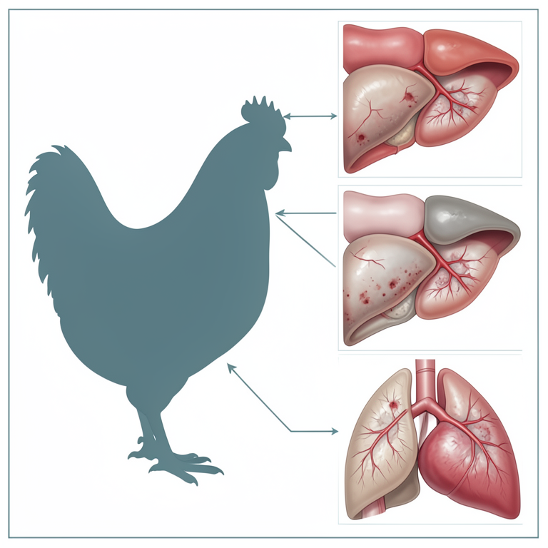
Tissue Impact Map
AI-generated conceptual illustration (domestic chicken (Gallus gallus domesticus)). It explains the idea and does not show study data.
View source evidence ↗Figures & tables
From the original paper Table 1
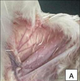 Figure 1
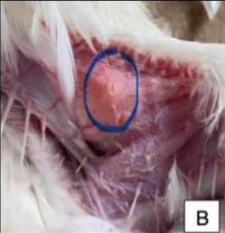 Figure 1
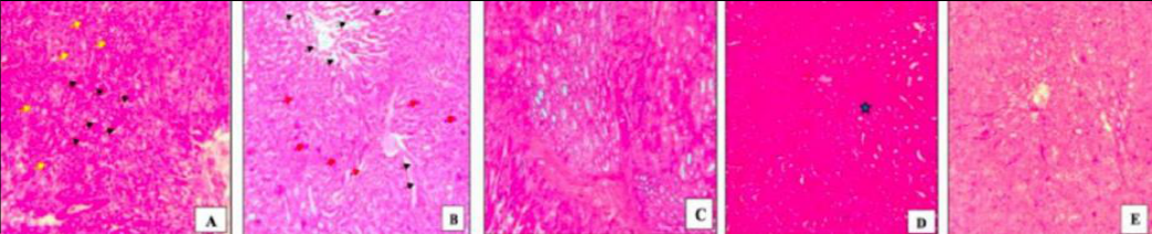 Figure 2
 Figure 3
 Figure 4
 Figure 5
Introduction
Fowl pox (FP) is a viral disease of both domesticated and wild birds [1] . The disease poses a significant challenge to the poultry industry that often leads to severe consequences like blindness, low egg production in laying hens, and increased mortality [2,3] . The Fowl pox virus is a cytoplasmic DNA virus categorized in the family Poxviridae, subfamily Chordopoxvirinae, and genus Avipoxviruses (APV) [4] . The virus causing FP in turkeys and chickens is predominantly transmitted to poultry via aerosols, biting insects, contaminated food and water [4] . Following viral penetration and systemic infection, two forms manifest in birds: cutaneous (dry form), and diphtheritic (wet form) [5] . The cutaneous variant is associated with lesions characterized by the presence of grey-white nodules located close to the eyes, skin, beak, and cloaca [1] whist the wet form appears in the upper digestive and respiratory tracts, thus complicating ingestion and inhibits hydration, ultimately resulting in starvation, dehydration and mortality [6] . A simultaneous manifestation of a mixed cutaneous and systemic variant of FP infection was observed in juvenile chickens [7] . Control measures for FP primarily rely on stringent biosecurity practices and vaccination with either attenuated FPV strains or antigenically similar Pigeon pox virus vaccine [5] . However most commercially available live FP vaccines, particularly in Nigeria, are produced using chorioallantoic membrane (CAM) of embryonated eggs [8,9] .
While CAM-based vaccines have been widely used, the present several drawbacks, including low titres, inability to illicit immune responses, high production costs, and laborintensive manufacturing processes, ultimately limiting vaccine availability [9,10] . These limitations have resulted in an inadequate supply of FP vaccines to meet the growing demands of the poultry industry. In view of the above shortcomings of CAM vaccines, the search for fowl pox virus vaccine adapted to a cell culture with high infectious and immunogenic properties becomes critical [9] . Given these challenges, there is an urgent need to develop FP vaccines using cell culture systems, which offer several advantages over CAM-based production. Cell culture-adapted vaccines can enhance viral replication efficiency, improve immunogenicity, produce vaccines with high titres, and streamline large-scale production, making them more reliable and cost-effective alternative [9] . Furthermore, cell culture technology has become a cornerstone of modern veterinary vaccinology, facilitating the production of safer and more effective vaccines [11] . In response to these needs, this study evaluated the safety of various locally produced cell cultureadapted live FPV vaccines in chickens, with the goal of optimizing vaccine efficacy and addressing the supply gap in FP prevention.
Methods
Authorization for this research involving the use of experimental animals (Specific pathogen free chickens), including eggs, was secured from the National Veterinary Research Institute, Vom Animal Use and Care Committee, under approval number: NVRI/AE/02/164/24. Five birds free of maternal antibodies randomly allocated into four groups after statistically calculated via Federer's formula were vaccinated with 10 times the standard field dose (≈ 0.12 mL per dose) of the four FP cell culture adapted vaccines propagated in four cell lines namely: chicken embryo fibroblast (CEF), duck embryo fibroblast (DEF), quail embryo fibroblast (QEF), and Vero cells with titres: CEF=10 9.3 TCID 50 /mL, QEF=10 8.6 TCID 50 /mL, DEF=10 7.6 TCID 50 /mL, and Vero cells=10 7.3 TCID 50 /mL. One group of five birds were kept as unvaccinated control group. The birds were vaccinated by pricking the wing web with a specialized applicator containing the vaccine, at five weeks of age and kept under the deep litter management system, while being feed and watered ad libitum. The experimental plan and treatment groups were as outlined in Table 1. The birds were then monitored daily for 7-10 days for evidence of 'takes', which is defined, "as a skin oedema or the development of a scab at the vaccination site". The birds were also monitored for the absence of adverse effects/reactions or disease, and/or mortality attributable to the vaccines [8,10] . On day 21 post vaccination at the termination of the experiment, two birds from each group were randomly selected and humanely euthanized. The euthanized chickens were necropsied and several tissues namely: kidney, liver, lungs, and heart were aseptically sampled and preserved in 10% buffered formalin, The fixed tissues were routinely processed for haematology-and-eosin staining (H&E) examination under a light microscope (Carl Zeiss Axio Imager A1 binocular) for histopathological changes [12] .
| Group (N=5Birds) | WEEK | — | — |
|---|---|---|---|
| Pre-vaccination | Vaccination | Post-vaccination (PV) | |
| Week 4 | Week 5 | Week 8 (21 DPV) | |
| B | Stabilise | CEF Vaccine – 10x Std field dose | Terminate Exp., Euthanize 2 birds |
| D | Stabilise | DEF Vaccine – 10x Std field dose | Terminate Exp., Euthanize 2 birds |
| F | Stabilise | Vero Vaccine – 10x Std field dose | Terminate Exp., Euthanize 2 birds |
| H | Stabilise | QEF Vaccine – 10x Std field dose | Terminate Exp., Euthanize 2 birds |
| K (Control) | Stabilise | No Vaccination | Terminate Exp., Euthanize 2 birds |
Table text extracted from the original PDF; superscript letters are shown inline. The original table image is below.
View original table image
Results
After vaccination, 100% (20/20) of the birds in the four groups that received 10 times the standard field dose developed "takes" (swelling at the injection site) within 7 days post-vaccination but the non-vaccinated control birds however did not develop "takes". The vaccinated birds that developed "takes" are shown in Figure 1 (a & In addition, all the vaccinated birds that received 10 times the standard field dose did not develop any undesirable/adverse symptoms or clinical disease related to the vaccine or vaccination with the 4 different vaccines. However, although these birds did not develop any undesirable/adverse symptoms or clinical disease or mortality related/associated with the vaccine or vaccination with the 4 different vaccines, there were minor histological changes including necrosis of the renal tubules and the renal corpuscles in the kidney tissues of CEF vaccinated birds (Figure 2B), while there were congestion of the renal vessels in QEF vaccinated birds (Figure 2D). The lung tissues on the other hand had congestion/ hemorrhages in blood/pulmonary vessels and clusters of polymorphonuclear cells in all the vaccinated birds (Figure 3G-J). The cardiac tissues have normal muscle and coronary vessels except the DEF vaccinated birds which had congested coronary vessels (Figure 4M). The hepatic tissues of the all the vaccinated birds had several histological changes including severe necrosis of the hepatocytes (CEF vaccinated birds-Figure 5Q) congestion of the central vein and necrosis of the hepatocytes (Figure 5R=DEF CEF=Chicken Embryo Fibroblast Adapted Vaccine; DEF=Duck Embryo Fibroblast Adapted Vaccine; QEF=Quail Embryo Fibroblast Adapted Vaccine; Vero=Vero Cells Adapted Vaccine; H&E=Haematoxylin and Eosin.
b) below
b) below
Discussion
Fowl pox remains a significant challenge in poultry production, leading to economic losses due to high morbidity, poor growth, and reduced egg production [13] . Since there are no standardized treatments, vaccination remains the primary control strategy to control FP. Traditionally, FPV vaccine are produced using the chorioallantoic membrane (CAM) of embryonated chicken eggs. However, this method has several drawbacks, including producing low titre vaccines, high production costs, and labor-intensive processes [8,9] . These limitations have necessitated the search for alternative vaccine production methods, particularly the adaptation of fowl pox virus to cell culture systems, which offer improved efficiency, high viral titres, and scalability [11] . This study evaluated the safety of Fowl pox vaccines derived from primary cell cultures (CEF, DEF, and QEF), and a secondary cell line (Vero cells). The results of this study demonstrate that cell culture-adapted fowl pox vaccines are both safe and effective. All vaccinated birds developed vaccine "takes," characterized by localized swelling at the inoculation site, indicating a strong immune response. Additionally, none of the vaccinated birds exhibited clinical disease, adverse reactions, or mortality, reinforcing the safety of these vaccines under laboratory conditions. Histopathological analysis revealed minor tissue changes in vaccinated groups, including renal tubular necrosis in CEF-vaccinated birds, vascular congestion in QEF-and DEF-vaccinated birds, and inflammatory cell infiltration in the lungs and liver across all vaccinated groups. However, these alterations did not translate into clinical illness, suggesting that the vaccines remain safe for use. When compared to findings from previous studies, these results align with earlier reports indicating that cell cultureadapted fowl pox vaccines induce a strong immune response while maintaining a favorable safety profile [14,15] . The presence of mild histopathological changes is also consistent with existing literature, which suggests that such tissue responses may be transient and not necessarily indicative of long-term adverse effects [16][17][18] . Importantly, the absence of clinical disease, mortality, or vaccine-associated complications in this study supports the growing body of evidence that cell culture-adapted vaccines are a viable alternative to traditional CAM-based vaccines. One of the primary advantages of cell culture-based vaccines is their efficiency in production. Unlike CAM-based vaccines, which are labor-intensive and yield relatively low virus titres, cell culture systems enable higher virus yields, improved scalability, and a more standardized manufacturing process [11] . This makes them a more practical and cost-effective alternative, particularly in regions with high demand for fowl pox vaccines. Furthermore, the ability to propagate the fowl pox virus in both avian and mammalian cell lines expands the potential for optimizing vaccine formulations with enhanced immunogenicity and safety [9] . Despite these promising findings, additional research is needed to fully validate the safety and efficacy of these vaccines in field conditions. Future studies should focus on molecular characterization of the vaccine strains to determine whether genetic mutations occur during cell culture adaptation. Advanced molecular techniques, such as quantitative PCR and immunohistochemistry, could provide further insights into vaccine virus distribution and persistence within host tissues. Additionally, large-scale field trials in commercial poultry farms would be essential in assessing the real-world efficacy and safety of these vaccines. Immunological studies examining cytokine responses and antibody production could further clarify the mechanisms of immune protection conferred by these vaccines.
Conclusion
This study provides strong evidence that cell culture-adapted Fowl pox vaccines are both safe and effective under laboratory conditions. Although minor histopathological changes were observed, they did not result in clinical disease or mortality, reinforcing the potential of these vaccines as viable alternatives to conventional CAM-based vaccines. Future research focusing on viral persistence, long-term safety, and immunogenicity will be critical in further validating these findings and optimizing vaccine formulations for widespread use in poultry production.
Acknowledgement
I acknowledge the African Union Commission (AUC) for the funding opportunity I had in carrying out this research and the staff members at Pan African University Life and Earth Sciences Institute (PAULESI).
Conflict of Interest Not available
Conflict of Interest Not available
Financial Support
Funding for this research was provided by the African Union Commission through the Pan African University Life and Earth Sciences Institute (including Health and Agriculture), Ibadan, Nigeria.
Figures and tables from the paper
References
18 in the original paper- Mohamed RI, Elsamadony HA, Alghamdi RA, Shemy ELA, Amer SAM, Safia MA, et al. Molecular and pathological screening of the current circulation of fowl pox and pigeon pox virus in backyard birds. Poultry Science. 2024;103(12):104249. https://doi.org/10.1016/j.psj.2024.104249.
- Minhas SK, Kumar P, Ritu P, Raman MYR, Sweety K, Gowthaman V. Fowl pox virus: A minireview. Worlds Poultry Science Journal, 2023, p. 1-19. https://doi.org/10.1080/00439339.2023.2273353.
- Ganapathy K, Parthiban S. Pros and cons on use of live viral vaccines in commercial chicken flocks. Avian Diseases. 2024;67(4):410-420. https://doi.org/10.1637/aviandiseases-D-23-99998.
- Umar BN, Adamu J, Ahmad MT, Ahmad KH, Sada A, Orakpoghenor O. Fowl pox virus: An overview of its classification, morphology and genome, replication mechanisms, uses as vaccine vector and disease dynamics. Worlds Poultry Science Journal. 2021;77(4):929-947. https://doi.org/10.1080/00439339.2021.1959278.
- Abd El Hafez MS, Shosha EAE, Ibrahim SM. Isolation and molecular detection of pigeon pox virus in Assiut and New Valley Governorates. Journal of Virological Methods. 2021;293:114142. https://doi.org/10.1016/j.jviromet.2021.114142.
- Alehegn E, Chanie M, Mengesha D. A systematic review of serological and clinicopathological features and associated risk factors of avian pox. Br. J. Poult Sci. 2014;3:78-87. https://doi.org/10.5829/idosi.bjps.2014.3.3.8553.
- Akanbi OB, Rimfa AG, Okewole PA. Comparative study on diphtheritic, cutaneous and systemic forms of natural avipoxvirus infection in chickens. Journal of World's Poultry Research. 2016;6(3). [Accessed 02 January 2025]. https://cyberleninka.ru/article/n/comparative-study-ondiphtheritic-cutaneous-and-systemic-forms-of-naturalavipoxvirus-infection-in-chickens.
- Khalili GM, Khalesi B, Ghaderi M, Taghizadeh M, Shahkarami MK, Karimi RH. Evaluation and optimization of chick embryo fibroblasts for production of a fowl pox vaccine based on cell culture. Iran J Virol. 2020;14(2):6-15. http://journal.isv.org.ir/article-1-394fa.html. [Accessed 02 January 2025].
- Yusifova K. The use of cell systems for the manufacture of vaccine against fowl pox. Fao.org, 2021. https://agris.fao.org/search/en/providers/123799/records/ 6474703e425ec3c088f1a6ea. [Accessed 02 January 2025].
- World Organization for Animal Health (WOAH). Fowl pox. In: WOAH Terrestrial Manual, 2023, Chapter 3.3.10. [Accessed 02 January 2025]. https://www.woah.org/en/what-we-offer/expertisenetwork/reference-laboratories/#ui-id-3.
- Soccol CR, Soccol VT, Dos SC, Piazenski IN, Candelario JPM. Introduction to animal cell culture. Springer, 2024, p. 7-27. https://doi.org/10.1007/978-3-031-55968-6_2.
- Bancroft's theory and practice of histological techniques. Elsevier. 2019. https://doi.org/10.1016/c2015-0-00143-5.
- Serbessa AT, Geleta GY, Terfa OI. Review on diseases and health management of poultry and swine. International Journal of Avian & Wildlife Biology. 2023;7(1):27-38. https://doi.org/10.15406/ijawb.2023.07.00187.
- Gilhare VR, Hirpurkar SD, Kumar A, Naik SK, Sahu T. Pock forming ability of fowl pox virus isolated from layer chicken and its adaptation in chicken embryo fibroblast cell culture. Veterinary World. 2015;8(3):245250. https://doi.org/10.14202/vetworld.2015.245-250.
- Sarma G, Kersting BA, Spina G. Field safety and efficacy of a unique live virus vaccine for controlling avian encephalomyelitis and fowl pox in poultry. Veterinary World. 2019;12(8):1291-1298. https://doi.org/10.14202/vetworld.2019.1291-1298.
- Mockett APA, Deuter A, Southee DJ. Fowl pox vaccination: Routes of inoculation and pathological effects. Avian Pathology. 1990;19(4):613-625. https://doi.org/10.1080/03079459008418717.
- Verma RK, Gangwar AK. Characterization of fowl pox virus. Advances in Experimental Medicine and Biology. 2024;1451:55-74. https://doi.org/10.1007/978-3-031-57165-7_4.
- Shalaby SM, Awadin WF, Hamed MF, Tholoth ELM, Ibrahim I, El AF. Pathological and ultrastructural characteristics of Newcastle and pox diseases in naturally infected pigeons in Egypt. Advances in Animal and Veterinary Sciences. 2021;9(11). https://doi.org/10.17582/journal.aavs/2021/9.11.1995.200