
Research article · 2025 · Full text
International Journal of Veterinary Sciences and Animal HusbandryRight hemithorax pneumonia in red-eared slider (Trachemys scripta elegans, Wied-Neuwied, 1839)
Volume 10(3) · Pages 262–265 · DOI: 10.22271/veterinary.2025.v10.i3d.2144 ↗
Research areasMicrobial infections and disease researchPhysiological and biochemical adaptationsTurtle Biology and Conservation
Abstract
Author-writtenA male red-eared slider (T. scripta elegans), weighing 779 g, was presented to the Clinic for Birds and Exotic Animals of the Faculty of Veterinary Medicine (FVM), University of Zagreb. The owner noticed that the turtle was floating on its right side, and it looked more swollen than usual. Clinical examination revealed that the shell was unevenly developed and moderately deformed, and edema was observed in the prefemoral fossas. The X-ray examination revealed irregular shape of tubular bones, the carapace was moderately porous, and the lungs were dotted with shadowing in the right hemithorax which indicated right hemithorax pneumonia and metabolic bone disease. Biochemical analysis and hemogram were performed. After 5 weeks of supportive therapy, fluid replacement and treatment with antimicrobial preparations (marbofloxacin and ceftiofur), the turtle fully recovered. The manuscript aimed to present a diagnosis, care and treatment of right hemithorax pneumonia in red-eared slider.
Summary
Source-checkedPlain-language reading of the full paper, prepared with AI and checked against the text. Each point links to the authors' own words.
Why it matters
- Aquatic and semi-aquatic turtles can develop respiratory infections due to poor hygiene, inadequate tank filtration, or low water temperatures. View evidence ↗
- A deficiency in vitamin A can lead to secondary inflammation in these animals. View evidence ↗
- Pneumonia in turtles is a lower respiratory tract disease that may be caused by bacteria, viruses, fungi, or parasites. View evidence ↗
The question
- The study aims to describe the diagnosis, care, and treatment of a red-eared slider with pneumonia in the right hemithorax. View evidence ↗
What they did
- The subject was a male red-eared slider weighing 779 g treated at the University of Zagreb. View evidence ↗
- Radiographic monitoring included an initial X-ray, a first control two weeks later, and a second control three weeks after that. View evidence ↗
- Blood was collected from the dorsal tail vein on the second day for hematological and biochemical analysis. View evidence ↗
- Urine analysis was conducted using Combur 10 Test M strips. View evidence ↗
What they found
- Clinical signs included a moderately deformed and unevenly developed carapace with peeling scales, a slightly swollen head, and edema in the prefemoral fossa. View evidence ↗
- Initial craniocaudal X-rays showed spotted shadowing in the right hemithorax lungs, while the left hemithorax remained transparent. View evidence ↗
- Radiography revealed irregularly shaped tubular bones and a moderately porous carapace, indicating metabolic bone disease (MBD). View evidence ↗
- Biochemical results showed low calcium (1.81 mmol/L), low uric acid (54 umol/L), and low total protein (25 g/L), alongside elevated AST (169 U/L). View evidence ↗
- Hematological tests revealed anemia characterized by low hemoglobin (5.6 g/dL) and low MCHC (23.3 g/dL). View evidence ↗
- By the second control X-ray on June 21, 2024, the lung parenchyma was transparent and free of infiltration. View evidence ↗
- The turtle achieved full recovery after five weeks of critical care and therapy. View evidence ↗
What it means
- The authors state that a combination of intensive care and five weeks of marbofloxacin and ceftiofur antibiotic therapy led to the total cure. View evidence ↗
Figures & tables
From the original paper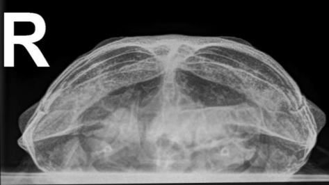 Figure 1
Introduction
The red-eared slider (Trachemys scripta elegans Wied-Neuwied, 1839) is a semiaquatic turtle originated from the Southeastern United States, around the Mississippi River and the Gulf of Mexico (Uwe and Havaš, 2007) [23] . As numerous individuals of the red-eared slider have been released or escaped into the wild in many parts of the world, wild populations of this invasive species are found in Australia, Europe, South Africa, the Caribbean Islands, Israel, Southeast and East Asia (Thomas and Hartnell, 2000; Lowe et al., 2004; Koren et al., 2018) [22,11,10] . This species is the most invasive, because it is more resistant to diseases, uses food better, earlier sexually mature and highly fertile compared to native species of turtles (Cadi and Joly, 2004; Lowe et al., 2004; Reshetnikov, 2023) [2,11,17] . In Europe, the breeding and sale of this species is prohibited, but in Croatia it is allowed to keep this species if it was acquired before the ban. Red-eared sliders are diurnal, omnivorous, and semi-aquatic animals, spending most of their time in the water or basking. They do not hibernate, but brumate, usually, at the bottom of ponds or shallow lakes when they become less active. They reach sexual maturity at the age of 5-6 years. Sexual dimorphism is present (Gibbons and Lovich, 1990) [7] . Males are usually smaller (10 cm) and have longer claws on their front legs, the cloaca is located closer to the tip of the tail, the plastron is slightly concave, and the tails are thicker and longer. Females are larger (15 cm) with flat plastron and shorter tail. Aquatic and semi-aquatic turtles are susceptible to respiratory infections if they are kept in unhygienic living conditions, the tanks do not have adequate filtration or the water temperature is too low (Silveira et al., 2014; Ciccarelli et al., 2020) [21,4] . Secondary inflammation can occur as a result of vitamin A deficiency. Pneumonia is a lower respiratory tract disease that can be caused by bacterial (eg Pseudomonas sp., Mycoplasma sp., etc), viral (eg ranavirus), fungal, or parasitic infections (Origgi and Jacobson, 2000; Marschang, 2011; McNally et al., 2021; Ebani, 2023) [16,12,15,5] . Clinical signs are excess mucus in the mouth, nasal discharge, sneezing, lethargy, loss of appetite, difficulty breathing and open-mouth breathing (Silveira et al., 2014; Ciccarelli et al., 2020; Morick et al., 2023) [21,4,14] . Turtles with pneumonia are sometimes seen swimming tilted to one side (because diseased lung tissue is heavier than normal lung tissue). For the assessment of the respiratory tract in chelonians, the most important radiographic views are the craniocaudal (Cr/Cd) and laterolateral (L/L) views (especially for the lungs), and for the interpretation of the trachea or bronchi, the dorsoventral (D/V) view is important (Gumpenberger, 2021) [8] . The manuscript aimed to present a diagnosis, care and treatment of right hemithorax pneumonia in red-eared slider.
Materials and Methods
Materials and Methods
Case history
A male red-eared slider (T. scripta elegans), weight 779 g was presented to the University Veterinary Hospital (Clinic for birds (and exotic) animals) at the Faculty of Veterinary Medicine (FVM), University of Zagreb. The owner noticed that the turtle was floating on its right side in the aquarium and looked more swollen than usual. The turtle was kept uner suboptimal conditions, fed with specialized pelleted food for turtles, dried shrimp and fish, meat, etc. He received Ca and vitamin D 3 supplements once a week. The turtle was adopted a few years ago, and the previous owners did not use UVB lamp.
Radiography
The first X-ray examination was performed at the FVM, Department of Radiology, Ultrasound Diagnostics And Physical Therapy, on the day of the first examination, the first control X-ray examination two weeks later, and the second control, three weeks later.
Blood sampling
On the second day, blood was taken from the dorsal tail vein in tubes with anticoagulant (heparin) and sent for biochemical and hematological tests. Biochemical analysis was done on the Catalyst One (Idexx Laboratories, USA), and hematological tests (hemogram and RBC) at the FVM, Department of Pathophysiology.
Urine test
The urine was tested with test strips (Combur 10 Test M, Roche, Mannheim, Germany).
Ethical approvement
The University Veterinary Hospital (FVM) provides services to patients, quality education for students, and conducts modern scientific research in accordance with prescribed ethical standards and animal welfare protocols in accordance with Directive 2010/63/EU of the European Parliament (2010) on the protection of animals used for scientific purposes.
Results
Results
Clinical examination
A clinical examination revealed that the carapace was unevenly developed and moderate deformed. Some scales were peeled off on the carapace. The head seemed slightly swollen. There were no characteristic changes within the oral cavity. Edema was observed in the prefemoral fossa. The skin was focally hyperemic (possible signs of hypothermia). The cloaca was clean without changes. There were no palpable changes in the coelomic cavity. After the clinical examination, the animal was sent for X-ray imaging.
Radiography 3.2.1 First X-ray examination (14 May 2024)
On D/V projection, a generalized homogeneous shadow could be observed in the coelomic cavity, which may correspond to generalized edema, and the other organs cannot be clearly differentiated. There are noticeable areas of lower shadow intensity that may correspond to gases in the digestive system. The tubular bones are irregularly shaped, and the carapace is moderately porous with signs of MBD. On the Cr/Cd projection, the lungs are shaded in spots in the right hemithorax, and the left hemithorax is transparent (Figure 1.). After the diagnosis (Pneumonia hemithoracis dex. susp. and Metabolic bone disease) inpatient treatment was suggested. On the L/L and Cr/Cd projections of the turtle in toto, the lung parenchyma was transparent, without infiltration.
Biochemical analysis
The results revealed: phosphate 1.26 mmol/L=3.906 mg/dL (0.74-3.71); calcium 1.81 mmol/L = 7.24 mg/dL (2.50-3.63); UA 54 umol/L = 0.908 mg/dL (71-167); TP 25 g/L=2.5 g/dL (30-70); ALT <10 U/L; AST 169 U/L (14-18);
Urine test
The specific gravity was 1.010; leukocytes 10-25; nitrites negative; pH 5; proteins 1/100; glucose negative and hemoglobin 50.
Hematological tests
Hematological tests
Treatment
Supportive therapy and fluid replacement with Ringer's solution and Duphalyte (4:1) 12 mL SC in prefemoral fossa for 7 days (every 24 hours), were obtained. Vitamin AD 3 E (Vitamin AD 3 E, Krka, Slovenia) in a dose of 0.15 mL/kg every three weeks and iron dextran (Anemin, Krka, Slovenia) every 24 h for a week 12 mg/kg. The animal was fed by gastroesophageal tube every other day in the first week with Emeraid Omnivore (Lafeber), first three ml, then 7.5 and 12 ml. Antibiotic therapy from the first day with marbofloxacin (Marbocyl, Krka, Slovenia) at a dose of 2 mg/kg every 24 h IM in the front limbs, and after 14 days ceftiofur (Ceftiomax, Laboratorios Calier, Spain) at a dose of 5 mg/kg IM was introduced in parallel with marbofloxacin, every 24 hours. After 5 weeks of critical care and therapy, the turtle fully recovered.
Discussion
Although red-eared turtles are an extremely invasive species (Lowe et al., 2004) [11] , those animals that are kept in captivity need certain care, so they should be given medical care if they are sick. Most diseases of reptiles in captivity result from improper care, housing conditions and feeding. In addition to the inadequate size and equipment of the aquaterrarium, the most common problem stems from inappropriate water temperatures and the space in which the semi-aquatic turtles are kept, as well as the lack of adequate UVA and UVB lamps, source of calcium and other nutrients (Mader, 2019) [6] . Many turtles with respiratory tract infections also have an underlying vitamin A deficiency, similar to our case. Due to the severity of the infection, daily medication, fluid replacement, and nutritional support, sick slider was hospitalized in intensive care. Respiratory infections in captive turtles are most commonly caused by opportunistic bacteria (Roberts et al., 2006) [18] . Treatment of bacterial respiratory tract infections usually involves the use of antibiotics, most often given parenterally.
A few antibiotics are commonly used in veterinary practice to treat bacterial infections in reptiles, including red-eared sliders are marbofloxacin and third generation cephalosporins. Both antibiotics commonly used to treat a variety of bacterial infections, including septicemia, respiratory and skin infections. Marbofloxacin is effective against Gram-positive and Gram-negative bacteria, while cephalosporin antibiotic used for serious infections, particularly those caused by resistant bacteria (Jacobson, 1999; Carpenter and Harms, 2023) [9,3] . Anemia in our case was normocytic-hypochromic (reduced hemoglobin and MCHC). The causes of this type of anemia in turtles can be depression of erythropoiesis, acute bleeding and chronic inflammation (Saggese, 2009; Martínez Silvestre, 2013) [19,13] . In our case, the cause of such anemia was pneumonia. Anemia therapy consisted of the administration of iron dextran and supportive therapy according to the animal's weight (Martínez Silvestre, 2013; Carpenter and Harms, 2023) [13,3] . Hypovitaminosis A is a common issue in turtles, particularly in species like the red-eared slider. This deficiency commonly cause squamous metaplasia and general decline in immunity often seen as ocular edema skin issues respiratory complications and increased susceptibility to secondary infections (Boro et al., 2022; Sahu and Dolai, 2024) [1,20] . Warming up the animal and exposing it to artificial ultraviolet radiation helps speed up metabolism, digestion, fluid replacement, and drug metabolism during admission to intensive care.
Conclusion
We can assume that improper thermal regime, too low water temperature, vitamin A deficiency and poor hygiene are prerequisites for the occurrence of pneumonia and edema in this turtle in our case. Intensive care and long-term antibiotic therapy with prescribed doses of marbofloxacin and ceftiofur combination for 5 weeks contributed to the total cure of right hemithorax pneumonia in the red-eared slider.
Conflict of Interest
Conflict of Interest
Financial Support
Financial Support
References
23 in the original paper- Boro PK, Yadav SN, Ahmed N. Diagnosis of hypovitaminosis A in a red-eared slider turtle (Trachemys scripta elegans) and its successful therapeutic management. Indian Journal of Animal Health. 2022;61(2):398-400. https://doi.org/10.36062/ijah.2022.04522.
- Cadi A, Joly P. Impact of the introduction of the redeared slider (Trachemys scripta elegans) on survival rates of the European pond turtle (Emys orbicularis). Biodiversity and Conservation. 2004;13:2511-2518.
- Carpenter JW, Harms CA. Exotic Animal Formulary. 6th ed. St. Louis: Elsevier Health Sciences; 2023. p. 104-106.
- Ciccarelli S, Valastro C, Di Bello A, Paci S, Caprio F, Corrente ML, et al. Diagnosis and Treatment of Pulmonary Disease in Sea Turtles (Caretta caretta). Animals. 2020;10:1355. https://doi.org/10.3390/ani10081355.
- Ebani VV. Bacterial Infections in Sea Turtles. Veterinary Science. 2023;10(5):333. https://doi.org/10.3390/vetsci10050333.
- Mader DR. Mader's Reptile and Amphibian Medicine and Surgery. 3rd ed. In: Divers SJ, Saunders SS, editors. Elsevier; 2019.
- Gibbons JW, Lovich JE. Sexual dimorphism in turtles, with emphasis on the slider turtle (Trachemys scripta). Herpetological Monographs. 1990;4:1-29. https://doi.org/10.2307/1466966.
- Gumpenberger M. Diagnostic Imaging of the Respiratory Tract of the Reptile Patient. Veterinary Clinics of Exotic Animal Medicine. 2021;24:293-320. https://doi.org/10.1016/j.cvex.2021.01.002.
- Jacobson ER. Antimicrobial Drug Use in Reptiles. In: Bonagura JD, editor. Current Veterinary Therapy XIII. Small Animal Practice. Philadelphia: Saunders; 1999. p. 581-592.
- Koren T, Štih A, Burić I, Koller K, Lauš B, Zadravec M. The current distribution of pond slider Trachemys scripta (Reptilia: Emydidae) in Croatia. Natura Sloveniae. 2018;20(1):33-44. https://doi.org/10.14720/ns.20.1.3344.
- Lowe S, Browne M, Boudjelas S, de Poorter M. 100 of the world's worst invasive alien species: A selection from the Global Invasive Species Database. The Invasive Species Specialist Group (ISSG) of the Species Survival Commission (SSC) (Report) (Updated and reprinted ed.). International Union for the Conservation of Nature (IUCN); 2004.
- Marschang RE. Viruses Infecting Reptiles: A Review. Viruses. 2011;3:2087-2126. https://doi.org/10.3390/v3112087.
- Martínez Silvestre A. How do I treat anemia in reptiles? Southern European Veterinary Conference, Barcelona, Spain, 17-19 October, 2013.
- Morick D, Levy Y, Davidovich N, Wosnick N, ZemahShamir Z, Tchernov D, et al. Pneumocoelom and secondary lung collapse treatment in a stranded loggerhead sea turtle (Caretta caretta) in the eastern Mediterranean Sea, Israel. Veterinary Record Case Report. 2023;11:e665. https://doi.org/10.1002/vrc2.665.
- McNally KL, Bowen JL, Brisson JO, Kennedy A, Innis CJ. Evaluation of the Respiratory Microbiome and the Use of Tracheal Lavage as a Diagnostic Tool in Kemp's Ridley Sea Turtles (Lepidochelys kempii). Animals (Basel). 2021;11(10):2927. https://doi.org/10.3390/ani11102927.
- Origgi FC, Jacobson ER. Diseases of the Respiratory Tract of Chelonians. Veterinary Clinics of North America: Exotic Animal Practice. 2000;3:537-549.
- Reshetnikov AN, Zibrova MG, Ayaz D, Bhattarai S, Borodin OV, Borzée A, et al. Rarely naturalized, but widespread and even invasive: the paradox of a popular pet terrapin expansion in Eurasia. NeoBiota. 2023;81(1):91-127. https://doi.org/10.3897/neobiota.81.90473.
- Roberts JF, Jacobson ER, Mitchell MA. Epizootic pneumonia in farmed red-eared slider turtles (Trachemys scripta elegans). Proceedings of the Association of Reptilian and Amphibian Veterinarians, Thirteenth Annual Conference, Baltimore, Maryland, USA, 23-27 April 2006. p. 71-72.
- Saggese MD. Clinical Approach to the Anemic Reptile. Journal of Exotic Pet Medicine. 2009;18(2):98-111. https://doi.org/10.1053/j.jepm.2009.04.003.
- Sahu H, Dolai M. Diagnosis and successful therapeutic management of conjunctivitis associated with hypovitaminosis A in a juvenile red-eared slider (Trachemys scripta elegans). International Journal of Veterinary Science and Animal Husbandry. 2024;9(6):03-06. https://doi.org/10.22271/veterinary.2024.v9.i6a.1817.
- Silveira MM, Morgado TO, Lopes ER, Kempe GV, Correa SHR, Godoy I, et al. Pneumonia bacteriana em jabuti-piranga (Chelonoidis carbonaria): aspectos clínicos, microbiológicos, radiológicos e terapêutica. Pesquisa Veterinária Brasileira. 2014;34(9):891-895.
- Thomas M, Hartnell P. An occurrence of a red-eared turtle (Trachemys scripta elegans) in the Waikato river at Hamilton, New Zealand. Herpetofauna. 2000;30:15-17.
- Uwe F, Havaš P. Checklist of Chelonians of the World. Vertebrate Zoology. 2007;57(2):207-208. https://doi.org/10.3897/vz.57.e30895.