
Research article · 2025 · Full text
International Journal of Veterinary Sciences and Animal HusbandryParasitic profiling of tigers and leopards of Maharashtra
Volume 10(3) · Pages 42–46 · DOI: 10.22271/veterinary.2025.v10.i3a.2107 ↗
Research areasViral Infections and VectorsZoonotic diseases and public healthVector-borne infectious diseases
Abstract
Author-writtenTigers (Panthera tigris) and leopards (Panthera pardus) in India are highly vulnerable to endoparasitic infections, which impact their health, fitness, and survival both in the wild and in captivity. This study, conducted at the Wildlife Research & Training Centre in Gorewada, Nagpur, aimed to assess the prevalence of gastrointestinal parasites, focusing on Sarcocystis spp., among tigers and leopards in Maharashtra. A total of 12 tigers and 14 leopards presented to the center between 2021 and 2022 were screened for parasitic infections using fecal analysis by flotation methods. Results identified a range of parasitic infections, including Sarcocystis spp., Toxocara cati, Spirometra spp., and Diphyllobothrium spp., with Sarcocystis spp. detected in one tiger. Parasitic prevalence showed variation based on species, age, and sex, with a higher infection rate in female tigers compared to males. The most common parasites detected were nematodes and cestodes, known to cause gastrointestinal issues that can lead to malnutrition and, in severe cases, death. The study results align with findings from other global studies, highlighting that wild carnivores are often asymptomatic carriers of Sarcocystis spp. Nevertheless, infections may cause chronic health impacts, especially under stressful conditions, affecting an animal's fitness and reproductive success. This study underscores the importance of monitoring and managing parasitic infections to support the conservation of India’s endangered big cats.
Summary
Source-checkedPlain-language reading of the full paper, prepared with AI and checked against the text. Each point links to the authors' own words.
Why it matters
- Tigers and leopards in India are susceptible to endoparasitic infections that can negatively affect their survival, fitness, and overall health in both captive and wild environments. View evidence ↗
- Endoparasites such as protozoa, trematodes, cestodes, and nematodes can cause severe health issues, including respiratory problems, nutritional deficiencies, and digestive disturbances, potentially leading to death. View evidence ↗
- In Maharashtra, the risk of parasite transmission between domestic animals and wildlife is increased due to human-wildlife interactions and habitat encroachment. View evidence ↗
The question
- The study aimed to determine the prevalence of gastrointestinal parasites in Maharashtra's tigers and leopards, with a specific focus on Sarcocystis spp. View evidence ↗
What they did
- Between 2021 and 2022, 14 leopards and 12 tigers from across Maharashtra were screened for gastrointestinal parasites using fecal examinations at the Wildlife Rescue Centre in Gorewada, Nagpur. View evidence ↗
- Fecal samples were analyzed in triplicate using a light microscope at 100X and 400X magnification, employing a flotation method with a saturated salt solution. View evidence ↗
What they found
- Sarcocystis spp. was detected in one tiger, confirming the presence of this parasite in the region's large felids. View evidence ↗
- In tigers, 50% of females tested positive for at least one parasite, while no male tigers showed any parasitic infections. View evidence ↗
- The identified parasites included Diphyllobothrium spp., Spirometra spp., Toxocara cati, and Sarcocystis spp. View evidence ↗
What it means
- The presence of Sarcocystis spp. and other parasites indicates a critical need for proactive health management for both wild and captive tiger and leopard populations in Maharashtra. View evidence ↗
- To ensure the survival of these species, conservation efforts should prioritize cross-species disease management, habitat protection, and health monitoring. View evidence ↗
- The authors state that long-term studies are required to clearly correlate health and fitness in wild felid populations with their parasitic load. View evidence ↗
Visual summary
2 visualsIllustrations explain the paper; charts are replotted from the paper's own table values.
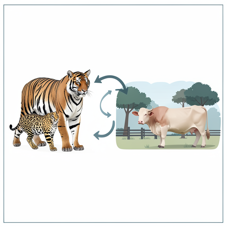
Parasite Transmission Cycle
AI-generated conceptual illustration (Bengal tigers, Indian leopards, and domestic cattle in a fragmented forest landscape). It explains the idea and does not show study data.
View source evidence ↗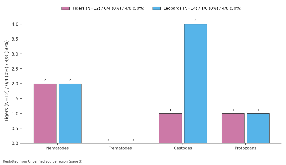
Tigers (N=12) / 0/4 (0%) / 4/8 (50%) and Leopards (N=14) / 1/6 (0%) / 4/8 (50%): Nematodes vs Trematodes vs Cestodes vs Protozoans
Replotted from a table (page 3) of the original paper. Every bar is the paper's own table value.
View source evidence ↗Introduction
In India, tigers (Panthera Tigris) and leopards (Panthera pardus) are vulnerable to a variety of endoparasitic infections, impacting their health, fitness, and survival in both wild and captive settings. Endoparasites, which include nematodes, cestodes, trematodes, and protozoa, often lead to digestive disturbances, nutritional deficiencies, respiratory problems, and, in severe cases, mortality. Nematodes, or roundworms, are some of the most frequently encountered parasites in tigers and leopards. Species such as Toxocara cati and Toxascaris leonina are gastrointestinal parasites that primarily infect felids through ingestion of contaminated prey or feces. Studies have documented significant nematode infections in both wild and captive felines, noting that these parasites can induce malnutrition, gastrointestinal distress, and, in severe cases, life-threatening obstruction of the intestines (Kumar & Khanna, 2008) [9] . Another nematode of concern is Ancylostoma spp., or hookworms, which anchor to the intestinal wall and feed on blood, leading to anemia, hypoproteinemia, and overall loss of body condition (Ravindran et al., 2012) [2] . Spirocerca lupi, a nematode that resides in the esophagus and stomach, has been found in captive tigers, causing esophageal nodules and granulomas that may even transform into malignant tumours if left untreated (Veeraselvam et al., 2019) [3] . Cestodes, or tapeworms, including Taenia spp. and Echinococcus spp., have also been reported in Indian big cats. These infections often result from consuming infected prey, highlighting the role of natural diets in parasite transmission. Tapeworm infestations may lead to chronic digestive issues, and in cases of Echinococcus infection, cysts form in organs like the liver and lungs, potentially leading to more severe health complications (Singh et al., 2010) [4] .
Although Echinococcus is more commonly associated with canids, instances in leopards underscore the zoonotic potential of this parasite, especially where wild felines come into contact with infected domestic animals or their carcasses (Jeyathilakan et al., 2008) [5] . Trematode, or fluke, infections are less common but still significant. The lung fluke Paragonimus spp., reported in various carnivores, including felines, affects the respiratory system, causing respiratory distress, coughing, and lung tissue damage due to cyst formation. In tigers and leopards, flukes such as Opisthorchis spp. and Dicrocoelium spp. may infect the liver and bile ducts, leading to jaundice and chronic liver disease (Babu et al., 2011) [6] . This is particularly concerning for wildlife rehabilitation centers where these infections can cause chronic health issues, often difficult to diagnose without detailed postmortem examinations. Protozoan infections are also frequently encountered in Indian big cats, with species such as Toxoplasma gondii known to cause toxoplasmosis. Toxoplasma, which is spread through ingestion of oocysts in contaminated environments or via consumption of infected prey, can lead to varied clinical presentations, from asymptomatic infections to severe neurological, respiratory, and gastrointestinal symptoms, particularly in immunocompromised animals (Sharma et al., 2013) [7] . Another significant protozoan is Sarcocystis spp., which forms cysts within muscle tissue and can induce fatigue, weakness, and muscle pain. Additionally, Giardia spp. and Cryptosporidium spp., two common intestinal protozoa, can cause severe diarrhoea, dehydration, and weight loss in infected animals (Gupta & Singla, 2012) [1] . These infections are particularly challenging to manage in young animals, which are more vulnerable to the nutritional deficits and dehydration caused by prolonged diarrhoea. Routine diagnostic techniques such as faecal analysis remain essential to identifying and quantifying parasite load in both captive and wild populations. Faecal flotation and microscopy allow for the identification of nematode eggs, tapeworm segments, fluke eggs, and protozoan oocysts. Polymerase Chain Reactions (PCR) based diagnostics and immunological assays have advanced the detection of protozoan infections, allowing for more accurate species identification (Sharma et al., 2013) [7] . Studies have highlighted the importance of understanding these infections' lifecycle and transmission dynamics to develop effective control strategies that can be applied across conservation and captive management settings. Continued research is necessary to refine diagnostic techniques, expand treatment options, and enhance preventive measures, ultimately contributing to the health and survival of these majestic big cats in India.
Materials and Methods
A total of 12 tigers and 14 leopards that were presented from various parts of Maharashtra state to Wildlife Rescue Centre, Gorewada, and Nagpur during 2021-2022 were screened for the presence of gastrointestinal parasites by faecal examination. The samples were collected on reception of tigers/leopards to Wildlife Research & Training Centre, Gorewada, and Nagpur. The samples were processed for parasitic screening by floatation method employing saturated salt solution as described in the reference. The samples were screened in triplicates and were examined under 100X and 400 X magnifications using a light microscope.
Results
The study conducted at the Wildlife Research & Training Centre (WRTC) in Gorewada, Nagpur, screened 12 tigers (Panthera Tigris) and 14 leopards (Panthera pardus) for gastrointestinal parasites, with a specific focus on identifying Sarcocystis spp. among other parasitic species. Parasitic infections were confirmed through fecal examinations using flotation methods, revealing a variety of endoparasitic infections among the sampled animals. Sarcocystis spp. was identified in one tiger, signifying its presence among large felids in the region.
Sr
Sr
Discussion
This finding aligns with other global studies that identify Sarcocystis spp. as a common parasite in wild carnivores, often asymptomatically infecting muscle tissue but potentially impacting fitness, particularly in stressed or immunecompromised animals (Dubey et al., 2023) [8] . In addition to Sarcocystis spp., the study detected Toxocara cati, Spirometra spp., Diphyllobothrium spp., and Isospora spp., indicating a diverse range of parasitic infections. The most frequently detected parasites were nematodes and cestodes, notably Toxocara cati and Spirometra spp., which are known to impact health by causing gastrointestinal disturbances and, in severe cases, obstructions (Kumar & Khanna, 2008) [9] . Among the tigers, 50% of females were positive for at least one parasitic infection, whereas no parasites were detected in male tigers. Among the leopards, both male and female animals showed evidence of infections, particularly [8,10] . However, infections may lead to muscular fatigue, weakness, and in some cases, inflammatory responses in heavily infected animals, as has been observed in captive studies (Máca, 2020) [11] . The diet and habitat of wild tigers and leopards significantly contribute to parasite exposure, especially in cases of Sarcocystis, which commonly spreads through the ingestion of infected prey or environmental exposure to contaminated feces. In regions where domestic animals overlap with wildlife, such as livestock areas near reserves, the risk of Sarcocystis transmission increases. This scenario has been noted in studies where Sarcocystis spp. spread from domestic animals to large wild carnivores in both India and Africa (Gjerde et al., 2020) [12] . In Maharashtra, India, habitat encroachment and the resulting human-wildlife interactions elevate the risk of parasite transmission between wildlife and domestic animals. Studies highlight that free-ranging wild animal, when captured or brought into care facilities, often present with higher parasitic loads, likely due to increased exposure to diverse pathogens in shared environments (Mahali et al., 2010; Pradhan et al., 2011) [13,14] . This finding suggests that conservation and management practices need to account for environmental factors contributing to parasite transmission, with a focus on minimizing wildlife and domestic animal overlap.
While Sarcocystis infections may remain asymptomatic, they could affect tigers' physical endurance and immune function, especially under stress, disease, or environmental challenges. Chronic infections may compromise animal fitness and reproductive success, factors that are crucial for the conservation of endangered species like tigers and leopards.
Research on the Sarcocystis life cycle reveals that the parasites form cysts within muscle tissues, often persisting long-term without immediate clinical effects but potentially affecting health over time (El-Kady et al., 2018) [15] . Given the wide range of symptoms associated with Sarcocystis in stressed or immunosuppressed animals, conservationists are increasingly concerned about the impact on endangered populations in high-risk environments. Long-term studies are needed to establish clear correlations between parasitic load, health, and fitness in wild felid populations. This can inform intervention measures, such as health monitoring and strategic deworming, in both captive and free-ranging settings (Varghese et al., 2016) [16] . [3,6] . Studies have shown that the immune response to one parasitic infection can influence susceptibility to others. This is particularly relevant in cases where wildlife is exposed to multiple pathogens through interaction with domestic animals or shared water and food sources (Jeyathilakan et al., 2008) [5] . Such interactions underscore the importance of integrated parasite management strategies, focusing on reducing coinfections to enhance the overall health of wildlife populations.
Conclusion
The detection of Sarcocystis spp. and other parasites in tigers and leopards in Maharashtra underscores the pressing need for proactive health management in both captive and wild populations. The study demonstrates that wild felids are susceptible to a wide array of parasitic infections that could impact their health, especially when exposed to stressors like habitat fragmentation and human-wildlife conflict. Conservation efforts should prioritize health monitoring, habitat protection, and cross-species disease management strategies to ensure the survival and well-being of these iconic species. Enhanced diagnostic and management practices will be essential to mitigate the risks associated with parasitic infections, contributing to the conservation of endangered felid populations in India.
Acknowledgement
Acknowledgement
Conflict of Interest Not available
Conflict of Interest Not available
Financial Support
Financial Support
References
16 in the original paper- Gupta N, Singla LD. Protozoan parasites in captive wildlife animals of Punjab, India. Tropical Biomedicine. 2012;29(3):537-545.
- Ravindran R, Lakshmanan B, Varghese T. Parasites of wildlife in India: Their ecology and zoonotic potential. Indian Journal of Animal Sciences. 2012;82(10):11021107.
- Veeraselvam M, John L, Selvaraj S. Surgical and medical management of spirocercosis in captive tigers. Journal of Veterinary Science and Technology. 2019;10:573.
- Singh A, Singh K, Khare S. Emergence of taeniasis and echinococcosis among wildlife and its implications. Journal of Zoo and Wildlife Medicine. 2010;41(2):265268.
- Jeyathilakan N, Raman M, Gomathinayagam S. Occurrence of echinococcosis in leopard. Journal of Veterinary Parasitology. 2008;22(1):63-64.
- Babu G, Ramachandran K, Ghosh S. Prevalence of helminth parasites in carnivores in captivity. Journal of Parasitic Diseases. 2011;35(2):203-205.
- Sharma R, Yadav A, Sharma S. Toxoplasmosis in felids: a review of the epidemiology and potential risk in India. Veterinary Parasitology. 2013;198(1-2):1-7.
- Dubey JP, Araujo DLS, Gupta A, Kwok OCH, Lovallo MJ. Sarcocystis and other parasites in feces of bobcats (Lynx rufus) from Mississippi. Journal of Parasitology. 2023;109(6):638-642.
- Kumar V, Khanna R. Gastrointestinal parasites of wildlife in India. Indian Journal of Animal Sciences. 2008;78(11):1283-1286.
- Chhabra MB, Samantaray S. Sarcocystis and sarcocystosis in India: Status and emerging perspectives. Journal of Parasitic Diseases. 2013;37:1-10.
- Máca O. Molecular identification of Sarcocystis from raccoon dog. Parasites & Vectors. 2020;13(1):231.
- Gjerde B, Fuente DLC, Alunda JM, Luzón M. Molecular characterization of five Sarcocystis species in domestic sheep (Ovis aries) from Spain. Parasitology Research. 2020;119(1):215-31.
- Mahali AK, Panda DN, Panda MR, Mohanty BN, Sahoo N. Incidence and seasonal variation of gastro-intestinal parasitic infections in captive carnivores in Nandankanan zoological park Orissa. Journal of Veterinary Parasitology. 2010;24(2):111-5.
- Pradhan S, Sharma D, Subba B, Chettri V. A preliminary investigation on the parasites of mammals at Padmaja Naidu Himalayan Zoological Park, Darjeeling. Zoo's PRINT. 2011;26(8):11-3.
- Kady ELAM, Hussein NM, Hassan AA. First molecular characterization of Sarcocystis spp. in cattle in Qena Governorate, Upper Egypt. Journal of Parasitic Diseases. 2018;42:114-121.
- Varghese K, Thomas D, Ranganathan R. Efficacy of deworming protocols in captive wildlife. Indian Journal of Animal Research. 2016;50(6):925-929.