
Research article · 2025 · Full text
International Journal of Veterinary Sciences and Animal HusbandryDiagnostic potential of purified bovine filarial antigens
Volume 10(3) · Pages 13–18 · DOI: 10.22271/veterinary.2025.v10.i3a.2099 ↗
Research areasParasitic Diseases Research and Treatment
Abstract
Author-writtenLymphatic filariasis is a vector borne parasitic diseases causing an important public health problem, affecting a significant proportion of individuals in terms of disease affliction, economy and sociological status in India. The Global Programme to Eliminate Lymphatic Filariasis (GPELF) launched in 1997 has covered over 570 million people in 48 countries with a motto of elimination of Lymphatic Filariasis as a public health problem by 2020. To monitor the elimination programme antigen detection tests using Og4C3 and immunochromatographic diagnostic test (ICT), developed in the recent past to detect the presence of infection are widely used. However, these tests may not be useful for assessment of transmission during post-MDA surveillance and hence there is a need for marker(s) that detect the exposure to filarial infection. In order to develop such marker(s), heterologous antigens from the filarial parasite Setaria digitata were explored by reacting with sera of individuals exposed to infection (antigen negative, antibody positive) of bancroftian filariasis. The results showed that one protein (~37 kDa) reacted with convalescent sera sample (antigen negative, antibody positive individuals) were isolated and characterized for possible application to be used in a diagnostic test for assessing the transmission of filarial infection during post-MDA surveillance.
Summary
Source-checkedPlain-language reading of the full paper, prepared with AI and checked against the text. Each point links to the authors' own words.
Why it matters
- Lymphatic Filariasis (LF), also known as elephantiasis, is a significant vector-borne parasitic disease caused by Wuchereria bancrofti, Brugia timori, and Brugia malayi. View evidence ↗
- As of 2018, over 51 million people were infected with LF globally, with 43 million experiencing irreversible chronic manifestations. View evidence ↗
- Current diagnostic tests detect active infection, but there is a need for markers that can detect exposure to the parasite during post-Mass Drug Administration (MDA) surveillance when transmission is low. View evidence ↗
The question
- The study aimed to identify a cross-reacting protein from the bovine filarial worm Setaria digitata to serve as a diagnostic marker for early-stage human filariasis. View evidence ↗
What they did
- Adult S. digitata parasites were harvested from the peritoneal cavity of cattle at the Greater Chennai Corporation abattoir. View evidence ↗
- The researchers used DEAE-Sepharose ion exchange chromatography to purify the somatic antigens from the parasite. View evidence ↗
- Protein profiles were analyzed using 12% SDS-PAGE under reducing conditions. View evidence ↗
- Immunoblotting was performed using a 1:50 dilution of convalescent sera from filaria-exposed individuals from JIPMER, Pondicherry. View evidence ↗
What they found
- SDS-PAGE analysis of the crude antigen revealed a prominent protein band at approximately 37 kDa and four weaker bands between 25 and 40 kDa. View evidence ↗
- The average total protein yield from the crude lysate of S. digitata was approximately 2.2 mg per worm (4.7 mg/ml). View evidence ↗
- Purified somatic antigens at 50 mM salt concentration (fractions 6 and 7) showed a prominent band between 23-40 kDa and a single band between 75-95 kDa. View evidence ↗
- Immunoblotting showed that the purified somatic antigen of approximately 37 kDa reacted with convalescent sera from patients infected with filarial parasites. View evidence ↗
- Sera from filaria-negative individuals showed no immunoreactivity against the purified somatic antigen of S. digitata. View evidence ↗
What it means
- The authors conclude that antigens from S. digitata cross-react with human sera from W. bancrofti infected individuals, specifically identifying a potential sero-reactive protein at ~37 kDa. View evidence ↗
- The identified antigen is suggested as a candidate for measuring individual exposure to filarial infection and assessing transmission during post-MDA surveillance. View evidence ↗
- The authors state that further purification and proteomic analysis of the antigen are needed to better understand its impact on the sensitivity of W. bancrofti diagnostic tests. View evidence ↗
Visual summary
1 visualsIllustrations explain the paper; they do not show study data.
Cross-Reactivity Concept
AI-generated conceptual illustration (Setaria digitata (bovine filarial worm) and human antibodies). It explains the idea and does not show study data.
View source evidence ↗Figures & tables
From the original paper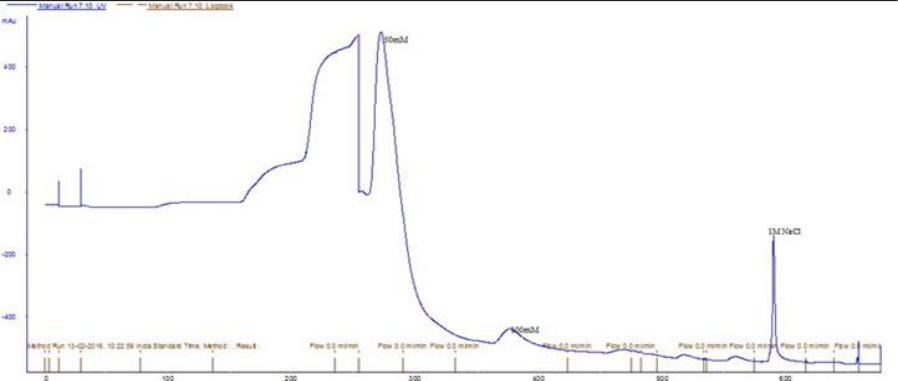 Figure 1
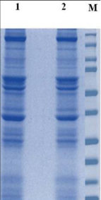 Figure 2
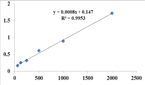 Figure 3
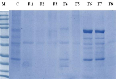 Figure 4A
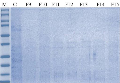 Figure 4B
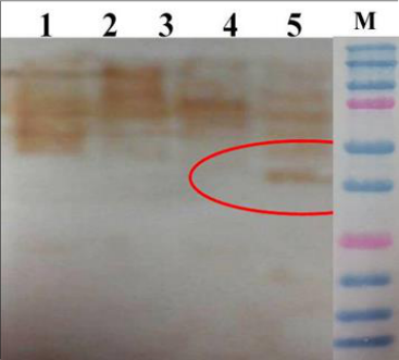 Figure 5A
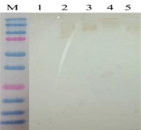 Figure 5B
Introduction
Lymphatic Filariasis (LF), caused by Wuchereria bancrofti, Brugia timori, and Brugia malayi, is a major vector-borne parasitic disease, commonly known as elephantiasis. It is transmitted by the mosquito vector Culex quinquefasciatus. Globally, 120 million people across 72 countries are affected by this disease, with over 51 million individuals infected as of 2018, of which 43 million suffer from irreversible chronic manifestations (WHO, 2024) [1] . LF causes significant physical and socio-economic problems for affected individuals (Durrheim et al., 2004 [2] ; Gbakima et al., 2005 [3] ; Sabesan et al., 2006 [4] ; Kreuels et al., 2008 [5] ; Streit and Lafontant, 2008 [6] ). In India, approximately 48.11 million people were infected with LF, primarily residing in 18 endemic states and Union Territories as of 2011 (Upadhyayula et al., 2012) [7] . The Global Programme to Eliminate Lymphatic Filariasis (GPELF), launched in 2000, aims to eliminate the disease through the Mass Drug Administration (MDA) program in co-endemic regions and to alleviate suffering by managing the disease's impact (WHO, 2017) [8] . In 2018, over 886 million people in 49 countries were at risk of LF (Collyer et al., 2020) [9] . The WHO reported a global decline of 74% in LF cases by 2018 due to active GPELF strategies. However, as stated in the GPELF progress report of 2019, India failed in 3 Implementation Units (IUs) during the Pre-transmission Assessment Survey (TAS), and 13 IUs failed in TAS while assessing the impact of MDA (Cromwell et al., 2020) [10] . Accurate and early detection tools, impact assessment, and post-MDA surveillance are critical for the success of LF elimination efforts. Recently, highly sensitive and specific immunodiagnostic kits have been developed, which have proven effective for monitoring the MDA program. Advanced techniques are also available for LF detection, such as W. bancroftispecific DNA probes (Hoti et al., 2002) [11] , molecular xenomonitoring protocols using mosquito vectors (Albuquerque et al., 2020) [12] , and polymerase chain reaction (PCR) assays using night blood samples from infected patients (McCarthy, 2000) [13] .
However, night blood sampling remains problematic in endemic countries, including India, due to its high cost, laborintensive nature, and the public's lack of compliance. Two of the immunological test kits such as the ICT card test (Weil et al., 1997; Kamkumo et al., 2015) [14,15] and Og4c3 ELISA kit (Hoti et al., 2002 [11] ; Wattal et al., 2007 [16] ; Mary et al. 2016 [17] ) are available currently commercially for the diagnosis of filarial infection that can detect the presence of circulating filarial antigens. These tests detect active infection, but during the post-MDA situation, when the transmission is expected to be very low tests that can detect the exposure are warranted. The commercially available antigen detection assays for bancroftian filariasis, namely the antigen test kit based on monoclonal antibody Og4C3 (Trop-Ag W. bancrofti, JCU Tropical Biotechnology, Pvt. Ltd, Townsville, Queensland, Australia) and the ICT card test (ICT Diagnostics, Balgowlah, New South Wales, Australia) CTK-Biotech-ICT, Encode Filariasis IgG/IgM Combo Rapid Test, PanLF Rapid-ICT, are very costly and therefore cannot be used for routine work including epidemiological surveys and control programs in India. Obtaining adequate amount of W. bancroftii antigens from human filarial infection is difficult. The cattle filarial parasite Seteria digitata, which resembles the human species in having microfilarial periodicity and chemotherapeutic response to known anti-filarial agents has been widely used as a model in various studies (Tandon et al., 1988) [18] including the immunological studies on diagnosis of W. bancrofti infection. Immunological studies on human bancroftian filariasis revealed that the sera of infected patients from high endemic region were showed higher specific antibody response than the sera collected from low endemic infected individuals (Jaoko et al. 2006) [19] . Setaria digitata, a filarial nematode dwelling in the peritoneal cavity of cattle has immunological determinants of filarial infection. The worm is quite similar with the human filarial parasite, W.bancrofti by morphology (Madathiparambil et al., 2011) [20] , histology (Decruse and Kaleysaraj, 1990) [21] and genome sequence (Senanayake et al., 2020) [22] . Hence it has been used as a model system in present study. The present study was carried out to identify the cross reacting protein of bovine filarial worm, S. digitata that could be used as a diagnostic marker to detect human filariasis at the early stage of infection by evaluating against the known human filarial sera sample.
Materials and Methods
Materials and Methods
Sample collection
Adult S. digitata parasites were collected from the peritoneal cavity of cattle slaughtered at the Greater Chennai Corporation abattoir and transferred to 1X Phosphate Buffered Saline (PBS, pH 7.2). The worms were transported to the laboratory, washed three times with sterile PBS, and stored at -20°C until use.
Preparation and purification of somatic antigens
Adult S. digitata worms were homogenized in sterile 1X PBS (pH 7.2) containing 0.1% of TritonX-100. The homogenate was dialysed against sterile 1XPBS (pH 7.2) at 4°C for overnight. The soluble material was extracted by centrifugation at 10,000 rpm for 20 min and the supernatant was stored at -20°C. Ion exchange chromatography using DEAE-Sepharose as an anion exchanger was performed for further purification. The DEAE-Sepharose column was equilibrated with 20 mM Tris-HCl buffer, pH 8.0. A 2.5 mL sample of S. digitata adult antigen extract was applied to the column at a flow rate of 0.5 mL/min. The proteins in the crude antigen were either eluted as a single fraction using 50 mM Tris-sodium chloride or fractionated with increasing concentrations of sodium chloride (50 mM, 100 mM, 150 mM, 200 mM, 250 mM, 300 mM). The column was washed with the same buffer at a flow rate of 1 mL/min, and 2 mL fractions were collected (Fig 1). Absorbance of the column fractions was measured at 280 nm, and fractions corresponding to the eluted peak were analyzed by SDS-PAGE. The protein concentration of various purified protein fractions was estimated using the Bicinchoninic Acid (BCA) method (Smith et al., 1985) [23] .
Fig 1:
Chromatogram output of the purification using wash buffer containing 50mM Tris, 50mM salt concentration and 5 ml column volume
Characterization and immunoblotting of purified somatic antigen of S. digitata
The protein profile of purified somatic antigen of S. digitata was analyzed by Sodium Dodecyl Sulfate-Polyacrylamide Gel Electrophoresis (SDS-PAGE) using 12% (w/v) gel under reducing condition as described by Laemmli (1970) [24] . Each well was loaded with 40µl of purified antigen. Electrophoresis was performed at 100V current in a Tris-glycine buffer system. The gel was stained with Coomassie brilliant blue R-250 (Sigma-Aldrich, USA). The reactivity of filarial antigens with known positive sera was analysed by immunoblotting technique as described by Towbin et al (1979) [25] . In brief, the setarial antigen was resolved by SDS-PAGE. The antigen components were electrophoretically transferred onto PVDF membrane with 0•6 amps current for 90 min at 4°C. After being blocked the PVDF membrane with 5% skimmed milk powder in 1X PBS, pH 7•2 for 1 hours at 37°C. The membrane was washed three times with PBST for 10 min and incubated further with a (1:50) dilution of convalescent sera of filaria exposed individual obtained from clinically suspected cases from JIPMER, Pondicherry for 1 hours at 37°C. The membrane was washed and incubated with a 1:8000 dilution of Recombinant Protein A\G Horse radish peroxidase-conjugate as secondary antibody and revealed with Diaminobenzidine (DAB) and 0.005% Hydrogen peroxide solution.
Results
Total protein from Setaria digitata was extracted using tissue homogenizer (Braun Biotech Homogenizer, Germany) followed by a freeze-thaw cycle. The protein sample was then run on a 12% SDS-PAGE gel, which revealed multiple bands of varying sizes. Four weak bands were observed in the range of 25 to 40 kDa, with one prominent band approximately at 37 kDa. Additionally, multiple sharp protein bands appeared in the range of 45 to 65 kDa (Figure 2). Western blotting using convalescent serum was done but the sera showed nonspecific binding (data not shown). The total protein concentration of crude lysate of Setaria digitata was quantified by using BCA method. The average total protein yield was approximately 2.2mg per worm (4.7mg/ml), (Figure 3). Additionally, four weaker protein bands were observed between 40-70 kDa (Figure 4a). In contrast, the purified fractions at a 100 mM salt concentration showed two faint peptide bands in the 50-70 kDa range in the first four fractions (Figure 4b).
Fig 4b:
12 percent SDS-PAGE analysis of purified protein fractions of Setaria digitata somatic antigen at 50mM and 100 mM salt concentration Immunoblot analysis of the purified somatic antigen from adult S. digitata (approximately 37 kDa) showed reactivity with convalescent sera samples collected from patients infected with filarial parasites (Figure 5a). However, the sera collected from filaria negative individual showed no immunoreactivity against purified somatic antigen of Setaria digitata (Figure 5b). Lane 1 to 5Reactivity of purified somatic antigen of S.digitata with negative antifilarial sera (Lane 1-5: 300, 200, 100, and 50 mM salt concentrations, respectively); MPrestained protein marker.
Discussion
Recent advances in various fields related to lymphatic filariasis (LF) have led to the development new approaches aimed at reducing the severity and impact of the disease (Ottesen et al., 1997) [26] . The introduction of novel diagnostic tools for detecting Wuchereria bancrofti infection in humans has opened new possibilities for interrupting transmission and, consequently, eliminating filariasis (WHO, 1994) [27] ; Crainey et al., 2016 [28] ; Sunish et al., 2015 [29] . This goal was reaffirmed in 1997 when the World Health Organization (WHO) set the target of eliminating filariasis worldwide by 2020 (Ottesen, 2000) [30] . The global program for LF elimination has now reached a stage where diagnostic paradigms are shifting. Microscopic examination of the microfilarial stage of the parasite is no longer feasible in the post-MDA (Mass Drug Administration) scenario, necessitating the use of alternative tools to detect filarialspecific antibodies, which indicate exposure to infection. Currently, there are no commercially available point-of-care kits for the early diagnosis of filariasis. Therefore, there is a need to develop rapid diagnostic serological tools to detect specific antigens in the early stages of infection. One of the major challenges in this area is the lack of sufficient quantities of filarial parasites and their antigens. This study aims to utilize heterologous antigens from the cattle parasite "Setaria digitata" as a diagnostic marker for human filariasis, to help prevent further disease transmission and enable early therapeutic intervention. Antigens were identified that reacted with the sera of individuals exposed to bancroftian filariasis. A 10% SDS-PAGE analysis of somatic antigens from Setaria cervi, purified using a preparative SDS-PAGE gel elution technique, revealed a prominent protein band at 37 kDa under reducing conditions, as visualized by silver staining (Priyadarshi et al., 2017) [31] . Sephacryl S-200 column-purified somatic antigen fractions of adult S. cervi revealed a prominent protein band in the 35-40 kDa range, while DEAE-cellulose-purified antigens displayed two major and one minor protein bands in the 65-75 kDa range in 10% SDS-PAGE analysis (Kaushal et al., 2009) [32] . Earlier studies on this parasite identified two adult worm antigens, 52 kDa and 130 kDa, which were used to detect Wuchereria bancrofti microfilariae in endemic areas (Wickremanayake et al., 2001) [33] . John and Raj (1998) [34] found that the heat-inactivated, detergent-soluble protein of S. digitata, with a molecular weight of 29 kDa, suppressed the host immune response and reported that this 29 kDa protein had diagnostic value in patients infected with bancroftian filariasis. Affinity-purified, E. coli-expressed recombinant protein of third-stage larvae of Brugia malayi (rBm-SL3) revealed a protein band at 37 kDa in 10% SDS-PAGE analysis. The rBm-SL3 protein showed a specific antibody response only with sera from individuals in endemic areas in immunoblot analysis (Dabir et al., 2006) [35] . Surface antigens from S. digitata isolated by EDTA extraction and purified by affinity chromatography using W. bancrofti infected sera demonstrated approximately six antigenic bands (ranging from 10-89 kDa) in immunoblotting (Theodore and Kaliraj, 1990) [36] . Two monoclonal antibodies, namely 13B4 and 15D6, have been developed against antigenic epitopes that are common to both S. cervi and B. malayi. These antibodies have proven useful for detecting circulating antigens in filarial-infected individuals (Kaushal et al., 1994) [37] , but not for measuring exposure. Known positive human anti-filarial sera (IgG antibody) collected from endemic regions reacted with adult somatic antigens from S. digitata and B. malayi, but not with their microfilarial antigens (Satapathy and Sahoo, 2019) [38] . In contrast, the secretory glycoprotein (200 kDa) of S. digitata dissociated into 15 polypeptide bands in the 11-66 kDa range under reducing conditions in SDS-PAGE analysis, with all proteins reacting with the mouse monoclonal antibody MK6BE3 in immunoblotting (Madathiparambil et al., 2009) [39] .
Conclusion
This present study demonstrates that cross-reactivity of antigens derived from S. digitata with human sera from W. bancrofti infected individuals occurs, highlighting the presence of a potential sero-reactive protein at ~37 kDa. Further purification and proteomic analysis of this antigen may provide valuable insights into how human infection with filarial parasites affects the sensitivity of diagnostic tests for W. bancrofti. The promising nature of this antigen suggests its potential utility in measuring individual exposure to filarial infection and in assessing transmission dynamics during post-MDA surveillance, making it an important candidate for improving diagnostic and surveillance methods for filarial diseases.
Acknowledgement
AcknowledgementThe authors are thankful to The Director and other technical staffs, CAHS, TANUVAS for the facilities and the authorities of the Greater Chennai Corporation slaughter house for the support provided for carrying out the work.
Figures and tables from the paper
References
39 in the original paper- World Health Organization. Elimination of lymphatic filariasis as a public health problem in Timor-Leste, 2024. Available from: https://www.who.int/news/item/08-10-2024-eliminationof-lymphatic-filariasis-as-a-public-health-problem-intimor-leste
- Durrheim DN, Wynd S, Liese B, Gyapong JO. Lymphatic filariasis endemicity -an indicator of poverty? Trop Med Int Health. 2004;9:843-845.
- Gbakima AA, Appawu MA, Dadzie S, Karikari C, Sackey SO. Lymphatic filariasis in Ghana: establishing the potential for an urban cycle of transmission. Trop Med Int Health. 2005;10(13):387-392.
- Sabesan S, Raju HK, Srividya A, Das PK. Delimitation of lymphatic filariasis transmission risk areas: A geoenvironmental approach. Filaria J. 2006;5:12-14.
- Kreuels B, Kobbe R, Adjei S, Kreuzberg C, Reden VC. Spatial variation of incidences in young children from a geographically homogeneous area with high endemicity. J Infect Dis. 2008;197:85-93.
- Streit T, Lafontant JG. Eliminating lymphatic filariasis: A view from the field. Ann NY Acad Sci. 2008;1136:53-63.
- Upadhyayula SM, Mutheneni SR, Kadiri MR, Kumaraswamy S, Nagalla B. A cohort study of lymphatic filariasis on socio-economic conditions in Andhra Pradesh, India. PLoS ONE. 2012;7(3):e33779.
- World Health Organization. Global programme to eliminate lymphatic filariasis: Progress report, 2016. Wkly Epidemiol Rec. 2017;92:594-607.
- Collyer BS, Irvine MA, Hollingsworth TD, Bradley M, Anderson RM. Defining a prevalence level to describe the elimination of Lymphatic Filariasis (LF) transmission and designing monitoring and evaluating (M&E) programmes post the cessation of mass drug administration (MDA). PLOS Negl Trop Dis. 2020;14(10):e0008644.
- Cromwell EA, Schmidt CA, Kwong KT, Pigott DM, Mupfasoni D, Biswas G, et al. The global distribution of lymphatic filariasis, 2000-18: A geospatial analysis. Lancet Global Health. 2020;8(9):e1186-94.
- Hoti SL, Elango A, Radjame K, Yuvaraj J, Pani SP. Detection of day blood filarial antigens by Og4C3 ELISA test using filter paper samples. Natl Med J India. 2002;15(5):263-266.
- Albuquerquea ALD, Araujoa TAD, Meloa DCTV, Paivaa MHS, Meloc FL, Oliveiraa CMF, et al. Development of a molecular xenomonitoring protocol to assess filariasis transmission. Exp Parasitol. 2020;215:1-7.
- McCarthy J. Diagnosis of lymphatic filarial infection. In: Lymphatic Filariasis, ed. Nutman TB, Imperial College Press, London, 2000, p. 127-41.
- Weil GJ, Lammie PJ, Weiss N. The ICT filariasis test: a rapid format antigen test for diagnosis of Bancroftian filariasis. Parasitol Today. 1997;13:401-440.
- Kamkumo RG, Djeunga NHC, Bopda J, Akame J, Tarini A, Kamgno J. Loss of sensitivity of Immunochromatographic Test (ICT) for lymphatic filariasis diagnosis in low prevalence settings: consequence in the monitoring and evaluation procedures. BMC Infect Dis. 2015;15:579.
- Wattal S, Dhariwal AC, Rhalhan PK, Tripathi VC, Regu K, Kamal S, et al. Evaluation of Og4C3 antigen ELISA as a tool for detection of bancroftian filariasis under lymphatic filariasis elimination programme. J Commun Dis. 2007;39(2):75-84.
- Mary KA, Krishnamoorthy K, Hoti SL. Scope of detectability of circulating antigens of human lymphatic filarial parasite Wuchereria bancrofti with smaller amount of serum by Og4C3 assay: Its application in lymphatic filariasis elimination programme. J Parasit Dis. 2016;40(4):1622-1626.
- Tandon A, Murthy PK, Saxena RP, Sen AB, Saxena KC. Dot ELBA for diagnosis of lymphatic filariasis. Indian J Med Res. 1988;87:429-431.
- Jaoko WG, Simonsen PE, Meyrowitsch DW, Benson B, Estambale A, Lazaro MMN, et al. Filarial-specific antibody response in East African bancroftian filariasis: effects of host infection, clinical disease, and filarial endemicity. Am J Trop Med Hyg. 2006;75(1):97-107.
- Madathiparambil MG, Raj NK, Linda J, Kaleysa R. Surface of the filarial parasite Setaria digitata and the structural changes on treatment with Triton X-100. Helminthologia. 2011;48:17-22.
- Decruse SW, Raj RK. Histological studies on female Setaria digitata (von Linstow 1906), a filaria of bovine. Proc Ani Sci. 1990;99:103-112.
- Senanayake KS, Derberg JS, Polajev A, Malmberg M, Karunanayake EH, Tennekoon KH, et al. The genome of Setaria digitata: A cattle nematode closely related to human filarial parasites. Genome Biol Evol. 2020;12(2):3971-3976.
- Smith PK, Krohn RI, Hermanson GT, Mallia AV, Gartner FH, Provenzano MD, et al. Measurement of protein using bicinchoninic acid. Anal Biochem. 1985;150:76-85.
- Laemmli UK. Cleavage of structural proteins during assembly of the head of T4 bacteriophage. Nature. 1970;227:630-634.
- Towbin H, Staehelin T, Gordon J. Electrophoretic transfer of proteins from polyacrylamide gels to nitrocellulose sheets: Procedure and some applications. Proc Natl Acad Sci. 1979;76(9):4350-4354.
- Ottesen EA, Duke BOL, Karam M, Behbehani K. Strategies and tools for the control/elimination of lymphatic filariasis. Bull World Health Organ. 1997;75:491-503.
- World Health Organization. Lymphatic filariasis infection and disease control strategies. Report of a WHO/CTD/TDR consultative meeting held at the Universiti Sains Malaysia, Penang, Malaysia, 1994, p. 22-24.
- Crainey JL, Ribeiro da Silva TR, Luz SLB. Historic accounts of Mansonella parasitaemias in the South Pacific and their relevance to lymphatic filariasis elimination efforts today. Asian Pac J Trop Med. 2016;9(3):205-10.
- Sunish IP, Kalimuthu M, Rajendran R, Munirathinam A, Kumar AV, Nagaraj J, et al. Decline in lymphatic filariasis transmission with annual mass drug administration using DEC with and without albendazole over a 10-year period in India. Parasitol Int. 2015;64(1):1-4.
- Ottesen EA. The global programme to eliminate lymphatic filariasis. Trop Med Int Health. 2000;5(9):591594.
- Priyadarshi P, Dravid P, Sheikh IH, Saxena S, Tandon A, Kaushal DC, et al. Characterization of immunoreactive proteins of Setaria cervi isolated by preparative polyacrylamide gel electrophoresis. Acta Parasitol. 2017;62(1):29-37.
- Kaushal NA, Srivastava N, Mustafa H, Tandon A, Singh SK, Kaushal DC. Immunological investigations: isolation of an antigen fraction from Setaria cervi adults having potential for immunodiagnosis of human filariasis. Immunol Invest. 2009;38:749-761.
- Wuchereria bancrofti: Detection of microfilariae in asymptomatic microfilaremic individuals with Setaria digitata antigens. Southeast Asian J Trop Med Public Health. 2001;32(2):230-234.
- John L, Raj RK. Immune response of electroeluted detergent soluble 29 kDa antigen from Setaria digitata (Von Linstow). Indian J Exp Biol. 1998;36(9):862-866.
- Dabir P, Dabir S, Krithika KN, Goswami K, Reddy MVR. Immunoprophylactic evaluation of a 37-kDa Brugia malayi recombinant antigen in lymphatic filariasis. Clin Microbiol Infect. 2006;12:361-368.
- Theodore JG, Kaliraj P. Isolation, purification and characterization of surface antigens of the bovine filarial parasite Setaria digitata for the immunodiagnosis of bancroftian filariasis. Helminthol. 1990;64(2):105-114.
- Kaushal NA, Kaushal DC, Ghosh S, Talwar GP. Monoclonal antibodies against antigenic epitopes common between Setaria cervi and Brugia malayi. Indian J Exp Biol. 1994;32:371-375.
- Satapathy AK, Sahoo PK. Species-specific and crossreactive antigen of filarial worm: Brugia malayi and Setaria digitata. J Vector Borne Dis. 2019;50(4):323329.
- Madathiparambil MG, Kavitharaj N, Kaleysa RR. A diagnostically useful 200-kDa protein is secreted through the surface pores of the filarial parasite Setaria digitata. Parasitol Res. 2009;105(4):1099-1104.