
Research article · 2025 · Full text
International Journal of Veterinary Sciences and Animal HusbandryFirst report of sarcoptic mange in a wild dog from India and its clinical management
Volume 10(3) · Pages 22–27 · DOI: 10.22271/veterinary.2025.v10.i3a.2101 ↗
Research areasDermatological diseases and infestationsInsects and Parasite InteractionsYersinia bacterium, plague, ectoparasites research
Abstract
Author-writtenSarcoptic mange, caused by the mite Sarcoptes scabiei, is an emerging global zoonosis that poses significant risks to wildlife conservation. This report documents the first known case of sarcoptic mange in a wild Asiatic wild dog (Cuon alpinus) in India, along with its clinical management. Wild canids, such as the endangered wild dogs, face severe threats from habitat fragmentation, human encroachment, and disease transmission. In this case, a wild dog exhibiting extensive alopecia and pruritus was captured from the Pench Tiger Reserve in Maharashtra, India. Clinical examination revealed alopecia concentrated on the hindquarters, forelimbs, and tail, with skin scrapings confirming the presence of sarcoptic mites. Blood and faecal samples were collected to rule out other parasitic infections, which identified co-infections with Taenia spp., Toxocara spp., and Sarcocystis spp. The treatment protocol included weekly administration of Doramectin, combined with antihistamines, vitamin supplements, and topical antifungal agents. A second round of tests post-treatment showed significant improvement, with complete resolution of alopecia by day 21. Sarcoptic mange has been implicated in population declines of endangered species across the world. However, data from India, particularly concerning wild dogs, is limited. The disease disrupts pack dynamics and poses challenges for treatment in free-ranging populations. This case highlights the critical need for improved disease surveillance, veterinary infrastructure, and timely intervention strategies to mitigate the effects of sarcoptic mange in endangered species. Enhanced research, coupled with conservation efforts, is essential to safeguard the remaining populations of wild dogs and ensure their long-term survival. The findings underline the importance of addressing wildlife diseases in conservation planning, especially in regions where endangered species live at the interface of human activities and natural habitats. The study calls for a holistic approach involving wildlife veterinarians, conservationists, and policymakers to address the broader implications of disease management in wildlife population.
Summary
Source-checkedPlain-language reading of the full paper, prepared with AI and checked against the text. Each point links to the authors' own words.
Why it matters
- Sarcoptic mange is a global zoonotic disease caused by Sarcoptes scabiei mites that can threaten wildlife conservation. View evidence ↗
- Asiatic wild dogs (Cuon alpinus) are an endangered species with fewer than 2,500 individuals remaining. View evidence ↗
- Wild dog packs often live near human areas and interact with domestic dogs, which increases the risk of disease transmission. View evidence ↗
The question
- The study aims to document the first known case of sarcoptic mange in an Asiatic wild dog in India and describe its clinical management. View evidence ↗
What they did
- The subject was an approximately six-year-old adult female wild dog captured using Xylazine-Ketamine anesthesia. View evidence ↗
- Mites were screened using light microscopy of skin scrapings after 10% Potassium hydroxide digestion. View evidence ↗
- Treatment included weekly Doramectin (0.6 mg/kg), daily Hydroxyzine hydrochloride, weekly Vitamin A (25,000 IU) and Vitamin D (60,000 IU), and topical antifungal powder. View evidence ↗
- Deworming was performed using Kiwoff® Plus XL, and Fluralaner (500 mg) was administered orally via chicken after the first week. View evidence ↗
What they found
- Microscopic examination of skin scrapings confirmed the presence of sarcoptic mites, diagnosing the animal with sarcoptic mange. View evidence ↗
- Faecal analysis revealed co-infections with Taenia sp., Toxocara sp., and Sarcocystis spp. View evidence ↗
- Alopecia began to improve after day 14 and was completely resolved by day 21. View evidence ↗
- A single dose of fluralaner caused the animal to experience anorexia and uneasiness for one day. View evidence ↗
What it means
- The authors conclude that there is a critical need for better disease surveillance and veterinary infrastructure to protect endangered species from sarcoptic mange. View evidence ↗
- The authors note that research gaps exist due to limited funding, trained manpower, and veterinary infrastructure in India. View evidence ↗
Visual summary
2 visualsIllustrations explain the paper; charts are replotted from the paper's own table values.
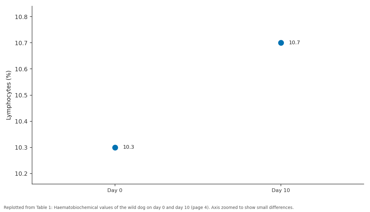
Lymphocytes (%): Day 0 vs Day 10
Replotted from Table 1 (page 4) of the original paper. Every bar is the paper's own table value.
View source evidence ↗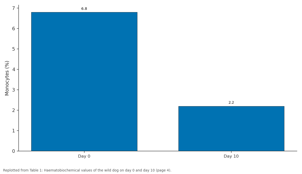
Monocytes (%): Day 0 vs Day 10
Replotted from Table 1 (page 4) of the original paper. Every bar is the paper's own table value.
View source evidence ↗Figures & tables
From the original paper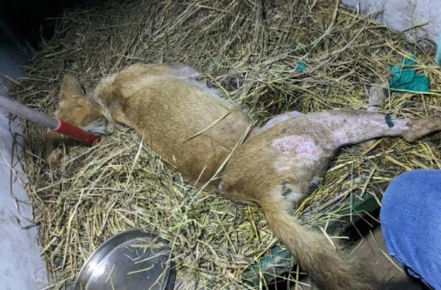 Figure 1
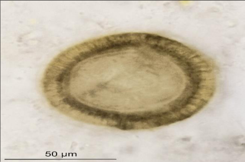 Figure 2
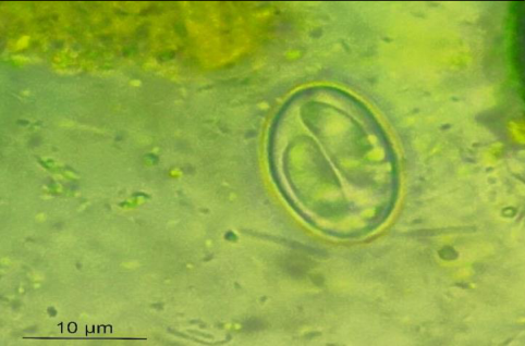 Figure 3
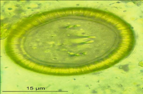 Figure 4
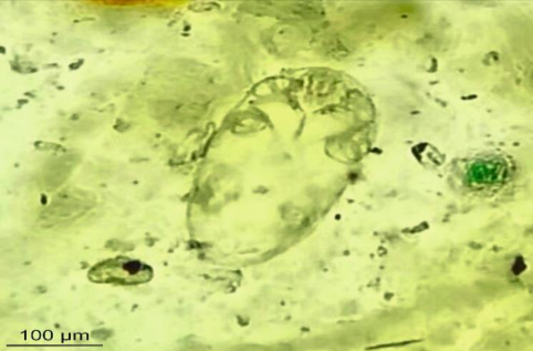 Figure 5
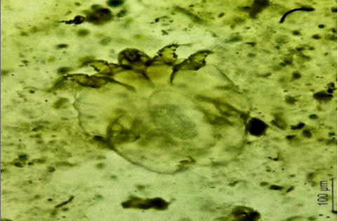 Figure 6
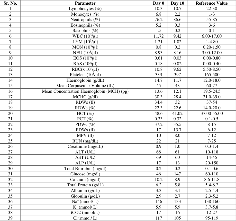 Table 1
Introduction
India is a biodiversity hotspot and is home to numerous species of wildlife. The mammalian taxa in the region is diverse and includes a variety of mammals inhabiting deserts, ice clad mountains, forests, marsh land etc. In the late 1970s the country saw a rapid industrial development leading to clearance of large area of forest for the development infrastructure like roads and railways. In preceding years, due to population explosion, a lot of forest land has been diverted to agriculture, development infrastructure and industries [1] . Loss of habitat has played a major role in reclining of population of many endangered species of the region. The government formulated policies to protect wildlife through the Wildlife Protection Act, 1972. The efforts of the government have led to stabilisation of many endangered population across the country. Diseases have been consistently neglected as a potential threat to endangered wildlife species. There have been evidences where diseases have led to bottle neck of the population or local extinction of a species [2] . Under the Indian context; stringent policy implementation, limited avenues to study wildlife and non-availability of the laboratories and facilities has been the greatest limitation in generating dependable data.
Canids are an important and diverse group of wild animals present throughout the country. Dhole or Asiatic wild dogs (Cuon alpinus) are endangered species found only in limited parts of the southern peninsular region of the country [3] . A typical pack size of these species in deciduous forests of central India is 12-14, however, the smaller packs of 2-4 individuals have also been noticed in the Pench Tiger Reserve, Maharashtra, India. The species are pack hunters and feed on spotted deers (Axis axis), rabbits, hares and birds. In recent times disease outbreaks have led to decline of the population in many regions of the country. In many of the protected areas, the wild dog pack thrive along the humanwildlife interface and interact with domestic dogs, leading to the risk of disease transmission [4] . Canine distemper is a serious concern among wild canids and can lead to serious illnesss among the affected individuals [5] . Ectoparasites are often neglected until they manifest clinical outcomes in the endangered species. Mange caused due to mites is a serious concern in endangered species across the world. Sarcoptic mange is caused due to Sarcoptes scabiei and has a global distribution across 150 species of wild and domestic animals [6] . The clinical manifestation of the infection is acute or chronic and transmission across species is a possibility. In the wild, the infection has been documented in wide array of species including the wild canids. Among the wild canids, the wild dogs, jackals and wolves have been found to be susceptible. Sarcoptic mange has been ranked among the top ten diseases leading to population decline [7] . The infection poses a threat to two endangered species Tatras chamois (R. rupicapra tatrica) and Northern hairy-nosed wombat (L.krefftii), while it also is a concern in many threatened species like the cheetah (A. jubatus), Mountain gorilla (G. beringei beringei) and Common chimpanzee (P. troglodytes). Reports of death due to sarcoptic mange and its complications have documented in cheetah. The study reported prevalence of 12.77% in Cheetahs and 4.7% in domesticated in Masai mara Kenya [8] . Reports of mange infection in endangered long-tailed goral (Naemorhedus caudatus) have raised concerns about the impact of the infection on the population dynamics [9] . Wild dogs are endangered as per the International Union for Conservation of Nature (IUCN) and fewer than 2500 individuals are the last survivors of the species [10] . The population of the species has declined due to fragmentation of the habitat, inbreeding, hunting, snaring etc. [11] . Incidences of mange in general have been reported from many parts of the world in a wide variety of species [12,13] including canids [14] . In dholes, the transmission of sarcoptic mange is attributed to prey species and domestic dogs at the human-wildlife fringes. There is no report on the matter from the Indian subcontinent, though interaction with forest rangers confirms the sporadic incidences of mange among wild dog packs.
Case Report Anamnesis & capture of wild dog
The forest department officials of the Sillari-Chourbahuli Range of Pench Tiger Reserve, Maharashtra, India noticed extensive alopecia in wild dog pack (Figure 1 & 2). The animals were restless and were found to scratch their body frequently. The alopecia was majorly limited to hind quarters, forelimbs and tail. In order to diagnose the etiology, it was decided to capture one animal from the pack with lesions and obtain samples for diagnosis. Accordingly, the pack of wild dogs was located through combing operations and deployment of trap cameras. An adult female, approximately six years was capture using Xyalazine-Ketamine anaesthesia using a darting gun (Dan-inject, Denmark). Blood, serum, faeces and skin scrapings were collected taking aseptic precautions. The dog was weighed using a spring balance and was immediately shifted to transport cage. The anaesthesia was reversed using Yohimbine. All precautions to minimize the human imprinting using visual barriers and minimising human presence in the locality.
Blood, Serum, Faecal, Skin scrapping and molecular examination
The blood was processed for a complete blood check and serum for serum biochemistry. The DNA was isolated from the blood (Blood & Tissue Kit, Qiagen) for screening for blood parasites. A PCR based test was used to screen for common blood protozoan including hepatozoonosis, erhlichiosis, babesiosis, theileriosis, anaplasmosis and trypanosomosis (Table 1). A blood smear was stained with Leishman's stain to screen for presence of haemoprotozoans. The flotation method using saturated salt solution as described in the reference was used for the detection of parasitic infection in the faecal sample [15] . The test was repeated after 14 days post-treatment to monitor the recovery. The skin scrapings were subjected to 10% Potassium hydroxide digestion by heating the slide on a flame followed by light microscopy (Mfg. Leica, Wetzlar, Germany) to screen for the presence of mites [15] .
| Sr. No. | Parameter | Day 0 | Day 10 | Reference Value |
|---|---|---|---|---|
| 1 | Lymphocytes (%) | 10.3 | 10.7 | 22-30 |
| 2 | Monocytes (%) | 6.8 | 2.2 | 1-3 |
| 3 | Neutrophils (%) | 76.2 | 86.6 | 55-85 |
| 4 | Eosinophils (%) | 5.2 | 0.3 | 3-6 |
| 5 | Basophils (%) | 1.5 | 0.2 | 0-1 |
| 6 | WBC (103/µl) | 11.72 | 9.42 | 6.00-17.00 |
| 7 | LYM (103/µl) | 1.21 | 1.02 | 1-4.80 |
| 8 | MON (103/µl) | 0.8 | 0.2 | 0.20-1.50 |
| 9 | NEU (103/µl) | 8.93 | 8.16 | 3.00-12.00 |
| 10 | EOS (103/µl) | 0.61 | 0.03 | 0.00-0.80 |
| 11 | BAS (103/µl) | 0.18 | 0.02 | 0.00-0.40 |
| 12 | RBC(x 106/µl) | 10.8 | 9.62 | 5.50-8.50 |
| 13 | Platelets (103/µl) | 333 | 397 | 165-500 |
| 14 | Haemoglobin (g/dL) | 14.7 | 11.7 | 12.0-18.0 |
| 15 | Mean Corpuscular Volume (fL) | 45 | 43 | 60-77 |
| 16 | Mean Concentration Haemoglobin (MCH) (pg) | 13.6 | 12.1 | 19.5-24.5 |
| 17 | MCHC (g/dl) | 30.3 | 28.4 | 31.0-39.0 |
| 18 | RDWs (fl) | 34.4 | 32 | 37-54 |
| 19 | RDWc (%) | 22.3 | 22.6 | 14.0-20.0 |
| 20 | HCT (%) | 48.6 | 41.02 | 37.00-55.00 |
| 21 | PCT (%) | 0.33 | 0.32 | 0.1-0.5 |
| 22 | PDWc (%) | 37.2 | 35.5 | 8-15 |
| 23 | PDWs (fl) | 17 | 13.7 | 6-12 |
| 24 | MPV (fl) | 10 | 8.0 | 7-12 |
| 25 | BUN (mg/dL) | 22 | 21 | 7-25 |
| 26 | Creatinine (mg/dL) | 0.9 | 1.0 | 0.3-1.4 |
| 27 | ALT (U/L) | 68 | 61 | 10-118 |
| 28 | AST (U/L) | 69 | 60 | 14-45 |
| 29 | ALP (U/L) | 17 | 13 | 20-150 |
| 30 | Total Bilirubin (mg/dl) | 0.2 | 0.2 | 0.1-0.6 |
| 31 | Glucose (mg/dl) | 46 | 147 | 60-110 |
| 32 | Calcium (mg/dl) | 10.2 | 8.9 | 8.6-11.8 |
| 33 | Total Protein (g/dL) | 6.2 | 5.8 | 5.4-8.2 |
| 34 | Albumin (g/dL) | 3.3 | 3.1 | 2.5-4.4 |
| 35 | Globulin (g/dL) | 2.9 | 2.7 | 2.3-5.2 |
| 36 | Na+ (mmol/ L) | 146 | 133 | 138-160 |
| 37 | K+ (mmol/ L) | 5.9 | 5.9 | 3.7-5.8 |
| 38 | tCO2 (mmol/L) | 17 | 16 | 12-27 |
| 39 | Cl-(mmol/ L) | 117 | 105 | 95-119 |
Table text extracted from the original PDF; superscript letters are shown inline. The original table image is below.
View original table image
Results
The Blood smear was negative for the presence of intracellular and extracellular haemoparasites. The same was confirmed by PCR test. The faecal sample was found to be positive for Taenia sp. Toxocara sp. and Sarcocystis spp (Figure 3,4,5). The 10% KOH digestion followed by microscopy revealed the presence of sarcoptic mites (Figure Thus, the case was diagnosed as sarcoptic mange with parasitic co-infection.
Treatment
The treatment consisted of injection Doramectin at a dosage of 0.6 mg/ kg at weekly intervals along with Tab. Atarax [Hydroxyzine hydrochloride] once a day, Vitamin A 25000 IU once a week, Vitamin D 60000 IU once a week and topical application of Candid powder. Deworming was carried out with half tablet Kiwoff ® Plus XL (Fenbental 150 mg, Praziquantel 50 mg and pyrantel pamoate 144 mg). After the first week, tablet Bravecto (Fluranalar 500 mg for 10-20 kg body weight) was administered orally by lacing the chicken (16). The wild dog was offered chicken with bones at the rate of 750 gms/day and was monitored using closed circuit camera. On day 14, the faecal samples were re-examined and found positive for spirometra sp (Figure 5). Blood sample was collected and analysed on day 14 to monitor the progress of the treatment. The wild dog showed improvement since day four with less scratching and consuming all the offered feed. The alopecia improved after the day 14 th and by day 21 the alopecia was completely resolved.
Discussion
Sarcoptic mange affects a wide range of mammalian species, including wild canids and humans [17] . The disease manifests with intense itching, alopecia, and secondary infections, severely impacting the health of affected animals. In wild dogs, particularly endangered species, sarcoptic mange can have devastating effects on individual survival and population dynamics, posing a significant threat to wildlife conservation [18] . The incidences of sarcoptic mange have been reported in many species of wildlife including African wild dogs (Lycaon pictus) [19] , Ethiopian wolf (Canis simensis) [20] , Red foxes (Vulpes vulpes) [21,22] , Gray wolves (Canis lupus), Coyotes (Canis latrans) [18] . The prevalence in some of the canid population has been significantly high. The African wild dogs reported a prevalence of 50-60% due to anthropogenic pressure and habitat loss in South Africa [19] . Similarly, Red foxes in Europe reported a prevalence of 80% in some areas, leading to significant declines in affected populations [23] . Sarcoptic mange poses a serious conservation challenge for endangered wild canid species on account of population decline [20] , loss of genetic diversity, disruption of pack structure [19] , overabundance of prey species and altered ecosystem dynamics [23] . Despite its global impact, there is a considerable lack of data on sarcoptic mange in wild canids, especially in India. India is home to many endangered wild canids like wild dogs, Indian wolf, Indian jackal etc. Parasitic infestation is commonly reported in wild canids however, manage and parasitic infestation has been previously reported from Europe [24] . Due to factors like limited veterinary infrastructure, stringent law enforcement, limited trained manpower and lack of funding for the research in these areas definite research gaps exists.
The diagnosis and treatment of the infection is critical in the pack living species as the infection quickly can spread to other members of the pack. The clinical presentation of all the infected animals was similar and consisted of uneasiness, scratching, redness at the infected parts, bruises due to scratching etc. The distribution of the lesions was majorly on the rump, hind limb and forelimbs with a few individuals exhibiting alopecia of neck and thorax. The diagnosis of confirms the observations of animals infected with sarcoptic mange. The treatment involves specific anti-infective, antiallergics, broad spectrum antifungals and vitamin supplementation [25] . The response to the therapy was noticed with the resolution of the alopecia and skin lesions. In the free range the capture and treatment of wild animals that are to be released in their natural habitat must be carried out in such a manner that there is minimal human imprinting. In the current case report, the possibility of the release of the animal on recovery was considered from day one and minimal human presence and naturalistic surroundings were provided during the treatment. The medications were majorly provided through the food thus bypassing the need to restrain the animal daily. Treatment of free-ranging populations is logistically challenging as pack dynamics are sensitive, repeated handling of individuals for treatment may lead to stress and social disruption, further complicating recovery efforts. The use of antiparasitic baits is projected as a more feasible solution for treating larger populations [19] . Ivermectin has been successfully used to treat mange in wildlife [20] , alongside Moxidectin [26,18] and doramectin. Topical Treatments with amitraz are also utilised for the treatment of the animals. In the current case, doramectin was employed with Fluranalar along with supportive therapy. The single dose therapy of fluranalar led to uneasiness and anorexia for a day. During the same time, Mudhumali Tiger Reserve in the Tamil Nadu state of India reported similar cases in wild dogs. However, the team could not capture any of the affected wild dogs to investigate the disease outbreak.
Acknowledgement
AcknowledgementsThe authors acknowledge the kind approval of the Principal Chief Conservator of Forest (Wildlife) & Chief Wildlife Warden, Maharashtra State, for carrying out the research work. Forest Development Corporation of Maharashtra Ltd., Nagpur and Office of the Divisional Manager, Gorewada Project, Nagpur, for the facilities provided for the research. Field Director, PTR, Nagpur for the permission and access to treat the wild dog. Shri. Kaustubh Gawande, (People for Animals, Wardha), Dr. Rohini Bawaskar, Veterinary Officer, APCCF, Nagpur, Dr. Nikhill Bangar, Veterinary Officer, PTR, Nagpur for the cooperation extended in treating the wild dog. Director, Wildlife Research & Training Centre to facilitate the research work at WRTC, Gorewada, Nagpur.
Figures and tables from the paper
References
26 in the original paper- Barve N, Kiran MC, Vanaraj G, Aravind NA, Rao D, Shaanker RU, Ganesan KN, Poulsen JG. Measuring and mapping threats to a wildlife sanctuary in southern India. Conservation Biology. 2005;19(1):122-30. Available from: https://repository.ias.ac.in/13212/1/335.pdf
- James TY, Litvintseva AP, Vilgalys R, Morgan JA, Taylor JW, Fisher MC, et al. Rapid global expansion of the fungal disease chytridiomycosis into declining and healthy amphibian populations. PLOS Pathogens. 2009;5(5):e1000458. Available from: https://journals.plos.org/plospathogens/article/file?id=10. 1371/journal.ppat.1000458&type=printable
- Srivathsa A, Puri M, Karanth KK, Patel I, Kumar NS. Examining human-carnivore interactions using a socioecological framework: Sympatric wild canids in India as a case study. Royal Society Open Science. 2019;6(5):182008. Available from: https://royalsocietypublishing.org/doi/full/10.1098/rsos.1 82008
- Nayak RM, Jadav KK, Rajput N, Gupta S, Rokde A, Singh KP. Surveillance of major canine pathogens in feral dogs and big cats at the domestic-wildlife interface in Panna tiger reserve, India. Journal of Animal Research. 2020;10(2):303-308. Available from: https://www.indianjournals.com/ijor.aspx?target=ijor:jar &volume=10&issue=2&article=020
- Deem SL, Spelman LH, Yates RA, Montali RJ. Canine distemper in terrestrial carnivores: A review. Journal of Zoo and Wildlife Medicine. 2000;31(4):441-51. Available from: https://bioone.org/journals/journal-ofzoo-and-wildlife-medicine/volume-31/issue-4/10427260_2000_031_0441_CDITCA_2.0.CO_2/caninedistemper-in-terrestrial-carnivores-areview/10.1638/10427260(2000)031[0441:CDITCA]2.0.CO;2.short
- Escobar LE, Carver S, Cross PC, Rossi L, Almberg ES, Yabsley MJ, et al. Sarcoptic mange: An emerging panzootic in wildlife. Transboundary and Emerging Diseases. 2022;69(3):927-42. Available from: https://onlinelibrary.wiley.com/doi/abs/10.1111/tbed.140 82
- Cypher BL, Rudd JL, Westall TL, Woods LW, Stephenson N, Foley JE, et al. Sarcoptic mange in endangered kit foxes (Vulpes macrotis mutica): Case histories, diagnoses, and implications for conservation. Journal of Wildlife Diseases. 2017;53(1):46-53. Available from: https://meridian.allenpress.com/jwd/article/53/1/46/1267
- Gakuya F, Ombui J, Maingi N, Muchemi G, Ogara W, Soriguer RC, et al. Sarcoptic mange and cheetah conservation in Masai Mara (Kenya): Epidemiological study in a wildlife/livestock system. Parasitology. 2012;139(12):1587-1595. Available from: https://www.cambridge.org/core/journals/parasitology/art icle/abs/sarcoptic-mange-and-cheetah-conservation-inmasai-mara-kenya-epidemiological-study-in-awildlifelivestocksystem/F0BBA5A9B1AC6B001F6A669D4D6F29C6
- Choi J, Kim HJ, Kim JY, Kim MH, Lee JY, Moon JC, et al. Two cases of mange mite (Sarcoptes scabiei) infestation in long-tailed goral (Naemorhedus caudatus) in Republic of Korea. The Korean Journal of Parasitology. 2022;60(6):423. Available from: https://pmc.ncbi.nlm.nih.gov/articles/PMC9806499/
- Kamler JF, Songsasen N, Jenks K, Srivathsa A, Sheng L, Kunkel K. Cuon alpinus. The IUCN Red List of Threatened Species 2015: e. T5953A72477893. Available from: https://www.researchgate.net/profile/ArjunSrivathsa/publication/286451829_Cuon_alpinus_The_IU
- Modi S, Mondol S, Nigam P, Habib B. Genetic analyses reveal demographic decline and population differentiation in an endangered social carnivore, Asiatic wild dog. Scientific Reports. 2021;11(1):16371. Available from: https://www.nature.com/articles/s41598021-95918-3
- Guillot J, Losson B, Delsart M, Briand A, Fang F, Rossi L. Sarcoptic mange in wild and domestic animals. In: Scabies. Cham: Springer International Publishing, 2023, p. 313-43. Available from: https://link.springer.com/chapter/10.1007/978-3-03126070-4_23
- Pérez JM, Granados JE, Espinosa J, Bravo RA, Olvera LJR, Rossi Let al. Biology and management of sarcoptic mange in wild Caprinae populations. Mammal Review. 2021;51(1):82-94. Available from: https://onlinelibrary.wiley.com/doi/abs/10.1111/mam.122 13
- Eo KY, Kwon OD, Shin NS, Shin T, Kwak D. Sarcoptic mange in wild raccoon dogs (Nyctereutes procyonoides) in Korea. Journal of Zoo and Wildlife Medicine. 2008;39(4):671-3. Available from: https://bioone.org/journals/journal-of-zoo-and-wildlifemedicine/volume-39/issue-4/2008-0034.1/SarcopticMange-in-Wild-Raccoon-Dogs-Nyctereutesprocyonoides-in-Korea/10.1638/2008-0034.1.short
- Taylor MA, Coop RL, Wall R. Veterinary Parasitology. 4th ed. John Wiley & Sons, 2016, p. 259-61.
- Taenzler J, Liebenberg J, Roepke RK, Frénais R, Heckeroth AR. Efficacy of fluralaner administered either orally or topically for the treatment of naturally acquired Sarcoptes scabiei var. canis infestation in dogs. Parasites & Vectors. 2016;9:1-5. Available from: https://link.springer.com/article/10.1186/s13071-0161670-1677.
- Moroni B, Rossi L, Bernigaud C, Guillot J. Zoonotic episodes of scabies: A global overview. Pathogens. 2022;11(2):213. Available from: https://www.mdpi.com/2076-0817/11/2/213
- Pence DB, Ueckermann E. Sarcoptic mange in wildlife. Revue Scientifique ET Technique-Office International des Epizooties. 2002;21(1):385-398. Available from: https://sanwildsite.wordpress.com/wpcontent/uploads/2005/10/sarcoptic-mange.pdf
- Woodroffe R, Groom R, McNutt JW. Outbreak of sarcoptic mange in a wild African wild dog (Lycaon pictus) population in northern Botswana. Journal of Wildlife Diseases. 2017;43(4):419-23.
- Laurenson K, Zubiri SC, Thompson H, Shiferaw F, Thirgood S, Malcolm J. Disease as a threat to endangered species: Ethiopian wolves, domestic dogs and canine pathogens. In: Animal Conservation Forum. 1998;1(4):273-280. Available from: https://www.cambridge.org/core/journals/animalconservation-forum/article/abs/disease-as-a-threat-toendangered-species-ethiopian-wolves-domestic-dogsand-caninepathogens/C4DF7E7335BF848D7049531556BF7FB6
- Moroni B, Valldeperes M, Serrano E, López-Olvera JR, Lavín S, Rossi L. Comment on: "The treatment of sarcoptic mange in wildlife: A systematic review". Parasites & Vectors. 2020;13:1-4. Available from: https://link.springer.com/article/10.1186/s13071-02004347-0
- Scott DM, Baker R, Tomlinson A, Berg MJ, Charman N, Tolhurst BA. Spatial distribution of sarcoptic mange (Sarcoptes scabiei) in urban foxes (Vulpes vulpes) in Great Britain as determined by citizen science. Urban Ecosystems. 2020;23(5):1127-40. Available from: https://link.springer.com/article/10.1007/s11252-02000985-5
- Lindström ER, Andrén H, Angelstam P, Cederlund G, Hörnfeldt B, Jäderberg L, et al. Disease reveals the predator: sarcoptic mange, red fox predation, and prey populations. Ecology. 1994;75(4):1042-9. Available from: https://esajournals.onlinelibrary.wiley.com/doi/abs/10.23 07/1939428
- Balestrieri A, Remonti L, Ferrari N, Ferrari A, Valvo TL, Robetto S, et al. Sarcoptic mange in wild carnivores and its co-occurrence with parasitic helminths in the Western Italian Alps. European Journal of Wildlife Research. 2006;52:196-201. Available from: https://link.springer.com/article/10.1007/s10344-0060036-4
- Dumitrache MO, Cadiergues MC. The most effective systemic treatment in dogs with sarcoptic mange: A critically appraised topic. BMC Veterinary Research. 2023;19(1):189. Available from: https://link.springer.com/article/10.1186/s12917-02303759-1
- Rowe ML, Whiteley PL, Carver S. The treatment of sarcoptic mange in wildlife: A systematic review. Parasites & Vectors. 2019;12:1-14. Available from: https://link.springer.com/article/10.1186/s13071-0193340-z