
Research article · 2025 · Full text
International Journal of Biology SciencesImpact of quinestrol on biochemical and reproductive parameters of adult male albino rats
Volume 7(2) · Pages 76–87 · DOI: 10.33545/26649926.2025.v7.i2b.301 ↗
Research areasBirth, Development, and HealthAdipose Tissue and MetabolismSelenium in Biological Systems
Abstract
Author-writtenThis study investigates the effects of quinestrol, a synthetic estrogen, on male reproductive and biochemical parameters in sexually mature male albino rats. A total of 40 rats were divided into two groups: a control group and a treatment group receiving 2 mg/kg of quinestrol for 10 days. Body weight was measured at intervals of 0, 5, and 10 days, revealing no significant differences between groups. However, significant reductions in reproductive organ weights (testis and epididymis) were observed in the quinestrol-treated group. Sperm parameters, including motility, viability, and concentration, were adversely affected, with a notable increase in sperm abnormalities. Biochemically, quinestrol treatment led to significant decreases in total lipid and phospholipid content while cholesterol levels increased in the testis and epididymis. Additionally, total soluble protein content decreased markedly in both reproductive tissues. These findings suggest that quinestrol disrupts the hypothalamic-pituitary-gonadal axis, leading to decreased testosterone synthesis and impaired spermatogenesis, highlighting its potential as a male contraceptive.
Summary
Source-checkedPlain-language reading of the full paper, prepared with AI and checked against the text. Each point links to the authors' own words.
Why it matters
- Male fertility is managed by the hypothalamic-pituitary-gonadal axis, which regulates the release of essential reproductive hormones. View evidence ↗
- Quinestrol is a synthetic estrogen derivative that can act as a potent antifertility agent by altering endocrine balance. View evidence ↗
- The structural and functional integrity of male reproductive cells depends on proteins, lipids, cholesterol, and phospholipids. View evidence ↗
The question
- The study aimed to investigate how quinestrol affects the biochemical and reproductive parameters of sexually mature male albino rats. View evidence ↗
What they did
- The treatment group was administered quinestrol via oral intubation for a period of 10 days. View evidence ↗
- Researchers measured body weight at three intervals, weighed reproductive organs after dissection, and analyzed total soluble protein using the Lowry method. View evidence ↗
What they found
- Quinestrol treatment did not cause any significant changes in the body weight of the rats at 0, 5, or 10 days compared to the control group. View evidence ↗
What it means
- The authors conclude that quinestrol likely acts as a male contraceptive by suppressing the hypothalamic-pituitary-gonadal axis, which reduces testosterone and impairs sperm production. View evidence ↗
- The authors state that more research is needed to determine the long-term effects of quinestrol and whether its impact on reproductive function is reversible. View evidence ↗
Visual summary
2 visualsIllustrations explain the paper; charts are replotted from the paper's own table values.
Control and Treatment: 0 vs 5 vs 10
Replotted from Table 1 (page 3) of the original paper. Every bar is the paper's own table value.
View source evidence ↗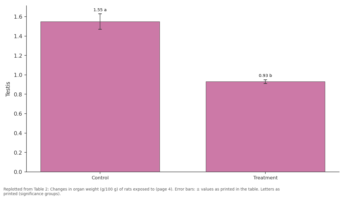
Testis: Control vs Treatment
Replotted from Table 2 (page 4) of the original paper. Every bar is the paper's own table value.
View source evidence ↗Figures & tables
From the original paper Table 1Page 3
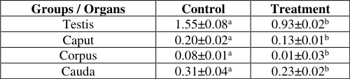 Table 2Page 4
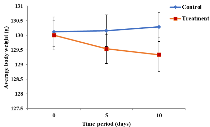 Figure 1Page 4
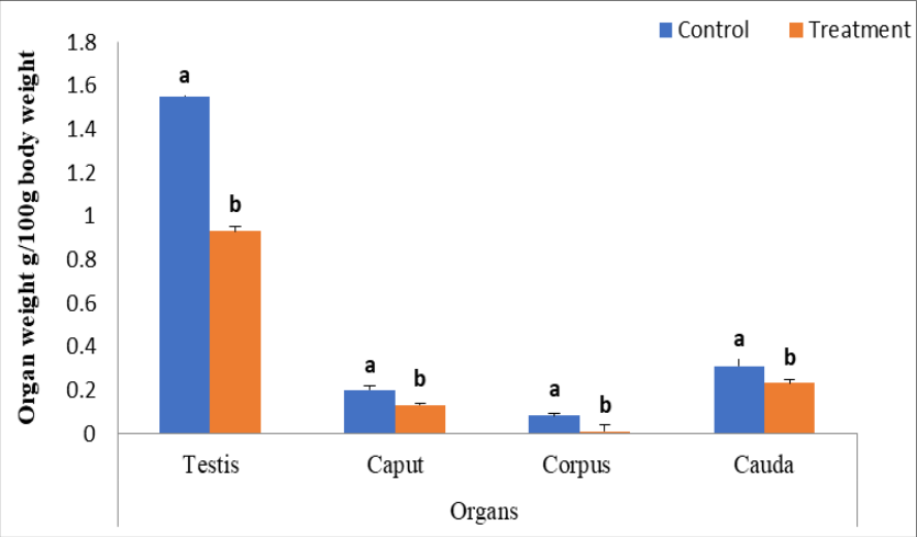 Figure 2APage 4
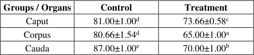 Table 3Page 5
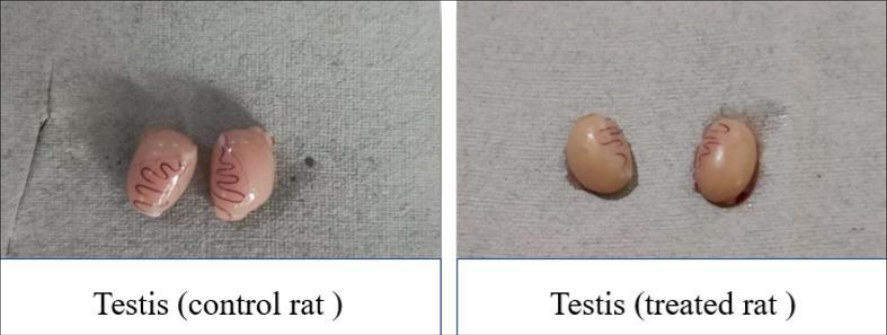 Figure 2BPage 5
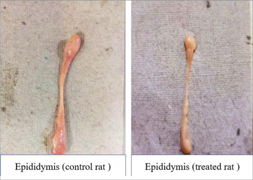 Figure 2CPage 5
 Table 4Page 6
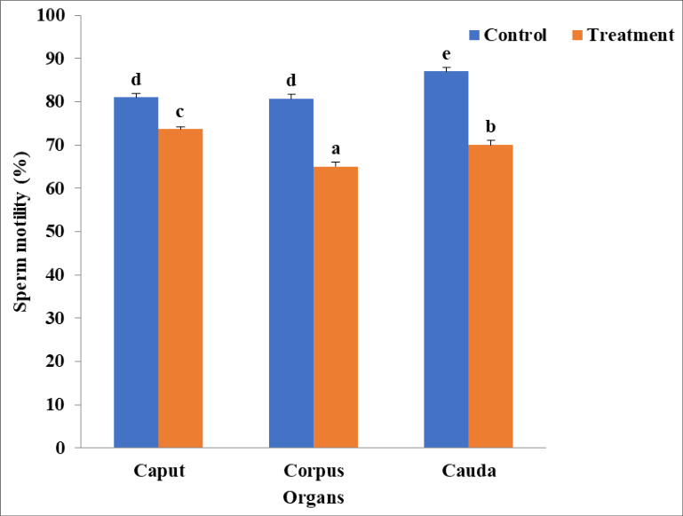 Figure 3Page 6
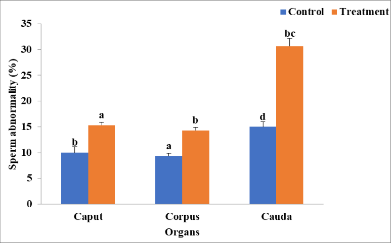 Figure 4Page 6
 Table 5Page 7
 Table 6Page 7
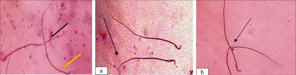 Figure 4APage 7
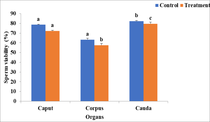 Figure 5Page 7
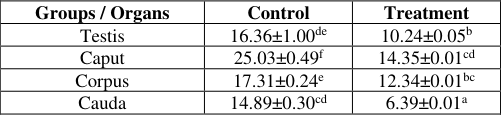 Table 7Page 8
 Table 8Page 8
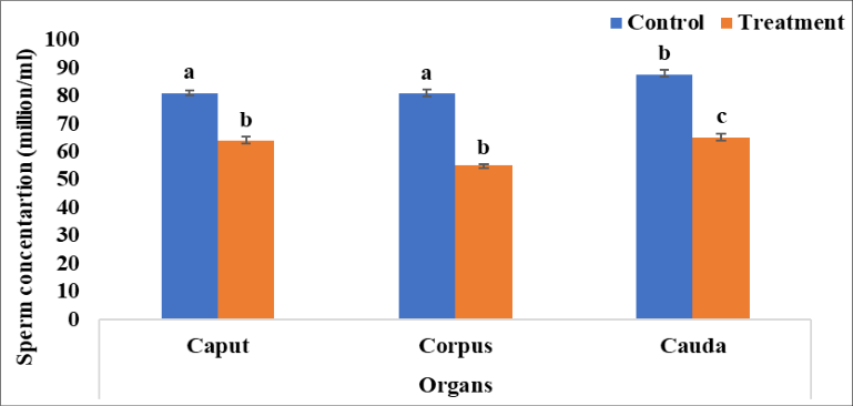 Figure 6Page 8
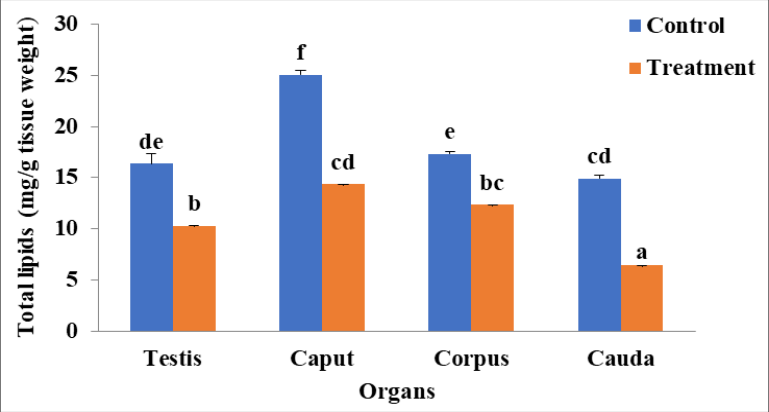 Figure 7Page 8
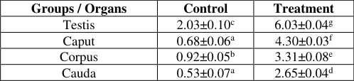 Table 9Page 9
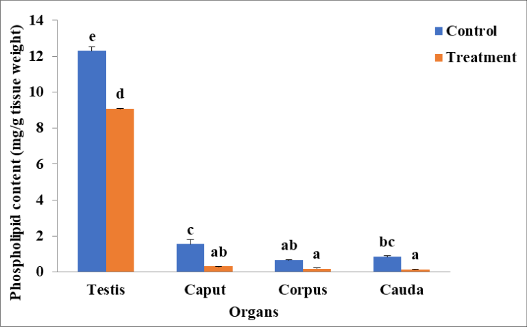 Figure 8Page 9
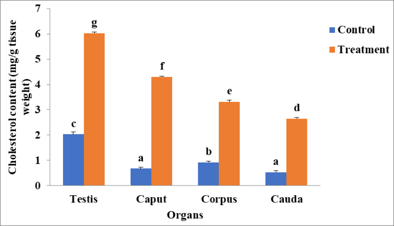 Figure 9Page 9
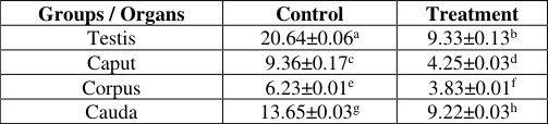 Table 10Page 10
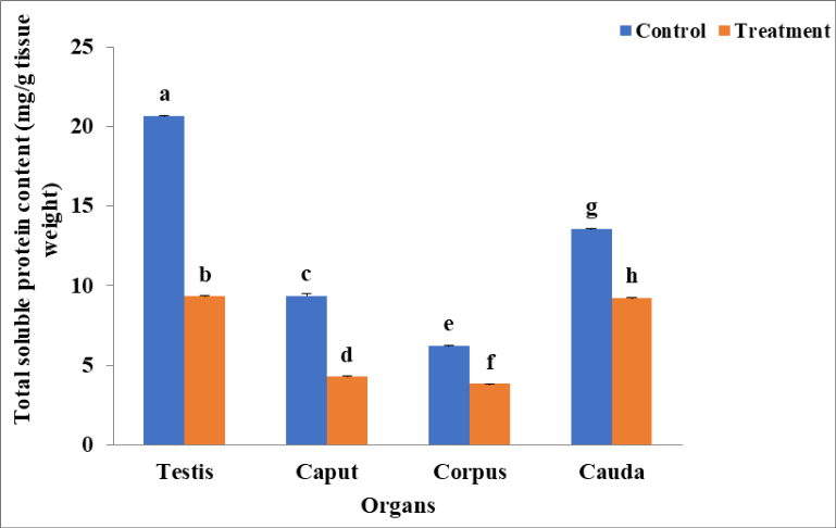 Figure 10Page 10
Introduction
Male fertility is a complex physiological process regulated by the hypothalamic-pituitarygonadal axis, which controls the secretion of reproductive hormones such as luteinizing hormone and follicle-stimulating hormone (Johnson et al., 2018) [17] . These hormones stimulate the Leydig and Sertoli cells in the testes, which are essential for testosterone synthesis and spermatogenesis (Smith & Walker, 2020) [29] . Any disruption in this endocrine axis can lead to infertility, making it a critical target for contraceptive research. Quinestrol, a long-acting estrogen derivative of ethinylestradiol, has gained attention as a potent antifertility agent due to its ability to alter endocrine homeostasis (Brown et al., 2017) [6] . Initially developed for female contraception, quinestrol has also shown efficacy in male fertility suppression by disrupting testosterone production and spermatogenesis (Garcia & Patel, 2019) [13] . Studies on male albino rats have demonstrated that quinestrol administration leads to significant biochemical and hormonal alterations, including decreased serum testosterone levels, increased estradiol concentrations, oxidative stress, lipid peroxidation, and enzymatic dysfunction (Lee et al., 2021) [19] . One of the primary mechanisms through which quinestrol exerts its antifertility effects is the suppression of gonadotropins. Quinestrol-induced negative feedback on the hypothalamicpituitary-gonadal axis inhibits the secretion of gonadotropin-releasing hormone, leading to reduced luteinizing hormone and follicle-stimulating hormone production (Zhang et al., 2016) [33] . This suppression results in decreased stimulation of Leydig cells, thereby reducing testosterone synthesis. Since testosterone is essential for spermatogenesis, its depletion leads to impaired sperm production, altered sperm morphology, and increased germ cell apoptosis (Anderson & Roberts, 2015) [4] . In addition to hormonal suppression, quinestrol has been found to induce oxidative stress in testicular tissues. Oxidative stress is a key factor in male infertility, as excessive reactive oxygen species can damage cellular structures, including lipids, proteins, and DNA (Gupta et al., 2018; Alrubaie and Khafaji, 2024) [15,20] . Studies have reported that quinestrol administration significantly reduces antioxidant enzyme activity, including superoxide International Journal of Biology Sciences https://www.biologyjournal.net dismutase, catalase, and glutathione peroxidase, leading to increased lipid peroxidation and testicular dysfunction (Wang & Li, 2020) [31] . Elevated oxidative stress levels contribute to mitochondrial damage, disrupted cellular metabolism, and apoptosis of germ cells, ultimately resulting in reduced sperm count and motility (Singh et al., 2019) [28] . Proteins, lipids, cholesterol, and phospholipids play crucial roles in the structural and functional integrity of male reproductive cells. Proteins are essential for sperm development, enzymatic regulation, and cellular signaling, while lipids and cholesterol contribute to sperm membrane fluidity and stability (Hernandez & Lopez, 2022) [16] . Phospholipids, particularly phosphatidylcholine and phosphatidylethanolamine, are key components of sperm membranes, influencing sperm motility and viability (Martinez et al., 2021) [23] . Quinestrol administration has been reported to cause significant alterations in these biochemical components. Studies have shown a marked reduction in total protein content in testicular tissues, likely due to decreased biosynthesis and increased proteolysis (Chen et al., 2017) [9] . This decline can impair spermatogenesis by affecting critical proteins involved in sperm maturation and function. Similarly, lipid metabolism is disrupted in quinestrol-treated rats, with significant decreases in testicular lipid and cholesterol levels (Zhou et al., 2018) [36] . Since cholesterol is vital for steroidogenesis and sperm membrane integrity, its depletion can negatively impact sperm viability and motility. Moreover, alterations in phospholipid composition have been observed, leading to reduced membrane stability and increased susceptibility to oxidative damage (Fernandez et al., 2020) [11] . Increased lipid peroxidation, as indicated by elevated malondialdehyde levels, further exacerbates membrane damage and functional impairments in spermatozoa (Park & Kim, 2019) [25] . By disrupting protein, lipid, cholesterol, and phospholipid homeostasis, quinestrol contributes to male infertility through biochemical pathways that impair sperm production, motility, and structural integrity. Investigating these biochemical alterations will provide deeper insights into the mechanisms of quinestrol-induced infertility and its potential as a contraceptive agent.
Materials and methods
Materials and methods
Procurement of animals
Sexually mature male albino rats weighing between 130-140 g were procured from the Disease-Free Small Animal House at Lala Lajpat Rai University of Veterinary and Animal Sciences, Hisar, after clearance of the proposal through the Institutional Animal Ethics Committee. A total of 40 adult male albino rats were procured. After being obtained, the rats were given a 15-day acclimatization period. In the animal house, the rats were kept in polypropylene cages and were exposed to an environment similar to their natural habitat. Wheat husk was utilized as bedding. Rats were fed a diet of cracked wheat and given adequate amounts of water. Water was provided in glass bottles, and feeders were equipped with rubber stoppers. Standard laboratory conditions were maintained, including ambient temperature control (22-25°C) and a 12-hour light/dark cycle during the treatment days. Before the experiment began, the rats were divided into two groups: control (n=20) and treatment (n=20). Permission was obtained at the 63rd meeting of the IAEC, which took place on January
Drug used for the experiment
To evaluate the effects of quinestrol, synthetic estradiol (17 alpha-Ethinylestradiol 3-cyclopentyl ether), a commercial formulation was acquired from CDH Chemicals and administered to the rats via oral intubation.
Experimental setup
Rats were acclimatized to lab conditions for 15 days before the experiment. After acclimatization, all 40 male rats were separated into two groups (n=20).
Groups
Treatment Group 1 (n=20) Control Group 2 (n=20) Quinestrol @2mg/Kg body weight Dimethyl sulfoxide, an organic solvent, was used to dissolve quinestrol for dose preparation. The second group received treatment via oral intubation for 10 days.
Effect on body weight
After 15 days of acclimatization and 10 days of treatment, the animals' body weights were recorded at 0, 5, and 10 days and compared.
Autopsy of rats
After completing the treatment, the rats were dissected. The testicles and epididymides were removed, and the adhering tissue was carefully excised to obtain precise weights. Tissue samples were stored at -20°C for biochemical examination.
Effect on reproductive organ weight
After the 10-day treatment period, the rats were sacrificed, and the reproductive organs (testis and epididymis (caput, corpus and cauda) of rats from both groups were dissected out and weighed.
Preparation of tissue homogenates of testis and epididymal tissues
The testis and epididymis were homogenized (caput, corpus, and cauda) by combining the tissue with known with a quantity of 0.1 M phosphate buffer saline (pH 7.6). The process was carried out in a homogenizer. The entire homogenate was spun in a chilled microcentrifuge for 10 minutes at 3000 rpm (825 rcf). In tissue homogenates of the testis and epididymis, the resulting supernatant was employed for biochemical parameters. [26] Immediately after dissection, a drop of caput, corpus, and cauda epididymal fluid was placed on a pre-warmed glass slide and observed under a microscope at 400X magnification. The number of motile and immotile spermatozoa was counted in various fields. [26] Sperm concentration was determined using a hemocytometer. The diluted caput, corpus, and cauda epididymal fluid was taken into a standard red cell dilution pipette, and small air bubbles were extracted. The dilution solution (0.2% eosin) was added to the 101-mark above the bulb. The mixture was shaken for 2-3 minutes and a drop was placed under a coverslip on a Neubauer chamber. The spermatozoa were allowed to settle for 2 minutes, and then counted at 400X magnification. The total spermatozoa were averaged across five squares on both sides of the chamber. The sperm concentration was calculated using the following formula:
Sperm Parameters Sperm Motility (Salisbury et al., 1978)
Sperm Parameters Sperm Motility (Salisbury et al., 1978)
Sperm Concentration (Salisbury et al., 1978)
Sperm Viability (Campbell et al., 1956) [37] A drop of sperm-containing epididymal fluid was mixed with eosin/nigrosin stain and incubated at 37°C for 2 minutes before preparing smears on glass slides. At 1000X magnification, the percentage of viable (unstained) and nonviable (pink-stained) spermatozoa was determined.
Sperm Abnormality
Sperm morphology was assessed under a light microscope. The abnormalities evaluated included head, midpiece, and flagellar defects. The percentage of sperm abnormalities was calculated based on 250-300 spermatozoa per rat.
Biochemical Parameters Quantitative Estimation of Total Soluble Protein Content:
The total soluble protein content was determined using the method of Lowry et al. (1951) [38] . Tissue homogenates were prepared, and 5.0 ml of a reagent mixture was added to 0.01 ml of the sample. The mixture was allowed to stand at room temperature for 10 minutes before the addition of 0.5 ml of diluted Folin's phenol reagent. After vigorous agitation, the sample was left at room temperature for 30 minutes. Absorbance was measured at 520 nm using a UV-Visible double beam spectrophotometer. The total soluble protein content in different tissue samples was calculated using a standard curve prepared with bovine serum albumin in the concentration range of 20-200 µg/ml. The results were expressed as mg protein per gram of wet tissue.
Extraction and Estimation of Total Lipids
Total lipids were extracted from testis and epididymal tissue (caput, corpus, and cauda) using the method of Folch et al. (1957) [12] . The tissue was homogenized using phosphatebuffered saline in a homogenizer, followed by the addition of an extraction medium containing chloroform and methanol in a 2:1 ratio. The mixture was subjected to intermittent shaking for 3-4 hours to ensure thorough mixing. After filtration through a G-2 sintered glass funnel, the filtrate was mixed with 0.85% saline solution and transferred to a separating funnel, where it was left overnight for complete phase separation. The bottom layer, containing lipids, was collected in a pre-weighed crucible, while the upper layer was rinsed with chloroform to ensure maximum lipid recovery. The crucible with lipids was placed in an oven at 60°C for vaporization, and after drying, it was weighed again to determine total lipid content. The lipid extract was further dissolved in chloroform for the quantitative analysis of lipids, cholesterol, and phospholipids. Lipid content was expressed as mg per gram of tissue weight.
Estimation of Total Phospholipids
Phospholipids were estimated in the testis and epididymal tissue (caput, corpus, and cauda) using the method of Ames (1966) [3] . Lipid samples and standard phosphate solutions were evaporated in a water bath at 60°C. A specific reagent was added to the test tubes containing the samples, which were then dried at 100°C in an oven. These tubes were subsequently heated over a strong flame until brown fumes ceased to emerge. Once cooled, hydrochloric acid was added, and the samples were vortexed before being placed in a boiling water bath for 15 minutes. After cooling, a color-developing reagent was added, followed by incubation at 45°C for 25 minutes. The absorbance was measured at 820 nm using a spectrophotometer, with reference to a blank reagent. A standard curve was prepared using potassium dihydrogen phosphate ranging from 2-6 µg/ml, and the phospholipid content was expressed as mg per gram of tissue weight.
Estimation of Total Cholesterol
Total cholesterol was estimated in the testis and epididymal tissue (caput, corpus, and cauda) using the method of Chiamori and Henry (1959) [8] . Lipid samples were evaporated at 60°C before the addition of a developing reagent. The contents were mixed thoroughly by vortexing before the addition of concentrated sulfuric acid. The samples were mixed again and left undisturbed for 10 minutes. Absorbance was measured at 560 nm using a spectrophotometer against a blank. A standard curve was created using cholesterol concentrations ranging from 20-200 µg/ml, and the results were expressed as mg per gram of tissue weight.
Results
Results
Effect on body weight
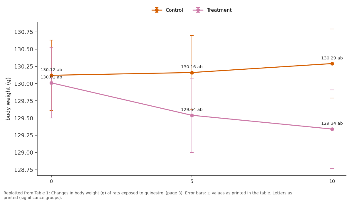
At 0, 5, and 10 days, measurements of each rat's body weight revealed variations, as indicated in table 1 and figure 1. The body weight of rats treated with quinestrol showed no alteration in body weight at 0, 5 and 10 days when compared with control rats. There was no statistically significant difference in the average body weight of the rats after quinestrol treatment at various intervals, showing that the treatment had no adverse effects on the body weight of the rats.
| Groups Days | Control | Treatment |
|---|---|---|
| 0 | 130.12±0.51ab | 130.01±0.51ab |
| 5 | 130.16±0.54ab | 129.54±0.54ab |
| 10 | 130.29±0.50ab | 129.34±0.57ab |
Table text extracted from the original PDF; superscript letters are shown inline. The original table image is below.
View original table image
Weight of reproductive organs Effect on reproductive organ weight
The weight of reproductive organs, testis and epididymis of all the control and treated rats were recorded after dissection at the end of treatment. The weight of the reproductive organ is represented as g/100 g body weight (table 2 and figure 2a, 2b and2c). The weight of testis and epididymal tissue i.e caput, corpus and cauda of treated rats showed a significant difference as compared to the control group. A remarkably significant decrease was seen in the weight of caput, corpus and cauda of rats exposed to quinestrol at the dose of 2 mg/Kg body weight for a period of 10 days as compared to control groups. Statistically, findings from the present study indicate that all the organs were affected by quinestrol treatment.
| Groups / Organs | Control | Treatment |
|---|---|---|
| Testis | 1.55±0.08a | 0.93±0.02b |
| Caput | 0.20±0.02a | 0.13±0.01b |
| Corpus | 0.08±0.01a | 0.01±0.03b |
| Cauda | 0.31±0.04a | 0.23±0.02b |
Table text extracted from the original PDF; superscript letters are shown inline. The original table image is below.
View original table image
Sperm parameters
In the present study, the fluid from three regions of the epididymis i.e caput, corpus, cauda was taken for sperm parameters like sperm motility (%), abnormalities (%) and concentrations (millions/ml) were evaluated and results were represented in table 3-6 and figure 3456. The sperm motility, viability and count were significantly decreased in quinestrol treated group in comparison to control group after 10 days of treatment whereas, the sperm abnormality (figure 4a) significantly increased in quinestrol group as compared to control group.
| Groups / Organs | Control | Treatment |
|---|---|---|
| Caput | 81.00±1.00d | 73.66±0.58c |
| Corpus | 80.66±1.54d | 65.00±1.00a |
| Cauda | 87.00±1.00e | 70.00±1.00b |
Table text extracted from the original PDF; superscript letters are shown inline. The original table image is below.
View original table image
Biochemical parameter in testis and epididymis Total lipids content (TLC)
The results of TLC in testis and epididymis were presented in table 7 and figure 7. In the testis, the TLC in control rats was observed to be 16.36±1.00mg/g tissue weight whereas a significant decline was observed in the TLC in the treatment group rats i.e., 10.24±0.05mg/g tissue weight as compared to control group. Similarly, the TLC in the epididymal tissue (caput, corpus, cauda) of control rats was 25.03±0.49, 17.31±0.24, 14.89±0.30 mg/g tissue weight. The TLC in the epididymis of rats treated with quinestrol were (caput, corpus, cauda) 14.35±0.01, 12.34±0.01, 6.39±0.01 mg/g tissue weight. This showed that there was a significant difference between the control and rats treated with quinestrol for dose of 2 mg/Kg body weight for a period of 10 days.
| Groups / Organs | Control | Treatment |
|---|---|---|
| Testis | 16.36±1.00de | 10.24±0.05b |
| Caput | 25.03±0.49f | 14.35±0.01cd |
| Corpus | 17.31±0.24e | 12.34±0.01bc |
| Cauda | 14.89±0.30cd | 6.39±0.01a |
Table text extracted from the original PDF; superscript letters are shown inline. The original table image is below.
View original table image
Phospholipid content
The phospholipid content was observed in the testis and epididymis of rats from control and treated groups and were presented in table 8 and figure 8. In the control rat testis, the phospholipid content was observed to be 12.29±0.20 mg/g tissue weight and in the rats, treated with quinestrol showed decrease in the phospholipid content i.e. 9.07±0.09 mg/g tissue weight as compared to control group. The data showed that there was a significant difference between the control and rats treated with quinestrol at the dose of 2 mg/Kg for a period of days.
| Groups / Organ | Control | Treatment |
|---|---|---|
| Testis | 12.29±0.20e | 9.07±0.09d |
| Caput | 1.55±0.27c | 0.31±0.01ab |
| Corpus | 0.67±0.02ab | 0.19±0.04a |
| Cauda | 0.85±0.05bc | 0.14±0.02a |
Table text extracted from the original PDF; superscript letters are shown inline. The original table image is below.
View original table image
Similarly, in the epididymis (caput, corpus, cauda) the phospholipid content was examined in the control group i.e. 1.55±0.27, 0.67±0.02, 0.85±0.05 mg/g tissue weight. A significant decrease was observed in the group treated with quinestrol with dose of 2mg/Kg body weight i.e.0.31±0.01, 0.19±0.04, 0.14±0.02 mg/g tissue weight as compared to control group.
Total Cholesterol
The cholesterol content was observed in the testis and epididymis of control and treated rats and were presented in table 9 and figure 9.
| Groups / Organs | Control | Treatment |
|---|---|---|
| Testis | 2.03±0.10c | 6.03±0.04g |
| Caput | 0.68±0.06a | 4.30±0.03f |
| Corpus | 0.92±0.05b | 3.31±0.08e |
| Cauda | 0.53±0.07a | 2.65±0.04d |
Table text extracted from the original PDF; superscript letters are shown inline. The original table image is below.
View original table image
In the testis, the cholesterol level in the control rats was 2.03±0.10 mg/g tissue weight the treatment group showed a significant increase in cholesterol level i.e. 6.03±0.04 mg/g tissue weight as compared to control groups. Similarly, in the epididymis the cholesterol level was examined among the control group i.e. 0.68±0.06, 0.92±0.05, 0.53±0.07mg/g tissue weight. In the rats treated with quinestrol the cholesterol content was 4.30±0.03, 3.31±0.08, 2.65±0.04 mg/g tissue weight which was significantly higher than control values. The results of present study indicated that after quinestrol administration the level of cholesterol increases. Total soluble proteins content Total soluble protein content was observed in testis and epididymis of control and treated group and were presented in shown in table 10 and figure 10. In the testis the total soluble protein content in control rats was 20.64±0.06 mg/g tissue weight whereas a significant decrease was observed in total soluble protein content in the treatment group i.e. 9.33±0.13 mg/g tissue weight, respectively. Similarly, in the epididymis of the control rats (caput, corpus, cauda) the total soluble protein content was 9.36±0.17, 6.23±0.01, 13.65±0.03 mg/g tissue weight whereas in the group treated with quinestrol it was 4.25±0.03, 3.83±0.01 and 9.22±0.03 mg/g tissue weight, respectively, which showed a significant difference in the Total soluble protein content between control and treated group. From the statistical data it was observed that all the organs were significantly affected by quinestrol.
| Groups / Organs | Control | Treatment |
|---|---|---|
| Testis | 20.64±0.06a | 9.33±0.13b |
| Caput | 9.36±0.17c | 4.25±0.03d |
| Corpus | 6.23±0.01e | 3.83±0.01f |
| Cauda | 13.65±0.03g | 9.22±0.03h |
Table text extracted from the original PDF; superscript letters are shown inline. The original table image is below.
View original table image
Discussion
The present study aimed to assess the impact of quinestrol treatment on body weight, reproductive organ weight, sperm parameters, total lipid content, cholesterol levels, and protein content in male albino rats. Our findings indicate that quinestrol treatment did not significantly alter the body weight of the rats throughout the study period (0, 5, and 10 days). This observation aligns with previous studies, such as those conducted by Li et al. (2014) [39] and Liu et al. (2013) [21] , which reported no significant changes in body weight in quinestrol-treated male rats. However, Kang et al. (2022) [18] observed a decrease in body mass index in rats treated with EP-1 and E groups, suggesting species or dose-specific variations in quinestrol's effects on body weight. Despite the lack of effect on overall body weight, quinestrol treatment significantly reduced the weight of reproductive organs, including the testis and epididymis (caput, corpus, and cauda). This reduction suggests that quinestrol disrupts normal reproductive physiology, likely through its impact on androgen production. The decrease in testis weight aligns with previous studies demonstrating the suppressive effects of estrogenic compounds on Leydig cell function and testosterone secretion (Zhang et al., 2005) [32] . Similar reductions in reproductive organ weights have been reported in various rodent species, including Brandt's voles (Zhang et al., 2004, 2005, 2006) [5,34] , greater long-tailed hamsters (Zhao et al., 2007) [35] , and male plateau pikas (Liu et al., 2011) [22] . The decline in testis weight following quinestrol treatment may be attributed to its suppressive effects on the hypothalamic-pituitary-gonadal axis, reducing luteinizing hormone and follicle-stimulating hormone secretion, thereby impairing testosterone production and spermatogenesis (Amroy et al., 2006; Chen et al., 2021) [1,10] . The disruption of the hypothalamic-pituitary-gonadal axis results in negative feedback inhibition, reducing testosterone synthesis and affecting sperm maturation in the epididymis. In addition to reductions in reproductive organ weight, sperm parameters were significantly affected by quinestrol treatment. Sperm motility and viability decreased, while sperm abnormalities increased, indicating that quinestrol disrupts spermatogenesis and leads to poor sperm quality. Carreau and Hess (2010) [7] demonstrated that excessive estrogen disrupts spermatogenesis and negatively impacts male fertility. Similarly, Adeoya-Osiguwa et al. (2003) [2] reported that 17-β estradiol and environmental estrogens impair mammalian sperm function. High levels of reactive oxygen species (ROS) are associated with increased apoptosis in spermatogenic cells, further contributing to poor sperm quality (Massawe et al., 2018) [24] . Su et al. (2019) [30] also confirmed that suppression of thehypothalamic-pituitary-gonadal axisis a key factor leading to reduced sperm concentration and motility.
Furthermore, the present study demonstrated that quinestrol administration significantly affects total lipid content, cholesterol levels, and protein content in the testis and epididymis of male albino rats. A notable decline was observed in total lipid and phospholipid content in the testis and epididymis of quinestrol-treated rats compared to the control group. The reduction in total lipid content can be attributed to oxidative stress which plays a crucial role in lipid peroxidation and cellular damage. The testicular and sperm plasma membranes, rich in polyunsaturated fatty acids, are highly susceptible to reactive oxygen species which disrupt lipid integrity, leading to cell damage and decreased lipid levels. These findings align with previous studies indicating that quinestrol induces oxidative damage in male reproductive organs, affecting sperm membrane composition and function. Additionally, the phospholipid decline in treated rats may indicate the degradation of cellular membranes, signifying loss of cellular integrity and function. Furthermore, cholesterol levels were significantly elevated in the testis and epididymis of quinestrol-treated rats. Cholesterol serves as a precursor for steroidogenesis, and its accumulation may suggest an impairment in steroid hormone synthesis due to oxidative stress-induced disruption of enzymatic activity, particularly 17β-HSD, which plays a key role in testosterone biosynthesis. This alteration in cholesterol metabolism is consistent with findings from prior research, suggesting that exogenous estrogen administration affects hepatic cholesterol processing and steroidogenesis, leading to an increase in cholesterol levels. Moreover, total soluble protein content exhibited a marked decline in the testis and epididymis of quinestrol-treated rats. This decrease may be associated with protein oxidation caused by excessive reactive oxygen species which disrupts protein synthesis and degrades structural proteins, further impairing reproductive function. The ability of quinestrol to induce testicular atrophy, sperm cell death, and damage to the testis and epididymis suggests that its contraceptive effects result from both direct impairment of testicular function and indirect suppression of gonadotropins. Quinestrol's role in suppressing the hypothalamic-pituitary-gonadal axishas been well documented (Gill-Sharma et al., 2001; Liu et al., 2013) [14,21] . It has also been shown to cause azoospermia by disrupting seminiferous tubule cytoarchitecture, leading to long-term infertility effects (Massawe et al., 2018) [24] . Sidhu et al. (2020) [27] similarly reported significant reductions in sperm motility, viability, and density in quinestrol-treated animals, accompanied by increased sperm abnormalities.
Conclusion
In conclusion, the present study confirms that quinestrol administration significantly affects reproductive organ weight, sperm parameters, total lipid content, cholesterol levels, and total soluble protein content without altering overall body weight. The suppression of the hypothalamicpituitary-gonadal axis leading to decreased testosterone synthesis and impaired spermatogenesis, is the likely mechanism underlying these effects. These findings reinforce quinestrol's potential as an effective male contraceptive. However, further research is required to assess the long-term effects of quinestrol treatment and its potential for reversibility in reproductive function.
Figures and tables from the paper
| Groups / Organs | Control | Treatment |
|---|---|---|
| Caput | 10.00±1.00b | 15.33±0.58a |
| Corpus | 9.33±0.57a | 14.33±0.58b |
| Cauda | 15.00±1.00d | 30.66±1.52bc |
Table text extracted from the original PDF; superscript letters are shown inline. The original table image is below.
View original table image
| Groups / Organ | Control | Treatment |
|---|---|---|
| Caput | 78.66±1.00a | 72.33±1.52a |
| Corpus | 63.33±0.09a | 57.66±1.52b |
| Cauda | 82.33±1.25b | 79.66±0.58c |
Table text extracted from the original PDF; superscript letters are shown inline. The original table image is below.
View original table image
| Groups / Organ | Control | Treatment |
|---|---|---|
| Caput | 81.00±1.00a | 69.00±1.54b |
| Corpus | 76.00±1.54a | 58.00±0.58b |
| Cauda | 89.36±1.54b | 71.00±1.54c |
Table text extracted from the original PDF; superscript letters are shown inline. The original table image is below.
View original table image
References
39 in the original paper- Amroyan E, Sargsyan A, Hovhannisyan L. Estrogen receptor-mediated reproductive toxicity in male rats. Endocrine Research. 2006;41(1):1-11.
- Adeoya-Osiguwa SA, Olagunju JA, Lasebikan AA. Effects of environmental estrogen on sperm function in mammals. Reproductive Biology and Endocrinology. 2003;1:102.
- Ames BN. Assay of inorganic phosphate, total phosphate, and phospholipids. In: Methods in Enzymology. Vol. 8. Academic Press; 1966. p. 115118.
- Anderson P, Roberts M. Mechanisms of testosterone depletion and its impact on spermatogenesis. Journal of Endocrinology and Reproductive Biology. 2015;12(3):245-259.
- Brandt NJ, et al. Effects of estrogen on male reproductive function in voles. Hormones and Behavior. 2004;46(5):623-630.
- Brown T, Smith K, Walker J. The role of quinestrol in fertility suppression: A review. Reproductive Endocrinology. 2017;25(4):312-328.
- Carreau S, Hess P. Estrogens and spermatogenesis: A review. Reproductive Biology and Endocrinology. 2010;8:169.
- Chiamori N, Henry JB. A new method for the determination of cholesterol. Clinical Chemistry. 1959;5(5):600-610.
- Chen H, Zhao Y, Liu W. Testicular protein metabolism in response to quinestrol exposure in rats. Molecular Andrology. 2017;19(2):120-132.
- Chen Y, et al. Mechanisms of reproductive endocrine disruption by environmental estrogens. Toxicology Letters. 2021;347:101-113.
- Fernandez R, Lopez D, Sanchez M. Phospholipid composition and its effects on sperm viability under oxidative stress. Journal of Lipid Research. 2020;28(1):67-79.
- Folch J, Lees M, Sloane Stanley GH. A simple method for the isolation and purification of total lipids from animal tissues. Journal of Biological Chemistry. 1957;226(1):497-509.
- Garcia L, Patel N. Quinestrol as an antifertility agent: Effects on male reproductive hormones and spermatogenesis. Contraceptive Research and Development. 2019;14(2):178-193.
- Gill-Sharma M. Estrogenic substances and their influence on the male reproductive system. The Journal of Sexual Medicine. 2001;30:1-9.
- Gupta R, Verma P, Singh S. Oxidative stress and male infertility: Role of reactive oxygen species. Andrology and Urology Research. 2018;10(3):99-112.
- Hernandez J, Lopez R. Lipid and protein interactions in sperm function and fertility. Molecular Reproduction. 2022;30(4):201-215.
- Johnson D, Miller C, Thompson A. Endocrine regulation of male reproductive function: Insights from animal models. Reproductive Physiology. 2018;22(1):45-63.
- Kang J, et al. Effects of estrogens on body mass index in rats: A focused study. Journal of Endocrinology. 2022;243(2):111-121.
- Lee K, Zhang Y, Chen J. Biochemical and hormonal alterations induced by quinestrol in male albino rats. International Journal of Biology Sciences https://www.biologyjournal.net Toxicological Reproductive Studies. 2021;18(3):256271.
- Alrubaie MFK, Khafaji SS. Impact of mancozeb on sperm quality and hormonal profiles in male rats at different periods. Zool Entomol Lett. 2024;4(2):54-59.
- Liu J, et al. The effects of estrogen treatment on male fertility in rats. BMC Urology. 2013;13:31.
- Liu X, et al. Steroid hormone synthesis and its role in reproductive physiology. Journal of Endocrinological Investigation. 2011;34(8):631-638.
- Martinez F, Gonzalez H, Ramirez T. The role of phospholipids in sperm membrane integrity and function. Biochemical Reproduction. 2021;27(2):143157.
- Massawe F, et al. The impact of estrogen exposure on male rat spermatogenesis. Asian Pacific Journal of Reproduction. 2018;7(5):185-191.
- Park J, Kim S. Lipid peroxidation and its impact on sperm function and male fertility. Reproductive Biochemistry. 2019;15(3):189-202.
- Salisbury GW, et al. The physiology of reproduction in the male. In: Reproduction in Domestic Animals. Academic Press; 1978. p. 301-344.
- Sidhu M, et al. The role of environmental estrogens in male fertility: A review. Reproductive Biology and Endocrinology. 2020;18:1-11.
- Singh R, Sharma P, Kumar V. Mitochondrial dysfunction and oxidative stress in male infertility. Cellular Reproduction and Metabolism. 2019;21(4):299-312.
- Smith K, Walker J. Leydig and Sertoli cell interactions in the regulation of testosterone and spermatogenesis. Endocrine Reviews. 2020;26(2):88-105.
- Su Y, et al. Estrogen-induced oxidative stress in the male reproductive system. Oxidative Medicine and Cellular Longevity. 2019;2019:Article ID 3470898.
- Wang H, Li P. Antioxidant enzyme activity and oxidative stress markers in quinestrol-treated rats. Toxicology and Reproductive Biology. 2020;16(1):6782.
- Zhang X, et al. Effects of phenolic compounds on male reproductive health: A review. Journal of Health Science. 2005;51(1):1-11.
- Zhang Y, Liu H, Chen W. The suppression of gonadotropins by quinestrol: Mechanisms and implications. Hormonal Research in Reproduction. 2016;13(2):99-114.
- Zhang L, et al. Impact of synthetic estrogens on reproductive health in rodents. Journal of Endocrinology. 2004;182(3):393-399.
- Zhao M, et al. Comparative effects of environmental estrogens on reproductive organs in male hamsters. Reproductive Toxicology. 2007;23(1):88-95.
- Zhou B, Wang X, Liu C. Lipid and cholesterol metabolism in male reproductive health: Effects of hormonal alterations. Andrology Research. 2018;19(1):77-89.
- Campbell DT. Enhancement of contrast as composite habit. The Journal of Abnormal and Social Psychology. 1956 Nov;53(3):350.
- Lowry OH, Rosebrough NJ, Farr AL, Randall RJ. Protein measurement with the Folin phenol reagent. J biol Chem. 1951 Nov 1;193(1):265-275.
- Li Y, et al. Effects of estrogen on reproductive parameters in male rats. Asian Journal of Andrology. 2014;16(2):223-229.