
Research article · 2025 · Full text
International Journal of Advanced Chemistry ResearchInduced hyperlipidaemia in adult male rates treated by using Salvia officinalis activity
Volume 7(1) · Pages 58–61 · DOI: 10.33545/26646781.2025.v7.i1a.258 ↗
Research areasHormonal and reproductive studies
Abstract
Author-writtenObjective of this study was to investigate the medicinal activities of Salvia officinalis extractions. Twenty adult male rats were randomly divided into four groups (A, B, C, and D), with five rats assigned to each group. Group A received no treatment (control group), Group B was induced with hyperlipidemia via an aqueous solution containing 0.5% H2O2 and 1% cholesterol in the feed for two months, Group C was induced with hyperlipidemia and treated with 50 mg/kg/day of Salvia officinalis extract, and Group D was induced with hyperlipidemia and treated with 100 mg/kg/day of the extract. Results showed significant increases (p< 0.05) in ALT and AST levels, cholesterol, triglycerides, and malondialdehyde (MDA), along with a significant decrease in HDL and glutathione (GSH) levels in Group B compared to Group A. Treatment with Salvia officinalis extract (Groups C and D) resulted in non-significant changes (p< 0.05) in liver enzymes, lipid profiles, MDA, and GSH levels compared to Group A.
Summary
Source-checkedPlain-language reading of the full paper, prepared with AI and checked against the text. Each point links to the authors' own words.
Why it matters
- Hyperlipidemia is characterized by elevated concentrations of lipoproteins in the blood. View evidence ↗
- Medicinal plants are studied for their ability to lower lipids by reducing cholesterol production and increasing its excretion. View evidence ↗
- Salvia officinalis is a plant known for various medicinal properties, including antioxidant and anti-inflammatory activities. View evidence ↗
The question
- The study aimed to investigate the medicinal activities of extracts from Salvia officinalis. View evidence ↗
What they did
- Hyperlipidemia was induced in Group B using a feed containing 1% cholesterol and 0.5% H2O2 for two months. View evidence ↗
- The researchers measured liver enzymes (ALT, AST), lipid profiles (TC, TG), and oxidative stress markers (MDA, GSH). View evidence ↗
What they found
- Inducing hyperlipidemia in Group B significantly increased AST (89.3 ± 7.32) and ALT (74.11 ± 5.4) levels compared to the control Group A (13.08 ± 3.8 and 11.93 ± 3.57). View evidence ↗
- Group B showed significantly higher total cholesterol (294.4 ± 12.93) and triglycerides (208.43 ± 13.2) than Group A (87.32 ± 9.4 and 68.2 ± 8.3). View evidence ↗
- Hyperlipidemia in Group B led to significantly higher MDA (2.57 ± 0.25) and lower GSH (0.283 ± 0.032) levels compared to Group A (1.74 ± 0.21 and 0.383 ± 0.032). View evidence ↗
- Treatment with Salvia officinalis extract in Groups C and D returned liver enzymes, lipid profiles, and oxidative stress markers to levels that were not significantly different from the control Group A. View evidence ↗
What it means
- The authors concluded that Salvia officinalis extract protects rats from liver damage and oxidative stress caused by hyperlipidemia. View evidence ↗
Figures & tables
From the original paper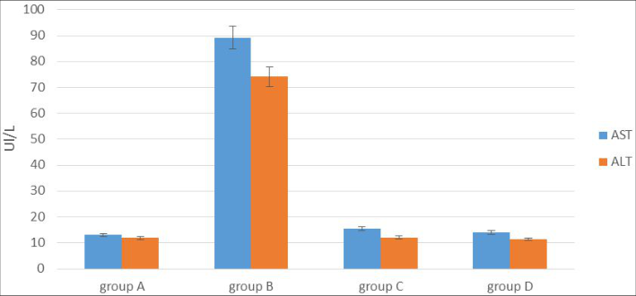 Figure 1
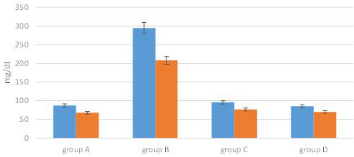 Figure 2
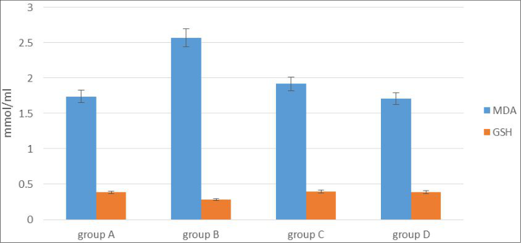 Figure 3
Introduction
Hypolipoproteinemia refers to elevated blood concentrations of lipoproteins and is often assessed through lipoprotein measurements [1][2] . Traditional medicinal plants have gained attention for their antihyperlipidemic properties due to their potential to reduce cholesterol biosynthesis and promote cholesterol excretion. The search for safer antihyperlipidemic drugs from natural sources remains a priority [3] . Salvia officinalis, a member of the mint family (Lamiaceae), comprises 700-900 species with reported medicinal properties, including anti-inflammatory, antimicrobial, hypoglycemic, antioxidant, and antitumor activities [4][5][6][7] . This plant is native to the Middle East and Mediterranean regions. Today, it has been widely naturalized across the globe, particularly in Europe and North America [8][9][10] . The leaves of Salvia officinalis contain bioactive compounds such as ursolic acid, carnosic acid, chlorogenic acid, caffeic acid, and flavonoids [11] . In traditional European medicine, Salvia officinalis (commonly known as sage) has been utilized to address mild digestive issues, such as heartburn and bloating, as well as excessive sweating, age-related cognitive decline, and inflammatory conditions affecting the throat and skin [12][13][14][15] .
Materials and Methods
Twenty adult male albino rats (weighing 200-250 g and aged 4-6 months) were procured from the College of Veterinary Medicine at Kirkuk University. The rats were maintained on a standard pellet diet for two weeks to ensure their health and absence of infections before the experiment. Fifty grams of dried Salvia officinalis leaves were combined with 1000 mL of distilled water and incubated at 35°C for 24 hours with continuous stirring. The mixture was then filtered and centrifuged at 2500 rpm for 10 minutes. The resulting filtrate was evaporated at 40°C until completely dry [16] .
Experimental Design
The rats were divided into four groups (n=5 per group):
International Journal of Advanced Chemistry Research 2025; 7(1): 58-61
• Group A: Standard pellet diet for seven days, followed by euthanasia. • Group B: Induced with hyperlipidemia using 0.5% H2O2 and 1% cholesterol in the feed for two months. • Group C: Induced with hyperlipidemia and treated with 50 mg/kg/day Salvia officinalis extract for one month.
• Group D: Induced with hyperlipidemia and treated with 100 mg/kg/day Salvia officinalis extract for one month.
Blood Sample Collection
Blood samples were collected via cardiac puncture then transferred to test tubes. After allowing the samples to clot, they were centrifuged for 10 minutes to separate the serum.
The serum was then stored at -20 °C for analysis.
Tissue Homogenization
Testis samples were washed with 0.9% NaCl buffer to remove blood. For oxidative stress analysis, 10% (w/v) of tissue was homogenized in buffer (pH 7.4) using a ceramic mortar, centrifuged for 10 minutes, and the supernatant was stored at -20°C [17] .
Measurements
Liver Enzymes and Lipid Profile: ALT, AST, total cholesterol (TC), and triglycerides (TG) were measured using Randox kits.
Oxidative Stress Markers: MDA was measured using a thiobarbituric acid (TBA) reaction, and GSH levels were determined using DTNB reagent [18,19] .
Statistical Analysis: Data were analyzed using the Minitab program. Differences between experimental groups were assessed using one-way ANOVA.
Results
Results
Liver Enzymes (ALT, AST)
Group B showed significantly elevated AST (89.3 ± 7.32) and ALT (74.11 ± 5.4) levels compared to Group A (13.08 ± 3.8 and 11.93 ± 3.57, respectively). Groups C and D demonstrated no significant differences compared to Group A (p< 0.05). as shown in figure (1).
Discussion
In the study demonstrated that Salvia officinalis extracts reduced hyperlipidemia-induced oxidative stress and liver damage. The findings align with Shahrzad et al. [20] , who noted that Salvia officinalis extracts reduce liver enzyme levels and exhibit antioxidant activities, likely due to flavonoid content. The extract's radical-scavenging ability and its phenolic constituents, such as rosmarinic acid and carnosic acid, contribute to its protective effects [21][22][23][24][25] . These compounds effectively scavenge free radicals, mitigate lipid peroxidation, and enhance endogenous antioxidant enzymes [26][27] .
Conclusion
We concluded that Salvia officinalis extract exhibits protective effects against hyperlipidemia-induced oxidative stress and liver damage in rats.
Acknowledgement
AcknowledgmentsThe authors acknowledge the Dean of the College of Science, Assistant Professor Dr. Neama Ali Ahmed, for his support and guidance
Figures and tables from the paper
References
27 in the original paper- Karam I, Ma N, Yang YJ, Li JY. Induce hyperlipidemia in rats using high fat diet investigating blood lipid and histopathology. J Hematol Blood Disord. 2018;4(1):104.
- Al-Tamimi H, Al-Dawood A, Awaishesh S, Abdalla T. Resveratrol mitigates hypercholesterolemia exacerbated hyperthermia in chronically heat-stressed rats. Vet World. 2019;12(2):337-344. DOI: 10.14202/vetworld.2019.337-344.
- Wang J, Wang Y, Xu C, Wang B, Yu J, Kang X, Liu X, Zhou L, Qin Y, Liao L, Zhang Y. Effects of total flavonoids extracted from Polygonum perfoliatum L. on hypolipidemic and antioxidant in hyperlipidemia rats induced by high-fat diet. Int J Clin Exp Med. 2018 Jan 1;11(7):6758-6766.
- Devansh M. Salvia officinalis Linn.: Relevance to modern research drive. Planta Activa. 2012 Jul 31;4:203-207.
- Garcia CS, Menti C, Lambert AP, Barcellos T, Moura S, Calloni C, Branco CS, Salvador M, Roesch-Ely M, Henriques JA. Pharmacological perspectives from Brazilian Salvia officinalis (Lamiaceae): antioxidant, and antitumor in mammalian cells. Anais Acad Bras Cienc. 2016 Feb 2;88:281-292. DOI: 10.1590/0001-3765201520150344.
- Lemle KL. Salvia officinalis used in pharmaceutics. In: IOP Conference Series: Materials Science and Engineering; 2018; Vol. 294, No. 1, p. 012037. IOP Publishing. DOI: 10.1088/1757-899X/294/1/012037.
- Mahmoodi G, Amini S. The effect of Salvia officinalis hydroalcoholic extract on scopolamine-induced memory impairment in adult male mice. J Basic Res Med Sci. 2019 Jan 10;6(1):12-20.
- Bisset NG, Wichtl M, editors. Herbal drugs and phytopharmaceuticals: a handbook for practice on a scientific basis. Stuttgart: Medpharm; 1994.
- Miura K, Kikuzaki H, Nakatani N. Apianane terpenoids from Salvia officinalis. Phytochemistry. 2001 Dec 1;58(8):1171-1175. DOI: 10.1016/s0031-9422(01)00341-7.
- Physicians' Desk Reference (PDR) for Herbal Medicines. 3 rd ed. Montvale, NJ: Thompson; 2004. p. 698-701.
- Miraj S, Kiani S. A review study of therapeutic effects of Salvia officinalis L. Der Pharmacia Lettre. 2016 Oct 8;6:299-303.
- Perry EK, Pickering AT, Wang WW, Houghton PJ, Perry NS. Medicinal plants and Alzheimer's disease: from ethnobotany to phytotherapy. J Pharm Pharmacol. 1999 May;51(5):527-534. DOI: 10.1211/0022357991772808.
- Adams M, Gmünder F, Hamburger M. Plants traditionally used in age-related brain disorders-A survey of ethnobotanical literature. J Ethnopharmacol. 2007 Sep 25;113(3):363-381. DOI: 10.1016/j.jep.2007.07.016.
- Wharf C. Community herbal monograph on Salvia officinalis L., Folium. Reprod. 2009:1-8.
- Kadhim SM, Mohammed MT, Ahmed OM, Jassim AM. Study of some Salvia officinalis L. (sage) components and effect of their aqueous extract on antioxidant. Int J Chem Sci. 2016;14(2):711-719.
- Alnidawi NA. Effect of adding Salvia officinalis oil to the ration in some blood traits of broiler Ross 308. J Kerbala Univ. 2017 (The third scientific conference of the College of Veterinary Medicine).
- Albir A. Histological changes in the liver of mice treated with cadmium. Iraqi J Sci. 2013;54(1):50-54.
- Ismail RM. The role of Coriandrum sativum in treatment of kidney lesions induced by Toxoplasma gondii in male rats. Tex J Agric Biol Sci. 2023 Mar 26;14:102-107.
- Mahmood NA. Glutathione-S-transferase enzyme and malondialdehyde (MDA) in colorectal cancer and in healthy control. Iraqi J Cancer Med Genet. 2010 Jun 1;3(1):21-26. DOI: 10.29409/ijcmg.v3i1.25.
- Shahrzad K, Mahya N, Fatemeh TB, Maryam K, Mohammadreza FB, Jahromy MH. Hepatoprotective and antioxidant effects of Salvia officinalis L. hydroalcoholic extract in male rats. Chin Med. 2014 May 20;5(2):130-136. DOI: 10.4236/cm.2014.52016.
- Janero DR. Malondialdehyde and thiobarbituric acidreactivity as diagnostic indices of lipid peroxidation and peroxidative tissue injury. Free Radic Biol Med. 1990;9(6):515-540. DOI: 10.1016/0891-5849(90)90131-2.
- Lima CF, Valentão PC, Andrade PB, Seabra RM, Fernandes-Ferreira M, Pereira-Wilson C. Water and methanolic extracts of Salvia officinalis protect HepG2 cells from t-BHP induced oxidative damage. Chem-Biol Interact. 2007 Apr 25;167(2):107-115. DOI: 10.1016/j.cbi.2007.01.020.
- Alkan FÜ, Gürsel FE, Ateş A, Özyürek M, Güçlü K, Altun M. Protective effects of Salvia officinalis extract against cyclophosphamide-induced genotoxicity and oxidative stress in rats. Turk J Vet Anim Sci. 2012;36(6):646-654. DOI: 10.3906/vet-1105-1136.
- Santos-Gomes PC, Seabra RM, Andrade PB, Fernandes-Ferreira M. Phenolic antioxidant compounds produced by in vitro shoots of sage (Salvia officinalis L.). Plant Sci. 2002 Jun 1;162(6):981-987. DOI: 10.1016/s0168-9452(02)00052-3.
- Grzegorczyk I, Matkowski A, Wysokińska HJ. Antioxidant activity of extracts from in vitro cultures of Salvia officinalis L. Food Chem. 2007 Jan 1;104(2):536-541. DOI: 10.1016/j.foodchem.2006.12.003.
- Ramos-Escudero F, Muñoz AM, Alvarado-Ortíz C, Alvarado A, Yáñez JA. Purple corn (Zea mays L.) phenolic compounds profile and its assessment as an agent against oxidative stress in isolated mouse organs. J Med Food. 2012 Feb 1;15(2):206-215.
- Pilarczyk B, Jankowiak D, Tomza-Marciniak A, Pilarczyk R, Sablik P, Drozd R, Tylkowska A, Skólmowska M. Selenium concentration and glutathione peroxidase (GSH-Px) activity in serum of cows at different stages of lactation. Biol Trace Elem Res. 2012 Jun;147:91-96. DOI: 10.1007/s12011-0119271-y.