
Research article · 2025 · Full text
International Journal of Agriculture and Food ScienceEffect of fucoidan extracted from the brown seaweed, Sargassum johnstonii on growth, survival, immune response and disease resistance to vibrio parahaemolyticus in Penaeus vannamei Boone, 1931
Volume 7(1) · Pages 258–267 · DOI: 10.33545/2664844x.2025.v7.i1d.266 ↗
Research areasAquatic life and conservationMarine and Coastal EcosystemsFood and Agricultural Sciences
Abstract
Author-writtenThe present study was conducted to investigate the effect of fucoidan extracted from the brown seaweed, Sargassum johnstonii on immunological parameters and disease resistance to Vibrio parahaemolyticus in Penaeus vannamei (Boone, 1931) [8]. Four experimental diets were formulated with a 35% protein level in all diets. T0 diet was considered as a control, T1, T2, and T3 diets contained Sargassum johnstonii fucoidan extract (0.5, 1, and 2% respectively). Penaeus vannamei (average weight 0.012g) were stocked at a density of 20 nos. in each of the 21 experimental tanks. At the end feeding trial, shrimps were challenged with 0.1 ml of Vibrio parahaemolyticus suspension at the rate of 1×10⁴ cfu ml-1 by selecting ten shrimps from each replication. The mortality was observed in all groups for 10 days. After the end of the experimental period, the result of this study shows that the highest prophenoloxidase activity, respiratory burst activity, and total haemocyte count were recorded in the 2% Sargassum johnstonii extracted fucoidan incorporated diet, while it was the lowest recorded in the control diet at the end of the experiment. In growth performance, mean weight gain, SGR and PER were higher in 2% Sargassum johnstonii extracted fucoidan incorporated diet, while lower in the control diet. FCR was better in the fucoidan extract-containing diet compared to the control diet. There was no significant difference in the survival rate of Penaeus vannamei in all treatments. After the challenge lower mortality rates were recorded in the 2% Sargassum johnstonii extracted fucoidan incorporated diet, while the mortality rate was higher in the control diet. Results of the study indicated that a diet prepared with Sargassum johnstonii extracted fucoidan could enhance the growth, immunological parameters, and disease resistance of Penaeus vannamei.
Summary
Source-checkedPlain-language reading of the full paper, prepared with AI and checked against the text. Each point links to the authors' own words.
Why it matters
- Penaeus vannamei, the white-leg shrimp, is a widely cultured species in global aquaculture. View evidence ↗
- Diseases in the shrimp farming industry cause significant economic losses, estimated at 3 billion USD annually and a 40% reduction in global production. View evidence ↗
- Strains of Vibrio parahaemolyticus are linked to acute hepatopancreatic necrosis disease (AHPND) or early mortality syndrome (EMS), causing severe losses in shrimp farming. View evidence ↗
The question
- The study aimed to investigate how fucoidan extracted from the brown seaweed Sargassum johnstonii affects the immune response and resistance to Vibrio parahaemolyticus in P. vannamei. View evidence ↗
What they did
- Two hundred and forty shrimps were divided into four groups: a control (T0) and three diets containing 0.5% (T1), 1% (T2), and 2% (T3) S. johnstonii fucoidan extract, all with 35% protein. View evidence ↗
- Post-larvae with an average weight of 0.012 g were stocked at 20 per tank for a 60-day experimental period. View evidence ↗
What they found
- There was no significant difference in the survival rate of P. vannamei among all treatment groups at the end of the feeding trial. View evidence ↗
What it means
- The authors concluded that a 2% S. johnstonii fucoidan extract diet enhances growth, improves immunological parameters, and reduces mortality during a V. parahaemolyticus challenge. View evidence ↗
Visual summary
1 visualsIllustrations explain the paper; they do not show study data.
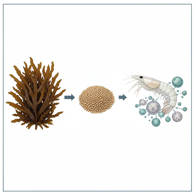
Conceptual Flow of Fucoidan Supplementation
AI-generated conceptual illustration (White-leg shrimp (Penaeus vannamei) and brown seaweed (Sargassum johnstonii)). It explains the idea and does not show study data.
View source evidence ↗Figures & tables
From the original paper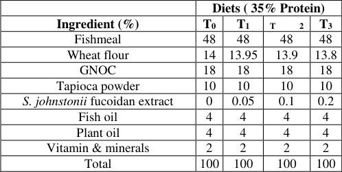 Table 1
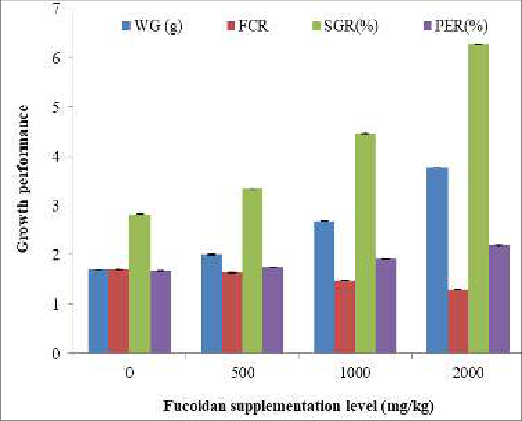 Figure 1
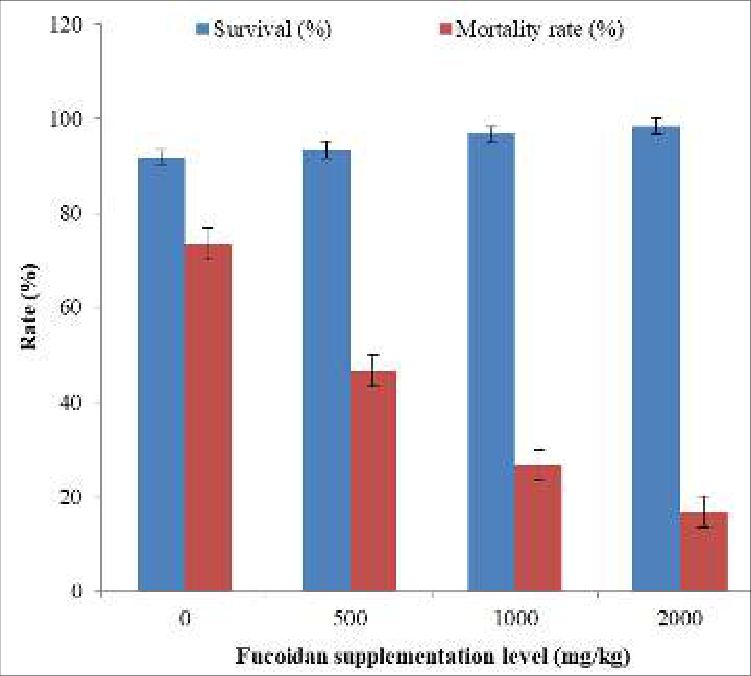 Figure 2
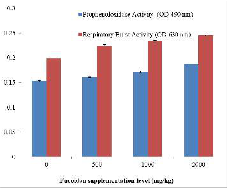 Figure 3
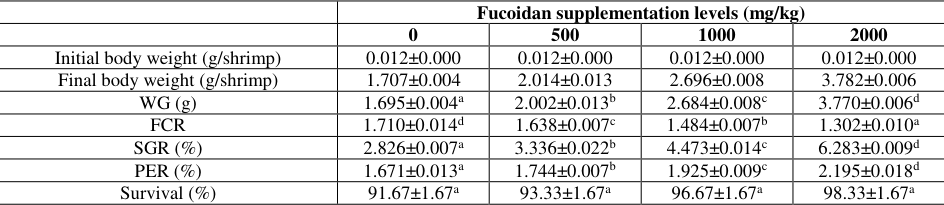 Table 2
 Table 3
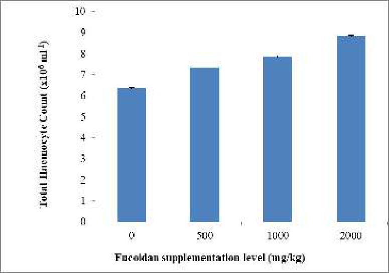 Figure 4
 Table 4
Introduction
Aquaculture is an underwater agriculture involving the propagation and cultivation of economically important aquatic organisms under controlled culture conditions. These culture organisms include fish, shellfish (crustaceans & molluscs), and aquatic plants. Among the aquaculture practices, shrimp culture plays a major role and it fetches high economy in certain countries. Since the 1970s, Penaeid shrimp culture has developed rapidly and become a major economic activity throughout the world (Wang et al., 2011) [49] . The white-leg shrimp Penaeus vannamei is one of the most common penaeid shrimp species currently being cultured worldwide. During shrimp culture practice, several associated stressors like overcrowding, transport, handling, grading, and poor water quality tend to adversely affect the cultured species' health (Li et al., 2004) [24] . These conditions produce a poor physiological environment increasing the susceptibility of shrimps to infectious and leading to substantial economic losses. It has been estimated that various diseases continued to lose the shrimp farming industry worldwide by about 3 billion USD per year, bringing down world shrimp production by 40% (HernándezRodríguez et al., 2001) [16] .
Shrimp diseases are mainly caused by biological agents like viruses, bacteria, fungi, epibiotic protozoans, and helminthic parasites and non-biological agents like nutritional and environmental factors (Sindermann, 1990; Lightner, 1993; Karunasagar et al., 1997) [37,26,22] . Recently, the global shrimp farming industry has suffered severe economic losses due to acute hepatopancreatic necrosis disease (AHPND) or early mortality syndrome (EMS) attributed to certain strains of V. parahaemolyticus being identified and suspected as the causative agents that resulted in total mortalities in penaeid shrimp (Tran et al., 2013) [48] . Prophylactic measures like vaccination, and the use of immunostimulants, probiotics, and prebiotics are very effective in controlling aquatic animal diseases. Vaccines can be used to reduce the disease risk, but no effective vaccines exist yet against several commercially important diseases in aquaculture (Raa, 1992) [33] . Crustaceans are primitive animals that lack memory cells. Hence, the vaccine does not work efficiently (Murthy et al., 2009) [30] . Because of the high cost, handling stress and undesirable side effects of adjuvants (Anderson, 1992; Mulvey et al., 1995; Midtlyng et al., 1996) [3,29,28] have resulted in a search for alternative approaches to achieve immune stimulation. Hence, there is growing interest in the development of immunomodulators that do not cause unwanted side effects on the host and can be given as a feed additive. Probiotics, prebiotics, and immunostimulants are good candidates to replace the use of chemicals and antibiotics in the development of sustainable aqua-farming systems (Murthy et al., 2009) [30] . Biological compounds called immunostimulants are available as alternative treatments, playing the role of alarm molecules that activate the immune system (Rodrıguez et al., 2003) [36] .
The potential of using medicinal plants such as seaweeds as natural immunostimulants in aquaculture as an alternative to antibiotics and immunoprophylaxis has been documented (Alghazeer et al [1,2,34,44] . Ulvaceae and Caulerpaceae (green algae); Sargassaceae (brown algae); and Gracilariaceae, Gelidiaceae, and Hypneaceae (red algae) are the most important seaweed families that have been used as immunostimulants in aquaculture (Hafezieh et al., 2014; Thanigaivel et al., 2016) [14,44] . Fucoidan, a cell-wall matrix polysaccharide, is one of the important polysaccharides present in brown seaweeds also called sulfated polysaccharides, fucoidin, fucan, fucosan, or sulphated fucan. It was first isolated from brown seaweeds by Kylin in 1913. It is named Fucoidan according to IUPAC rules. Fucoidan backbone consists of repeating subunits of L-fucose with alternating α (1-3) and α (1-4) glycosidic bonds (Berteau and Mulloy, 2003; Li et al., 2008) [7,25] . It has various biological activities like anti-thrombotic, anticoagulant (Chevolot et al., 1999; Wang et al., 2010; Nagumo, 2017) [10,50,31] anticancer, anti-proliferative, antiviral (Li et al., 2004) [24] anti-inflammatory and anticomplementary agent (Baba et al., 1988; Riou et al., 1996; Ponce et al., 2003) [6,35,32] these activities differ according to linkage pattern, the content of ester sulfate, sugar component and molecular weight (Berteau and Mulloy, 2003) [7] and which vary for each seaweed species. Fucoidans are L-fucose polysaccharides containing sulfate groups and minor monosaccharides, like D-galactose, Dxylose, D-glucose, D-mannose, and D-uronic acid. The monosaccharide composition and structure of algal fucoidans depend on algal species.
Materials and Methods
Materials and Methods
Materials
The brown seaweed S. johnstonii was collected from Porbandar coast, Gujarat, and the experiment was conducted in College of Fisheries Science, Kamdhenu University, Veraval, and Gujarat, India.
Extraction of Fucoidan
The collected seaweed was washed thoroughly and dried under shade at room temperature. The dried seaweed was ground well by using a mixer grinder and sieved using a nylon sieve to remove seaweed fiber. The extraction of fucoidan was carried out as described by Yang et al. (2008) [51] with a slight modification. Twenty grams (20 g) of shade-dried, powdered seaweed were mixed with 1 ml of 85% ethanol and stirred continuously for 12 hours at room temperature to remove proteins and pigments. After ethanol treatment, the seaweed was washed with acetone, centrifuged at 10 000 rpm for 10 minutes, and then air-dried. Five grams of this dried material were then extracted twice with 100 ml of distilled water at 65 °C for 1 hour each time, with continuous stirring. The resulting extracts were combined. The combined extract was centrifuged at 10 000 rpm for 20 minutes, and the resulting supernatant was treated with 1% CaCl₂ and stored overnight at 4 °C to precipitate alginic acid. After a second centrifugation at 10 000 rpm for 20 minutes, the new supernatant was collected. Ethanol was added to this supernatant to reach a 30% concentration, and the solution was chilled at 4 °C for 4 hours. Following another centrifugation at 10 000 rpm for 20 minutes to remove remaining impurities, the supernatant was treated with additional ethanol to achieve a final concentration of 70% and left overnight at 4 °C to precipitate the fucoidan. Finally, the precipitated fucoidan was collected by centrifugation at 10 000 rpm for 15 minutes, washed with ethanol and acetone, and then airdried. The yield was calculated based on the following formula: Yield (%) = Weight of the obtained fucoidan (g)/ Weight of the dried biomass (g) ×100
Experimental Animal
White-leg shrimp Penaeus vannamei post larvae with an average weight of 0.012 g were selected for this experiment. P. vannamei post larvae were brought to the Department of Aquaculture, College of Fisheries Science, Kamdhenu University, and Veraval from a commercial shrimp hatchery. PLs were packed in oxygenated polythene bags during transportation. The PLs were stocked in plastic tanks of 500 L capacity and fed a commercial diet for 7 days to acclimatize them to the experimental conditions. The ingredients used in the formulation of different experimental diets were fishmeal, rice bran, groundnut oil cake, tapioca flour, vitamin, and mineral premix. All the ingredients were purchased from the local market.
Experimental Design
Two hundred and forty shrimps were randomly distributed into four groups. Four experimental groups namely Control (T0) (Basal diet with no fucoidan extract), T1 (0.5% S. johnstonii fucoidan extract), T2 (1% S. johnstonii fucoidan extract), and T3 (2% S. johnstonii fucoidan extract) with 35% protein level were formulated using the square method (Hardy, 1980). The dietary treatments were tested in triplicate following a completely randomized design (CRD). Uniform-sized post-larvae of P. vannamei with an average weight of 0.012 g were stocked at 20 numbers/ tank. The experiment was carried out for 60 days. P. vannamei were fed two times a day: morning (8.00 a.m.) and evening (6.00 p.m.) at the rate of 10% of body weight during the experimental period. Continuous aeration was provided throughout the experimental period to maintain the dissolved oxygen level in each aquarium. Every morning and evening experimental tanks were cleaned manually and siphoning was done every day to remove excess feed pellets and the remaining fecal matter. Around 10% filtered and disinfected seawater was replaced the siphoned water. This was carried out throughout the experimental period of 60 days.
Experimental Diets
The composition of the diet is given in Table 1. The required quantities of ingredients were weighed accurately, mixed, and hand-kneaded to require consistency with just a sufficient quantity of water to get smooth dough (ball). The dough so obtained was cooked under steam in an autoclave at 121 °C and 15 lbs. pressure for 10-15 minutes. The cooked feed was cooled to room temperature rapidly by spreading in an enamel tray and the required dose of fucoidan extract and vitamin-mineral premix were added, mixed, and blended. The dough was extruded through a pelletizer having a 2 mm diameter. The pellets were spread on a plastic sheet and sun-dried till the moisture content was reduced to less than 10%. Diets were packed separately in high-density polythene bags, labeled, and stored on a wooden shelf at room temperature for further use.
| — | Diets ( 35% Protein) | — | — | — |
|---|---|---|---|---|
| Ingredient (%) | T0 | T1 | T 2 | T3 |
| Fishmeal | 48 | 48 | 48 | 48 |
| Wheat flour | 14 | 13.95 | 13.9 | 13.8 |
| GNOC | 18 | 18 | 18 | 18 |
| Tapioca powder | 10 | 10 | 10 | 10 |
| S. johnstonii fucoidan extract | 0 | 0.05 | 0.1 | 0.2 |
| Fish oil | 4 | 4 | 4 | 4 |
| Plant oil | 4 | 4 | 4 | 4 |
| Vitamin & minerals | 2 | 2 | 2 | 2 |
| Total | 100 | 100 | 100 | 100 |
Table text extracted from the original PDF; superscript letters are shown inline. The original table image is below.
View original table image
Growth Parameters
Shrimps were weighed at the beginning of the experiment and every 15-day interval. The growth performance of shrimp was assessed in terms of weight attained.
Mean weight increment = Final average body weight-Initial average body weight
Mean weight increment = Final average body weight-Initial average body weight
Final weight -Initial weight Experimental period * 100
FCR =
FCR =
PER =
PER =
No.of fish survived after rearing
No.of fish stocked * 100 At the end of the feeding trial, the animals were used to measure immune parameters and resistance to Vibrio parahaemolyticus.
Immune Parameters of P. vannamei After the 60-day feeding trials, 100 µl haemolymph was collected from the ventral sinus of 10 shrimps of all treatments into a 1 ml syringe (26 gauge) containing 900 µl anticoagulants solution (0.114 M trisodium citrate and 0.1 M sodium chloride, pH 7.45) for analysis of prophenoloxidase activity, respiratory burst activity and total haemocyte count.
Prophenoloxidase Assay (PPO)
The test was performed as described by Lopez et al. (2003) [27] . A sample of 50 µl was incubated for 3 min at 25 ºC with 50 µl of trypsin (0.1 mg ml -1 ). Then, 50 µl of L-DOPA (3 mg ml -1 in Cac buffer) was added and incubated for 10 min. The absorbance at 490 nm was measured (Herna´ndez-Lo´pez et al., 1996) [15] .
Respiratory Burst Activity (NBT assay)
The test was performed as described by Song and Hsieh (1994) [42] . The test was performed in flat-bottomed microtitre plates. 100 µl of HBSS was added to the first two wells as blank. 100 µl of haemolymph was added into separate wells and incubated for 1 h at 37 ºC for adherence of cells. Then, the supernatant was removed and 100 µl NBT solution was added to each well and incubated for 30 min at 37 ºC. NBT solution was removed after 30 min and 100 µl absolute methanol was added. Absolute methanol was removed and cells were washed gently 3 times with 100 µl of 70% methanol. All methanol was removed and 120 µl KOH and 140 µl DMSO were added to dissolve cytoplasmic formazan. The optical density was measured at 630 nm in an ELISA reader.
Total Haemocytes count
Haemocyte counting was done with a microscope and a Neubauer chamber.
Challenge Experiment of V.parahaemolyticus to P. vannamei
After the end of the experiment for 60 days ten shrimp from each treatment were used for the infection study. The study was conducted by stocking ten shrimp in each tank with continuous aeration. Ten shrimp were reared separately and fed with a controlled diet.
Preparation of V. parahaemolyticus Stock Culture
The bacterial strain of V. parahaemolyticus was purchased from the College of Fisheries Science, Guru Angad Dev Veterinary and Animal Sciences University, Ludhiana, Punjab. The V. parahaemolyticus was cultured on tryptic soy agar plates (TSA supplemented with 2.5% NaCl) for 24 h at 25 ºC. Then the culture was transferred to 10ml of tryptic soy broth (TSB supplemented with 2% NaCl) and the broth was incubated at 25 ºC for 24h as a stock culture for the experiment.
Preparation of Required Density of V. parahaemolyticus V. parahaemolyticus culture was spun in a refrigerated centrifuge at 3000 rpm for 10 minutes at 4 ºC. The liquid was removed, and the remaining solid was resuspended in PBS to an optical density of 0.8 at 620 nm, equivalent to 6.4 × 10⁸ cfu/ml. This standardized bacterial suspension was centrifuged at 3000 rpm for 10 minutes and the pellet was resuspended in 50 µl of PBS. From this, 7.81 µl was made into 50 µl using PBS to get 1× 10 8 cfu/50 µL was made into 50 µL using PBS to get 1 x 10 8 cfu/50 µL. These bacterial suspensions were serially diluted using the standard dilution technique with PBS to get the required concentration of 10 8 to 10 4 cfu/50 µL.
Injection of V. parahaemolyticus to Penaeus vannamei P. vannamei were challenged with V. parahaemolyticus intramuscularly between the second and third abdominal segment by the injection (1 ml capacity syringe with 26 gauge needle) of 100 µl of bacterial suspension containing 10 4 cfu/ ml PBS (LD50 dose) after the feeding trial of 60 days. Penaeus vannamei juvenile treatments (T0-T6) along with positive controls (injected same concentration of V. parahaemolyticus to control treatment feed without immunostimulant) and negative controls (injected same concentration of PBS to control treatment feed without immunostimulant) in triplicate. The susceptibility was conducted for 10 days.
Assessment of Mortality
After injection of V. parahaemolyticus, the survival of shrimp was monitored at regular till 10 days. Shrimp not reacting to gentle mechanical stimulation with a soft paintbrush were considered to be dead. The dead shrimp were removed from the respective tanks during each observation day and recorded.
Statistical Analysis
Mean growth, survival, immune responses, and disease resistance of shrimp achieved in response to different formulated test diets were analyzed statistically by using one-way analysis of variance technique (ANOVA) in all the treatments (Snedecor and Cochran, 1994) [40] . For comparison of different treatments, Duncans's multiple range tests were used.
Results and Discussion
Growth and survival of Penaeus vannamei fed S. johnstonii fucoidan extract supplemented diets Weight Gain (WG), Feed Conversion Ratio (FCR), Specific Growth Rate (SGR) and Protein Efficiency Ratio (PER) were significant difference (p<0.05) between the treatment groups in Table 2 (Figure 1).There was no significant survival rate difference found among the treatment group in Table 2 (Figure 2).
| — | Fucoidan supplementation levels (mg/kg) | — | — | — |
|---|---|---|---|---|
| 0 | 500 | 1000 | 2000 | |
| Initial body weight (g/shrimp) | 0.012±0.000 | 0.012±0.000 | 0.012±0.000 | 0.012±0.000 |
| Final body weight (g/shrimp) | 1.707±0.004 | 2.014±0.013 | 2.696±0.008 | 3.782±0.006 |
| WG (g) | 1.695±0.004a | 2.002±0.013b | 2.684±0.008c | 3.770±0.006d |
| FCR | 1.710±0.014d | 1.638±0.007c | 1.484±0.007b | 1.302±0.010a |
| SGR (%) | 2.826±0.007a | 3.336±0.022b | 4.473±0.014c | 6.283±0.009d |
| PER (%) | 1.671±0.013a | 1.744±0.007b | 1.925±0.009c | 2.195±0.018d |
| Survival (%) | 91.67±1.67a | 93.33±1.67a | 96.67±1.67a | 98.33±1.67a |
Table text extracted from the original PDF; superscript letters are shown inline. The original table image is below.
View original table image
Immune Parameters and resistance against V. parahaemolyticus infection of Penaeus vannamei
Prophenoloxidase activity in P. vannamei fed Sargassum johnstonii fucoidan extract supplemented diet was significantly higher (p<0.05) than those animals fed the control diet in Table 3. The highest phenoloxidase activity was recorded in prawns fed 2% Sargassum johnstonii fucoidan extract supplemented diet (Figure 3). Respiratory burst activity in P. vannamei fed brewer's Sargassum johnstonii fucoidan extract incorporated diets showed a significant (p<0.05) difference among the treatment groups in Table 3. The highest respiratory burst activity was recorded in animals fed 2% Sargassum johnstonii fucoidan extract incorporated diet (Figure 3). Total haemocytes were also significantly different in the control and the treatment groups fed graded levels of Sargassum johnstonii fucoidan extract in Table 3. The highest total haemocytes count of 8.827±0.0328 was recorded in shrimp fed a 2% Sargassum johnstonii fucoidan extract supplemented diet (Figure 4). The mortality of P. vannamei after the challenge test in the various treatments at the end of the experiment is detailed in Table 4. The highest mortality (%) was observed in the control group which is significant (p<0.05) difference among the treatment groups and the lowest mortality (%) was observed in the 2% Sargassum johnstonii fucoidan extract incorporated diet which is not significant (p>0.05) difference among the 1% Sargassum johnstonii fucoidan extract incorporated diet treatment groups (Figure 2).
| — | Fucoidan supplementation levels (mg/kg) | — | — | — |
|---|---|---|---|---|
| 0 | 500 | 1000 | 2000 | |
| Mortalityrate (%) | 73.33±3.33c | 46.67±3.33b | 26.67±3.33a | 16.67±3.33a |
Table text extracted from the original PDF; superscript letters are shown inline. The original table image is below.
View original table image
| — | Fucoidan supplementation levels (mg/kg) | — | — | — |
|---|---|---|---|---|
| 0 | 500 | 1000 | 2000 | |
| Prophenoloxidase Activity (OD 490 nm) | 0.153±0.0012a | 0.161±0.0009b | 0.171±0.0015c | 0.187±0.0013d |
| Respiratory Burst Activity (OD 630 nm) | 0.199±0.0006a | 0.225±0.0012b | 0.233±0.0012c | 0.245±0.0012d |
| Total Haemocyte Count (x106 ml-1) | 6.350±0.0416a | 7.337±0.0481b | 7.860±0.0513c | 8.827±0.0328d |
Table text extracted from the original PDF; superscript letters are shown inline. The original table image is below.
View original table image
Discussion
The P. vannamei post-larvae were reared in four different concentrations (0, 500, 1000, and 2000 mg kg-1) of fucoidan-incorporated diets for 60 days and the growth performance was studied. The weight gain and SGR of a control group of shrimp were comparatively lower than the experimental groups of shrimp fed on different concentrations (500-2000 mg/kg) of fucoidan-incorporated diets. The growth performance of all the tested groups of shrimp post-larvae was significantly (p<0.05) high with increasing concentrations of fucoidan. Similarly, Traifalgar et al. (2009) [47] reported the effect of fucoidan on growth performance in shrimp P. monodon after 30 days of feeding experiment. They reported that the weight gain (899.1-1049.7%) and SGR (7.90-8% per day) were significantly (p<0.05) increased with increasing concentrations of dietary fucoidan supplementation than the control treatment (weight gain: 562.2% and SGR: 6.4% per day). Following these, Immanuel et al. (2010) [20] stated the effect of hot water extracts of brown seaweeds S. wightii and Sargassum duplicatum on the growth performance of shrimp P. monodon post-larvae. In their result, they observed that the growth performance of all the tested groups was increased with increasing concentrations of seaweed extracts. In the lowest concentration (250 mg/l) of S. wightii and S. duplicatum extracts, the growth performance was noted very low (0.274 & 0.295 g weight gain and 16.27 & 16.62% SGR). But in higher concentrations (750 mg/l) of both the seaweed extracts enriched Artemia nauplii fed P. monodon post-larvae exhibited higher weight gain (0.294 and 0.323 g) and SGR (16.61 and 17.06%). Invariably, Immanuel et al. (2004) [21] suggested that P. indicus juveniles fed with seaweed products (crude extracts) of Ulva lactuca and S. wightii enriched Artemia nauplii exhibited much weight gain after 30 days of feeding experiment. Enhancement of nutrient digestibility, resulting in efficient protein utilization and improvement of growth rate has also been reported in juvenile P. vannamei fed diets supplemented with polysaccharides from Macrocystis pyrifera (Cruz-Suarez et al., 2000) [13] . The growth enhancement effects of dietary seaweed polysaccharides might be attributed to the efficient nutrient digestion and assimilation caused by the activation of fixed phagocytes in the hepatopancreas that secrete hydrolytic enzymes in the digestive gland (Azad et al., 2005) [5] . Traifalgar et al. (2010) [46] reported that the FCR and PER of shrimp after the feeding trial was affected by dietary fucoidan supplementation. FCR and PER were significantly enhanced by dietary fucoidan supplementation. The result obtained in the present investigation is also supported by several researchers such as Immanuel et al. (2004, 2012) [21,19] ; Traifalgar et al. (2009) [47] . Traifalgar et al. (2010) [46] reported that the survival of shrimp after the feeding trial was not affected by dietary fucoidan supplementation. The result obtained in the present investigation is also supported by several researchers such as Immanuel et al. (2004) [21] , Chotigeat et al. (2004) [11] , Traifalgar et al. (2009) [47] , Immanuel et al. (2012) [19] , Traifalgar et al. (2012) [45] , Kitikiew et al. (2013) [23] , Sinurat et al. (2016) [38] . PO is recognized as one of the most important humoral immune responses in crustaceans. This enzyme is synthesized by hemocytes and exists as a proenzyme termed prophenol oxidase (proPO). The activation of proPO to PO involves a series of proteolytic reactions that also activate other cellular defense reactions including opsonization, encapsulation, phagocytosis, and generation of microbicidal compounds (Soderhall and Cerenius, 1998) [41] . Oral administration of immunostimulants has been known to enhance PO activity in shrimp. Takahashi et al. (2000) [43] reported that dietary supplementation of Pantoe aagglumerans lipopolysaccharide in M. japonicus enhanced PO and phagocytic activities, resulting in improved protection against penaeid acute viremia virus. Enhanced PO activity and improved survival against artificial infection with white spot syndrome virus were also documented in P. monodon fed diets supplemented with β-glucan and Indian immunostimulant herbs (Chang et al., 2003, Citarasu et al., 2006) [9,12] . Immanuel et al. (2012) [19] reported that the Prophenoloxidase activity (PO) of experimental shrimp was significantly (p<0.05) increased than control shrimp during the WSSV challenge test. At the beginning (0 days) the PO activity of the control group was 0.1418, but it was 0.1725-0.1820 in the experimental groups. The result obtained in the present investigation is also supported by several researchers such as Huang et al. (2006) [17] , Traifalgar et al. (2010) [46] , Kitikiew et al. (2013) [23] , Sivagnanavelmurugan et al. (2014) [39] , Arizo et al. (2015) [4] , Sinurat et al. (2016) [38] . Comparable to these findings, all treatment groups receiving fucoidan supplementation had an enhanced PO activity with the highest response observed in the treatments receiving 2000 mg supplementation. This suggests that the PO immune function of P. vannamei could also be enhanced by oral administration of S. johnstonii fucoidan. Immanuel et al. (2012) [19] reported that the respiratory burst activity (superoxide anion activity) of experimental groups of shrimp was significantly (P<0.05) higher than the control group after the WSSV challenge experiment. The result obtained in the present investigation is also supported by several researchers such as Huang et al. (2006) [17] , Huynh et al. (2011) [18] , Kitikiew et al. (2013) [23] , Sivagnanavelmurugan et al. (2014) [39] , Sinurat et al. (2016) [38] . Immanuel et al. (2012) [19] reported that at the beginning of the challenge experiment, the THC significantly (P<0.05) increased with increasing concentrations of fucoidan than the control group. The result obtained in the present investigation is also supported by several researchers such as Huang et al. (2006) [17] , Traifalgar et al. (2010) [46] , Kitikiew et al. (2013) [23] , Sivagnanavelmurugan et al. (2014) [39] , Arizo et al. (2015) [4] , Sinurat et al. (2016) [38] . Traiflgar et al. (2012) [45] investigated the efficacy of U. pinnatifida fucoidan supplementation on the survival of P. japonicus against V. harveyii. They observed the post-challenge survival of P. japonicus larvae increased with increasing levels of dietary fucoidan supplementation. The highest survival was observed in dietary treatment with 1000 mg/kg fucoidan supplementation, followed by the treatment with 500 mg/kg supplementation. The authors attributed these protective effects to the enhancement of hemocyte phagocytic activity and the inhibition of pathogen adsorption to the host. Recent evidence suggests that shrimp larvae are capable of producing antibacterial peptides in their hemocytes as a response to the presence of infectious and immune-stimulating agents (Traiflgar et al., 2012) [45] . The mode of action of fucoidan attribute that it may enhance shrimp immune response by the activation of a phagocytosis-activating protein, known to initiate and enhance hemocyte phagocytic activity (Chotigeat et al., 2004) [11] . The result obtained in the present investigation is also supported by several researchers such as Immanuel et al. (2004) [21] , Huang et al. (2006) [17] , Traiflagar et al. (2009) [47] .
Conclusion
Based on results obtained from the present study, it was concluded that feeding Penaeus vannamei with a diet containing 2% Sargassum johnstonii fucoidan extract results in increased weight gain, Specific Growth Rate (SGR) (%), Protein Efficiency Ratio (PER), and immunological parameters (phenoloxidase activity, respiratory burst activity, total haemocyte count) and reducing Feed Conversion Ratio (FCR). It also, reduces the mortality rate in shrimp during the Vibrio parahaemolyticus challenge. However, there was no significant difference (p>0.05) in survival rate among all the treatments at the end of 60 days.
Acknowledgement
AcknowledgementThe authors are grateful to the Principal and Dr. P. V. Parmar, College of Fisheries Science, Kamdhenu University, Veraval, Gujarat for given the support, and assistance for providing the necessary facilities.
References
49 in the original paper- Alghazeer R, Whida F, Abduelrhman E, Gammoudi F, Naili M. In vitro antibacterial activity of alkaloid extracts from green, red and brown macroalgae from western coast of Libya. African Journal of Biotechnology. 2013;12(51):7086-7091.
- Al-Saif SS, Abdel-Raouf N, El-Wazanani HA, Aref IA. Antibacterial substances from marine algae isolated from Jeddah coast of Red Sea, Saudi Arabia. Saudi Journal of Biological Sciences. 2014 Jan 1;21(1):57-64.
- Anderson DP. Immunostimulants, adjuvants, and vaccine carriers in fish: applications to aquaculture. Annual Review of Fish Diseases. 1992 Jan 1;2:281-307.
- Arizo MA, Simeon EC, Layosa MJ, Mortel RM, Pineda CM, Lim JJ, et al. Crude fucoidan from Sargassum polycystum stimulates growth and immune response of Macrobrachium rosenbergii against white spot syndrome virus (WSSV). Aquaculture, Aquarium, Conservation & Legislation. 2015 Aug 1;8(4):535-43.
- Azad IS, Shanker KM, Mohan CV. Defense mechanism in shrimps and other crustaceans. Fish. Chimes. 1995;160:17-18.
- Baba M, Snoeck R, Pauwels R, De Clercq E. Sulfated polysaccharides are potent and selective inhibitors of various enveloped viruses, including herpes simplex virus, cytomegalovirus, vesicular stomatitis virus, and human immunodeficiency virus. Antimicrobial Agents and Chemotherapy. 1988 Nov;32(11):1742-5.
- Berteau O, Mulloy B. Sulfated fucans, fresh perspectives: structures, functions, and biological properties of sulfated fucans and an overview of enzymes active toward this class of polysaccharide. Glycobiology. 2003 Jun 1;13(6):29R-40R.
- Boone L. Anomuran, Macruran crustacea from Panama and Canal Zone. American Museum of Natural History. 1931 Dec 30.
- Chang CF, Su MS, Chen HY, Liao IC. Dietary β-1, 3glucan effectively improves immunity and survival of Penaeus monodon challenged with white spot syndrome virus. Fish & Shellfish Immunology. 2003 Oct 1;15(4):297-310.
- Chevolot L, Foucault A, Chaubet F, Kervarec N, Sinquin C, Fisher AM, et al. Further data on the structure of brown seaweed fucans: relationships with anticoagulant activity. Carbohydrate Research. 1999 Jun 30;319(1-4):154-65.
- Chotigeat W, Tongsupa S, Supamataya K, Phongdara A. Effect of fucoidan on disease resistance of black tiger shrimp. Aquaculture. 2004 Apr 26;233(1-4):23-30.
- Citarasu T, Sivaram V, Immanuel G, Rout N, Murugan V. Influence of selected Indian immunostimulant herbs against white spot syndrome virus (WSSV) infection in black tiger shrimp, Penaeus monodon with reference to haematological, biochemical and immunological changes. Fish & Shellfish Immunology. 2006 Oct 1;21(4):372-84.
- Cruz-Suárez E, Ricque-Marie D, Tapia-Salazar M, Guajardo-Barbosa C. Uso de harina de kelp (Macrocystis pyrifera) en alimentos para camarón. Avances en Nutrición Acuícola, 2000.
- López HJ, Galván GT, Albores VF. Activation of the prophenoloxidase system of the brown shrimp Penaeus californiensis Holmes. Comparative Biochemistry and Physiology Part C: Pharmacology, Toxicology and Endocrinology. 1996 Jan 1;113(1):61-6.
- Rodríguez HA, Oliviero AC, Sanchez R, Jory D, Vidal L, Franco CLF. Aquaculture development trends in Latin America and the Caribbean. Aquaculture in the Third Millennium. Subasinghe R, Bueno PB, Phillips, MJ, Hough, C., McGladdery, SE y Arthur, JR (eds.), 2001, p. 317-40.
- Huang X, Zhou H, Zhang H. The effect of Sargassum fusiforme polysaccharide extracts on vibriosis resistance and immune activity of the shrimp, Fenneropenaeus chinensis. Fish & Shellfish Immunology. 2006 May 1;20(5):750-757.
- Huynh TG, Yeh ST, Lin YC, Shyu JF, Chen LL, Chen JC. White shrimp Litopenaeus vannamei immersed in seawater containing Sargassum hemiphyllum var. chinense powder and its extract showed increased immunity and resistance against Vibrio alginolyticus and white spot syndrome virus. Fish & Shellfish Immunology. 2011 Aug 1;31(2):286-93.
- Immanuel G, Sivagnanavelmurugan M, Marudhupandi T, Radhakrishnan S, Palavesam A. The effect of fucoidan from brown seaweed Sargassum wightii on International Journal of Agriculture and Food Science https://www.agriculturaljournals.com WSSV resistance and immune activity in shrimp Penaeus monodon (Fab). Fish & Shellfish Immunology. 2012 Apr 1;32(4):551-564.
- Immanuel G, Sivagnanavelmurugan M, Balasubramanian V, Palavesam A. Effect of hot water extracts of brown seaweeds Sargassum spp. on growth and resistance to white spot syndrome virus in shrimp Penaeus monodon postlarvae. Aquaculture Research. 2010 Sep;41(10):e545-53.
- Immanuel G, Vincybai VC, Sivaram V, Palavesam A, Marian MP. Effect of butanolic extracts from terrestrial herbs and seaweeds on the survival, growth and pathogen (Vibrio parahaemolyticus) load on shrimp Penaeus indicus juveniles. Aquaculture. 2004 Jun 14;236(1-4):53-65.
- Karunasagar I, Otta SK, Karunasagar I. Histopathological and bacteriological study of white spot syndrome of Penaeus monodon along the west coast of India. Aquaculture. 1997 Jun 15;153(1-2):9-13.
- Kitikiew S, Chen JC, Putra DF, Lin YC, Yeh ST, Liou CH. Fucoidan effectively provokes the innate immunity of white shrimp Litopenaeus vannamei and its resistance against experimental Vibrio alginolyticus infection. Fish & Shellfish Immunology. 2013 Jan 1;34(1):280-90.
- P, Lewis DH, Gatlin III DM. Dietary oligonucleotides from yeast RNA influence immune responses and resistance of hybrid striped bass (Morone chrysops× Morone saxatilis) to Streptococcus iniae infection. Fish & Shellfish Immunology. 2004 May 1;16(5):561-569.
- Li B, Lu F, Wei X, Zhao R. Fucoidan: structure and bioactivity. Molecules. 2008 Aug 12;13(8):1671-95.
- Lightner DV. Diseases of cultured penaeid shrimp. CRC Handbook of Mariculture. 1993 Apr 26;1:393486.
- López N, Cuzon G, Gaxiola G, Taboada G, Valenzuela M, Pascual C, Sánchez A, Rosas C. Physiological, nutritional, and immunological role of dietary β 1-3 glucan and ascorbic acid 2-monophosphate in Litopenaeus vannamei juveniles. Aquaculture. 2003 Jun 30;224(1-4):223-43.
- Midtlyng PJ, Reitan LJ, Speilberg L. Experimental studies on the efficacy and side-effects of intraperitoneal vaccination of Atlantic salmon (Salmo salar L.) against furunculosis. Fish & Shellfish Immunology. 1996 Jul 1;6(5):335-50.
- Mulvey B, Landolt ML, Busch RA. Effects of potassium aluminium sulphate (alum) used in an Aeromonas salmonicida bacterin on Atlantic salmon, Salmo salar L. Journal of Fish Diseases. 1995 Nov;18(6):495-506.
- Murthy HS, Li P, Lawrence AL, Gatlin III DM. Dietary β-glucan and nucleotide effects on growth, survival and immune responses of pacific white shrimp, Litopenaeus vannamei. Journal of Applied Aquaculture. 2009 Aug 21;21(3):160-168.
- Nagumo T, Nishino T. Fucan sulfates and their anticoagulant activities. Polysaccharides in Medicinal Applications. 2017 Oct 19:545-574.
- Ponce NM, Pujol CA, Damonte EB, Flores ML, Stortz CA. Fucoidans from the brown seaweed Adenocystis utricularis: Extraction methods, antiviral activity and structural studies. Carbohydrate Research. 2003 Jan 20;338(2):153-165.
- Raa J. The use of immunostimulants to increase resistance of aquatic organisms to microbial infections. Dis. Asian A, 1992, p. 1.
- Reverter M, Bontemps N, Lecchini D, Banaigs B, Sasal P. Use of plant extracts in fish aquaculture as an alternative to chemotherapy: current status and future perspectives. Aquaculture. 2014 Sep 20;433:50-61.
- Riou D, Jouault CS, Sel PDD, Bosch S, Siavoshian S, Bert LV, et al. Antitumor and antiproliferative effects of a fucan extracted from Ascophyllum nodosum against a non-small-cell bronchopulmonary carcinoma line. Anticancer Research. 1996 May 1;16(3A):1213-8.
- Rodríguez A, Cuesta A, Ortuno J, Esteban MA, Meseguer J. Immunostimulant properties of a cell wallmodified whole Saccharomyces cerevisiae strain administered by diet to seabream (Sparus aurata L.). Veterinary Immunology and Immunopathology. 2003 Dec 15;96(3-4):183-92.
- Sindermann CJ. Principal Diseases of Marine and Shellfish. Gulf Professional Publishing, 1990 Feb 28.
- Sinurat E, Saepudin E, Hudiyono S. Immunostimulatory activity of brown seaweed-derived fucoidans at different molecular weights and purity levels towards white spot syndrome virus (WSSV) in shrimp Litopenaeus vannamei. Journal of Applied Pharmaceutical Science. 2016 Oct 29;6(10):082-91.
- Sivagnanavelmurugan M, Thaddaeus BJ, Palavesam A, Immanuel G. Dietary effect of Sargassum wightii fucoidan to enhance growth, prophenoloxidase gene expression of Penaeus monodon and immune resistance to Vibrio parahaemolyticus. Fish & Shellfish Immunology. 2014 Aug 1;39(2):439-449.
- Snedecor GW, Cochran WG. Statistical Methods. IOWA. Iowa State University Press. Starkstein, SE, & Robinson, RG (1989). Affective disorders and cerebral vascular disease. The British Journal of Psychiatry. 1980;154:170-182.
- Söderhäll K, Cerenius L. Role of the prophenoloxidaseactivating system in invertebrate immunity. Current Opinion in Immunology. 1998 Feb 1;10(1):23-28.
- Song YL, Hsieh YT. Immunostimulation of tiger shrimp (Penaeus monodon) hemocytes for generation of microbicidal substances: analysis of reactive oxygen species. Developmental & Comparative Immunology. 1994 May 1;18(3):201-209.
- Thanigaivel S, Chandrasekaran N, Mukherjee A, Thomas J. Seaweeds as an alternative therapeutic source for aquatic disease management. Aquaculture. 2016 Nov 1;464:529-536.
- Traifalgar R, Koshio S, Ishikawa M, Serrano AE, Corre VL. Fucoidan supplementation improves metamorphic survival and enhances vibriosis resistance of Penaeus japonicus larvae. Journal of Fisheries and Aquaculture. 2012 Jan 1;3(1):33.
- Traifalgar RF, Kira H, Tung THA, Michael RF, Laining A, Yokoyama S, et al. Influence of dietary fucoidan International Journal of Agriculture and Food Science https://www.agriculturaljournals.com supplementation on growth and immunological response of juvenile Marsupenaeus japonicus. Journal of the World Aquaculture Society. 2010 May;41:23544.
- Traifalgar RF, Serrano AE, Corre V, Kira H, Tung HT, Michael FR, et al. Evaluation of dietary fucoidan supplementation effects on growth performance and vibriosis resistance of Penaeus monodon postlarvae. Aquaculture Science. 2009 Jun 20;57(2):167-74.
- Tran L, Nunan L, Redman RM, Mohney LL, Pantoja CR, Fitzsimmons K, Lightner DV. Determination of the infectious nature of the agent of acute hepatopancreatic necrosis syndrome affecting penaeid shrimp. Diseases of Aquatic Organisms. 2013 Jul 9;105(1):45-55.
- Wang PH, Wan DH, Gu ZH, Deng XX, Weng SP, Yu XQ, et al. Litopenaeus vannamei tumor necrosis factor receptor-associated factor 6 (TRAF6) responds to Vibrio alginolyticus and white spot syndrome virus (WSSV) infection and activates antimicrobial peptide genes. Developmental & Comparative Immunology. 2011 Jan 1;35(1):105-14.
- Wang J, Zhang Q, Zhang Z, Song H, Li P. Potential antioxidant and anticoagulant capacity of low molecular weight fucoidan fractions extracted from Laminaria japonica. International Journal of Biological Macromolecules. 2010 Jan 1;46(1):6-12.
- Yang C, Chung D, You S. Determination of physicochemical properties of sulphated fucans from sporophyll of Undaria pinnatifida using light scattering technique. Food Chemistry. 2008 Nov 15;111(2):503507.