
Research article · 2025 · Full text
International Journal of Veterinary Sciences and Animal HusbandryCauliflower, canine papillomatosis, lithium antimony thiomalate, thuja 300cc
Volume 10(1S) · Pages 68–70 · DOI: 10.22271/veterinary.2025.v10.i1sb.2023 ↗
Research areasCancer and Skin LesionsCancer Diagnosis and TreatmentMedical Imaging and Pathology Studies
Abstract
Author-writtenFive dogs exhibited multiple cauliflower-like growths on the skin, primarily located on the face, ventral mandible, areas around the oral cavity, as well as on the legs and ventral abdomen. Similar growths were also observed on the dorsal and ventral surfaces of the tongue. These growths were grayish-white in appearance and were clinically diagnosed as canine papillomatosis. Initial treatments, including thuja 30cc (a homeopathic remedy), autoimmune therapy using serum, and topical applications such as Podowart and apple cider vinegar, were found to be ineffective. However, subsequent treatment with lithium antimony thiomalate successfully resolved the condition.
Summary
Source-checkedPlain-language reading of the full paper, prepared with AI and checked against the text. Each point links to the authors' own words.
Why it matters
- Papilloma viruses cause warts in dogs, which typically appear on the skin, conjunctival membranes, vulva, and penis. View evidence ↗
- While multiple papillomas in dogs are uncommon, they often indicate that more than one type of papilloma virus is involved. View evidence ↗
- Existing treatment options for canine papillomatosis range from surgical removal and cryotherapy to medications like azithromycin, thuja, and lithium antimony thiomalate. View evidence ↗
The question
- The study aims to report on the management of a refractory case of canine papillomatosis characterized by multiple mucocutaneous tumors. View evidence ↗
What they did
- The study involved five dogs (non-descriptive males and Chippiparai male and female) aged 6 months to 2 years, weighing 10 to 15 kg, all presenting with cauliflower-like masses for two weeks. View evidence ↗
- Physical exams showed grayish-white growths on the face, ventral mandible, oral cavity, legs, ventral abdomen, and the tongue across all five dogs. View evidence ↗
- Initial treatment included Thuja 30cc drops, apple cider vinegar, Cefixime (10mg/kg), a herbal immunomodulator (Stenot), vitamins, liver extract, and two doses of autologous serum injections. View evidence ↗
What they found
- After seven days of the initial treatment (Thuja, vitamins, and autoimmune therapy), there was a slight reduction in the number of warts, but some increased in size. View evidence ↗
- Hematology on the seventh day of initial treatment showed normal parameters except for eosinophilia, which ranged from 10 to 17%. View evidence ↗
- The dogs experienced rapid recovery and complete regression of the lesions following three doses of Anthiomalin combined with Thuja 30cc drops. View evidence ↗
What it means
- The authors conclude that combining lithium antimony thiomalate and Thuja 30C drops may be an effective approach for managing resistant cases of canine papillomatosis. View evidence ↗
- The authors note that there is currently limited literature regarding the use of anthiomaline for treating papillomatosis in dogs. View evidence ↗
Visual summary
1 visualsIllustrations explain the paper; they do not show study data.
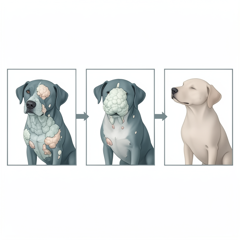
Treatment Progression Flowchart
AI-generated conceptual illustration (dogs with canine papillomatosis). It explains the idea and does not show study data.
View source evidence ↗Figures & tables
From the original paper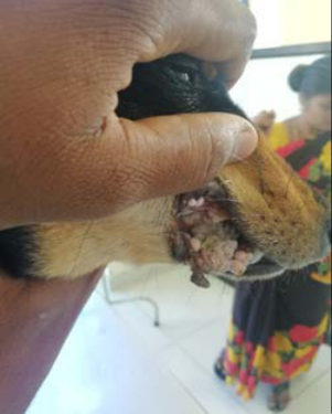 Figure 1
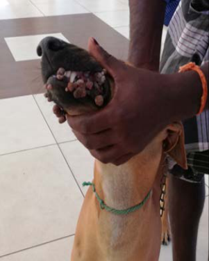 Figure 1
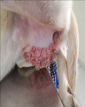 Figure 1
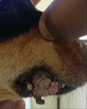 Figure 2
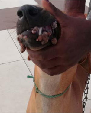 Figure 2
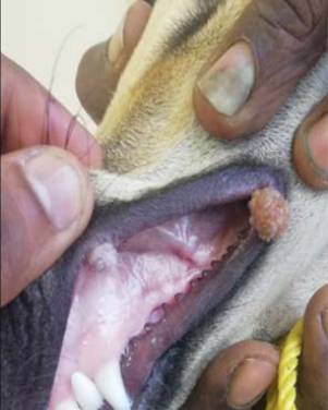 Figure 2
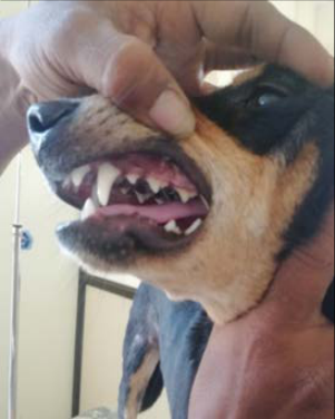 Figure 3
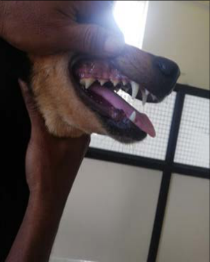 Figure 3
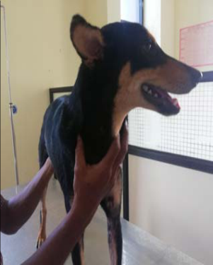 Figure 3
Introduction
Papilloma viruses are known to cause warts in dogs with these warts commonly appearing on the penis, vulva, skin, and conjunctival membranes (Sansom et al., 1996) [10] . Various forms of papilloma associated with papilloma virus infection include cutaneous squamous papilloma (Campbell et al., 1988) [2] , cutaneous inverted papilloma (Shimada et al., 1993) [12] , and canine pigmented epidermal nevus (Nagata et al., 1995) [7] . Although multiple canine papillomas are relatively rare, their occurrence often suggests the involvement of multiple papilloma virus types. Treatment options for canine papillomatosis include autogenous vaccination for both prevention and treatment (Nicolls and Stanley, 1999) [8] , spontaneous regression, autoimmune therapy (Ghim et al., 2000) [4] , surgical removal, cryotherapy, crushing of warts to enhance immune response, and the use of medications such as azithromycin (Yaggci et al., 2008) [15] , lithium antimony thiomalate (Dileepkumar and Ansari, 2012) [3] , vincristine sulfate, and thuja (Singh and Bhardwaj, 2014) [13] . This report focuses on a refractory case of canine papillomatosis involving multiple mucocutaneous tumors in dogs.
Materials and Methods
Five intact non-descriptive male and chippiparai (a male and female) and all the dogs were age group around 6 months to 2yrs, weighing 10 to 15 kgs were presented with the history of number of small cauliflower like mass noticed for past 2 weeks. The owner also reported that the dogs were not dewormed and vaccinated regularly. Physical examination revealed rectal prolapse and abdominal palpation of two male non-descriptive and a male growth on skin of face, ventral mandible, around oral cavity and also on skin of legs and ventral abdomen. Such growth was also present on dorsal and ventral areas of tongue and same as such growth in all other four dogs. These warts were greyish-white in appearance. As reported, warts started appearing on the body two weeks back with gradual increment in size. Based upon the clinical examination, it was diagnosed as a case of canine papillomatosis. The dog had marked halitosis with mild discharge from the eyes. Blood was collected in EDTA for haematological examination. Haemoglobin content, total leucocyte count, neutrophils, lymphocytes and monocytes were 9.6 to 15 g%, 12000 to 14600/cm, 56 to 82%, 13 to16% and 1 to2%, respectively.
The haematology was indicative of neutrophillia and leukocytosis. The dog was treated with drops Thuja® 30cc for one week with topical application of Apple cidar vinegar. Oral medication included tab. Cefixime @10mg/kg, orally for three days, Stenot® (Natural remedies) (a herbal immunomodulator) orally, supplements like tablets Vitamin C, Vitamin B-complex for a week and liver extract for days orally in recommended doses along with eye drops containing Oflxacin. Autoimmune therapy was undertaken by injecting intramuscularly with 2 ml of serum from the same bitch and was repeated every three days for a total of two doses. The dog was examined after 7 days of treatment. There was mild improvement reduced in numbers but there was an increase in the size of the some of the warts. Haematology on seventh day revealed that all the parameters were normal except eosinophilia (10 to 17%). The dogs was not dewormed for a considerable period of time and skin infection which could be the possible reason for the increase in the eosinophil count and anaemia.
Results and Discussion
After that the dogs were treatment with inj. lithium antimony thiomalate (Anthiomalin® each ml contains 60 mg of lithium antimony thiomalate) was started with a total dose of 0.5 ml deep intramuscularly and along with daily drops of Thuja 30cc TID and a total of three doses on alternate days were given with 0.5 ml inj. lithium antimony thiomalate increment on subsequent dosing. The dogs were showed fast recovery and there was complete regression of lesions after three doses of Anthiomalin with drops Thuja 30cc. The case was successfully resolved following the initiation of a combined therapy involving lithium antimony thiomalate and oral administration of Thuja 30 °C. Thuja occidentalis, a medicinal plant, has long been utilized for managing various ailments. In traditional medicine, extracts from its dried twig tips have been used to address conditions such as bronchial catarrh, enuresis, cystitis, psoriasis, uterine carcinomas, amenorrhea, and rheumatism. The therapeutic potential of T. occidentalis is attributed to its active compounds, including dextro-pinene, dextro-thujone, thujine, laevo-fenchone, and pinpierin. These compounds have demonstrated the ability to enhance interleukin 1, interleukin 6, and tumor necrosis factor alpha, while locally activating cytokine-producing cells for priming without inducing a systemic increase. Additionally, they promote T-helper cell induction, leading to elevated interleukin 2 production (Kalita et al., 2022) [5] . Various other methods have also been reported for lesion removal, including cryotherapy, surgical excision, cauterization, application of local anesthesia, autologous or heterologous vaccination, and the use of immunomodulators to stimulate the immune system non-specifically (Sharun et [11,9,5] .
Conclusion
Oral papillomatosis is a common, contagious, and typically self-limiting condition in dogs. However, the cases discussed here were resistant to standard treatment approaches. There is limited literature available on the use of anthiomaline for managing papillomatosis in dogs. Nevertheless, the observations in these cases align with those of Kavithaa et al. (2014) [9] , who reported that anthiomaline successfully treated 81% of papilloma cases in Jersey cattle compared to 70% effectiveness with Thuja. These findings suggest that a combination of lithium antimony thiomalate and Thuja 30C drops may yield favorable outcomes in managing such cases.
Conflict of Interest Not available
Conflict of Interest Not available
Financial Support
Financial Support
Figures and tables from the paper
References
15 in the original paper- Agnihotri A, Kushwah M, Kumar T, Kumar P, Kumar A, Srikant, et al. Successful therapeutic management of a case of canine papillomatosis. Haryana Vet. 2015;54(1):79-80.
- Campbell KL, Sundberg JP, Goldschmidt MH, Knupp C, Reichmann ME. Cutaneous inverted papillomas in dogs. Vet Pathol. 1988;25:67-71.
- Dileepkumar KM, Ansari MM. Clinico-therapeutic management of cutaneous papillomatosis in a buffalo calf. Intas Polivet. 2012;13(I):67-69.
- Ghim S, Newsome J, Bell J, Sundberg JP, Schlegel R, Jenson AB. Spontaneously regressing oral papillomas induce systemic antibodies that neutralize canine oral papillomavirus. Exp Mol Pathol. 2000;68:147-151.
- Kalita J, Verma P, Jakhar J, Patidar S. Case report on therapeutic management of canine oral papillomatosis. 2022;7:385-389. https://doi.org/10.35629/7781-0701385389.
- Kavithaa NV, Rajkumar NV, Jiji RS. Papillomatosis in Jersey cows and its medical treatment. Int J Sci Environ Tech. 2014;3(2):692-94.
- Nagata M, Nanko H, Moriyama A, Washizu T, Ishida T. Pigmented plaques associated with papillomavirus infection in dogs: Is this epidermodysplasia verruciformis? Vet Dermatol. 1995;6:179-86.
- Nicholls PK, Stanley MA. Canine papillomavirus-A centenary review. J Comp Pathol. 1999;120:219-233.
- Richman AW, Kirby AL, Rosenkrantz W, Muse R. Persistent papilloma treated with cryotherapy in three dogs. Vet Dermatol. 2017;28(6):625-e154.
- Sansom J, Barnett KC, Blunden AS, Smith KC, Turner S, Waters L. Canine conjunctival papilloma: A review of five cases. J Small Anim Pract. 1996;37:84-86.
- Sharun K, Kalaiselvan E, Sindhoora K, At FR, Khan A, Pawde AM, et al. Oral papillomatosis in a dog: Surgical management and histopathological findings. Adv Anim Vet Sci, 2020, p. 8.
- Shimada A, Shinya K, Awakura T, Narama I, Maeda H, Umemura T. Cutaneous papillomatosis associated with papillomavirus infection in a dog. J Comp Pathol. 1993;108:103-107.
- Singh J, Bhardwaj Y. Clinical management of oral papillomas in a dog. Intas Polivet. 2014;15(I):34-35.
- Sundberg JP, Smith EK, Herron AJ, Jenson AB, Burk RD, Ranst VM. Involvement of canine oral papillomavirus in generalized oral and cutaneous verrucosis in a Chinese Shar Pei dog. Vet Pathol. 1994;31:183-7.
- Yaggci BB, Ural K, Ocal N, Haydardedeoglu AE. Azithromycin therapy of papillomatosis in dogs: A prospective, randomized, double-blinded, placebocontrolled clinical trial. Vet Dermatol. 2008;19(4):194198.