
Research article · 2025 · Full text
International Journal of Biology SciencesEtiology of anemia in children: A comprehensive review
Volume 7(1) · Pages 164–170 · DOI: 10.33545/26649926.2025.v7.i1c.285 ↗
Research areasIron Metabolism and Disorders
Abstract
Author-writtenAnemia, a condition characterized by a reduction in the quantity of hemoglobin in the blood. This condition is one of the most prevalent public health problems all over the world and it affects both developing and developed countries. It has been estimated that at a global level, anemia affects 1.62 billion people, corresponding to a prevalence of 24.8% in the population. This situation is even more dramatic in the pediatric population, especially in preschool and school-age children. In 2011, the World Health Organization estimated that there are about 293 million preschool children with anemia worldwide, and their figures show that 47.4% of the preschool children of 6-59 months are anemic. In a study carried out in Brazilian children from two to six years old, the global prevalence of anemia was 20.4%, and there is a significant difference in both sexes, with a prevalence of 30.9% in males and 9.2% in females
Summary
Source-checkedPlain-language reading of the full paper, prepared with AI and checked against the text. Each point links to the authors' own words.
Why it matters
- Anemia is a global public health issue defined by a reduction in blood hemoglobin levels, affecting populations in both developed and developing nations. View evidence ↗
- Globally, anemia is estimated to affect 1.62 billion people, which represents a prevalence rate of 24.8%. View evidence ↗
- The pediatric population is severely impacted; WHO 2011 estimates indicate 293 million preschool children are anemic, with a 47.4% prevalence in children aged 6-59 months. View evidence ↗
The question
- The aim of this review is to provide a comprehensive overview of the various causes of anemia in children due to its high prevalence. View evidence ↗
- The review specifically seeks to highlight common and unique causes of pediatric anemia and suggest differential diagnoses and laboratory tests for effective clinical diagnosis. View evidence ↗
What they did
- The authors conducted a review of several studies focusing on anemia etiologies, consequences, and diagnostic pathways, with a particular emphasis on children aged 6 months to 5 years. View evidence ↗
- Anemia is classified by red blood cell size (MCV): macrocytic (> 82 fL), normocytic (82-88 fL for <2 years; 82-100 fL for >2 years), and microcytic (< 82 fL). View evidence ↗
What they found
- Anemia is estimated to contribute to over 370,000 deaths, representing one-third of the 1.1 million deaths classified under other causes of mortality. View evidence ↗
- Iron deficiency anemia is estimated to affect approximately 20-25% of the global population. View evidence ↗
- Average iron requirements are 7-8 mg/day for children up to age 10, increasing to 15 mg/day for teenage girls during pubertal growth. View evidence ↗
- A study in Yogyakarta found that genetic factors contributed to up to 25.5% of childhood anemia cases. View evidence ↗
- Symptoms of anemia typically appear when hemoglobin drops below 11 g/dL, while physical findings often remain absent until levels fall below 9 g/dL. View evidence ↗
- WHO recommends 60 mg/day of elemental iron for infants from 6 months of age, increasing to approximately 120 mg/day for those with confirmed iron deficiency anemia. View evidence ↗
What it means
- The authors conclude that nutritional interventions are the cornerstone of anemia control and are generally safe and cost-effective. View evidence ↗
- The authors emphasize that identifying the specific cause of anemia is crucial, as treatment plans must be tailored to the causative disease and severity. View evidence ↗
Visual summary
1 visualsIllustrations explain the paper; they do not show study data.
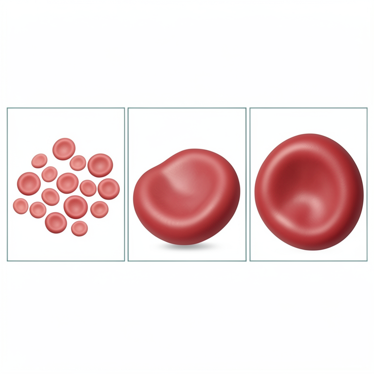
Classification of Anemia by RBC Size
AI-generated conceptual illustration (human red blood cells). It explains the idea and does not show study data.
View source evidence ↗Introduction
Anemia, a condition characterized by a reduction in the quantity of hemoglobin in the blood. This condition is one of the most prevalent public health problems all over the world and it affects both developing and developed countries. It has been estimated that at a global level, anemia affects 1.62 billion people, corresponding to a prevalence of 24.8% in the population. This situation is even more dramatic in the pediatric population, especially in preschool and school-age children. In 2011, the World Health Organization estimated that there are about 293 million preschool children with anemia worldwide, and their figures show that 47.4% of preschool children of 6-59 months are anemic. In a study carried out in Brazilian children from two to six years old, the global prevalence of anemia was 20.4%, and there is a significant difference in both sexes, with a prevalence of 30.9% in males and 9.2% in females [1][2][3][4] . The etiology of anemia in children is multifactorial and the most common cause of anemia is nutritional in origin, especially iron deficiency anemia. However, other systemic and structural diseases can present anemia as a symptom, such as genetic variations in hemoglobin cellular structure that produce inherited hemolytic anemia and can be aggravated by impairment of the production of erythrocyte cells. With this in mind, physicians should consider anemia as a serious symptom and always need a systematic approach to characterize, classify, identify the etiology and decide upon the most appropriate therapy for the case. In view of the high prevalence of anemia and its various origins, this review provided an overview of the causes of anemia in children. Such information should contribute to the Sumaré Medical School teaching process, providing complementary information to pediatrics medical students [5][6][7][8] .
Anemia is a global public health problem affecting both developing and developed countries with major consequences for human health as well as social and economic development. It is a common problem in children, and infants and preschool children are at increased risk of anemia due to requirements for growth and the development of an expanded blood volume. In children, mild anemia may lack significant clinical symptoms, and the reduction and elimination of iron deficiency anemia may demonstrate potential benefits in infants with delayed development or psychological questionnaire scores lowered physical growth, which are remediable, once they are identified. It is estimated that anemia contributes to 1/3 of the 1.1 million deaths classified as other causes of mortality, totaling more than 370,000 deaths [11] .
Classification of anemia in children
Anemia is classified according to the pathophysiological process that causes the disease and includes hypoproliferative anemia, hemolytic anemia, and hemoglobinopathy. According to the clinical manifestations and the size of red blood cells (RBCs), the disease is divided into the following: (1) macrocytic anemia: MCV > 82 fL; (2) normocytic anemia: MCV = 82-88 fL (<2 years old), MCV = 82-100 fL (>2 years old); and (3) microcytic anemia: MCV < 82 fL [12] . In children, iron deficiency anemia (IDA) is the most common nutritional anemia, followed by other anemia caused by a deficiency of vitamins such as B12 and folic acid. Multiple micronutrient deficiencies coexist in the anemic children leading to the concurrence of different anemias of etiologies. Severe malaria is a common cause of anemia in children living in malaria-endemic areas, and other infections can also lead to anemia or erythropoiesis disorders. In premature infants, the incidence of anemia is high, but most of them are mild in severity and have a good prognosis. In conclusion, the etiologies of anemia in children are complicated [13] .
Iron deficiency anemia
Iron deficiency anemia is the most common etiology of hypochromic microcytic anemia. After a period of significant body growth, the incidence of anemia in children increased significantly. It has been estimated that about 20-25% of the global population are experiencing iron deficiency anemia. However, there are great differences in the incidences of iron deficiency anemia among geographical regions. For example, iron deficiency anemia is infrequently seen in Northern Europe, Australia and New Zealand, while developing countries, particularly in Africa, South and Southeast Asia are suffering widespread iron deficiency. Recent data also suggest that a large number of American children are experiencing iron deficiency anemia. Since it has been recognized that iron deficiency anemia can result in poor neurodevelopment, impaired immune function, and longlasting effects on growth, it is important to recognize the etiology of anemia in children, particularly in those in developing countries, and to provide them with effective treatments. In this review, we will clearly demonstrate the possible etiologies of anemia in different stages of growth and discuss efficient and effective treatments for these conditions according to the mechanisms underlying these etiologies [14] .
Vitamin deficiency anemias
Anemia due to a deficiency in folic acid or cobalamin is rare, with a reported annual incidence at 3.9 and 7.6 per 100,000, respectively, compared to idiopathic anemia in children. During childhood, folic acid plays an important role in the birth and development of hematopoietic stem cells and the synthesis of myeloblasts, and it is very rare to present only by folic acid intake in adulthood. Traditionally, causes of deficiency grew before birth of the fruits of nutrition and folic acid-deficient diets, severe malabsorption syndrome, and hemodialysis patients. Currently, in economically advanced countries, in which folic acid-fortified food or folic acid is provided to pregnant women, this incidence has been reduced, and it has been known to be caused by malabsorption diseases, anticonvulsants, or methotrexate use. Cobalamin acts as a coenzyme in the methionine synthesis reaction that is necessary for the synthesis of nucleic acids, nerve fibers, cell division, and maturation of the erythroid cell, and helps to build a nerve fiber membrane, myelin sheath, and nervous system. The main cause of cobalamin deficiency in children is the intake of diet, severe malabsorption diseases, infection of the small intestine of the meat tapeworm of newborns, and intractable hereditary disorders of cobalamin metabolism. Types of anemia in children. However, nutritional anemia is still common in developing countries as a result of deficient intake of essential nutrients. Better understanding of the etiology of anemia in the pediatric population might lead to expedited diagnosis and treatment. The aim of our comprehensive review is to highlight the common and unique causes of pediatric anemia and to suggest differentials and available laboratory tests to help the clinician to diagnose anemia effectively. Furthermore, shared interests in the pediatric and hematology community might improve etymology-based focal education for anemia among pediatricians and family physicians [15,16] . Anemia is still a common symptom in children and is diagnosed by a decrease in the normal range of hemoglobin concentration, of which the age-gender and ethnicity are known as reference ranges in each country. Since the 1960s, it has been recognized that approximately 10% of children aged 1-36 months have anemia, which is common during infancy and childhood, and a rediscovery of the importance of iron in growth and development has occurred. Throughout the world, more than 2.0 billion people have anemia, and the etiology has been described as three main categories based on mechanisms: (1) Nutritional anemia caused by the body's iron stores being depleted before iron intake is adequate; (2) anemia caused by chronic infections that reduce the iron stores of the body; and (3) anemia secondary to chronic diseases. Given the common occurrence of these anemia in children, the need for knowledge of the causes of these types of anemia by disorders and deficiency syndromes, which are probably the most common etiologies, is necessary. Hematologists are involved in diagnostic evaluation and management in cases of unexplained anemia, while it remains uncommon for a pediatrician to consult with a hematology specialist for guidance on routine anemia. For this reason, pediatric hematologists must be aware and track the causes of anemia observed in children and understand the nuances of the differential diagnostic approach for the causes of anemia. The hematologic system in humans is a host-genetic product, induced by gene-environment interaction, with cells constantly maturing, aging, and dying at a rate that allows for International Journal of Biology Sciences https://www.biologyjournal.net the replacement of blood cells at a rate consistent with the cell's clearance. Changes in environmental factors can decrease rates of cells being cleared, and these extrinsic factors are what recognized as the cause of anemia [17,18] .
Hemolytic anemias
Hereditary spherocytosis (HS) is the most common cause of inherited chronic hemolysis in patients of Northern European descent. HS is an autosomal dominant disorder caused by mutations in the genes for the erythrocyte cytoskeletal proteins spectrin, ankyrin, band 3, and protein 4.2. The major clinical manifestations of HS include anemia, jaundice, splenomegaly, and gallstones (pigment stones rather than the cholesterol stones seen in beta-thalassemia). Children often present with pallor, jaundice, and intermittent dark urine that results from the passage of red urine (hemoglobinuria) when unduly stimulated, such as by a viral illness or after exercise. Family histories frequently reveal prior splenectomies for similar characteristics in affected relatives. Fortunately, most children with HS do well, with a few hospitalizations for transfusions and occasional encounters with biliary colic [19] .
The fragmentation (schizocyte) picture that occurs with increased RDW is not as pronounced as the amount of chronic hemolysis and is due to the ability of the spherocytes to flex in the microcirculation, which spares the release of all of the hemoglobin within the vessels. Other morphological abnormalities in the RBCs may appear in acute infections, aplastic crises, or rare susceptibilities to the paroxysmal nocturnal hemoglobinuria (PNH) antigen. However, HS is the most likely cause in children with a family history of HS.
The degree of anemia in HS is related to the severity of the reticulocytosis. Rapid reticulocytosis replaces the removed red cells at a slower rate than they are destroyed, which usually promotes progressively increasing anemia. In most cases, after the red cell mass stabilizes, the spleen maintains the reticulocytosis during the entire patient's lifetime. Lymphoid hyperplasia in the medullary sinuses sequesters the hemolyzed debris and forms the basis for splenomegaly that will shrink after a red cell count decreasing with splenic sequestration reduction. Although reticulocytes alone identify relatively severe hemolysis over short time intervals, they do not correlate well with the decrease in Hgb levels over longer time periods. Posttransfusion hyperbilirubinemia can also complicate the evaluation of patients 3-5 weeks after transfusion therapy. Instead, the percentage of spherocytes is the earliest detection method of HS relapse after red cell transfusions, but the percentage may be decreased by the remainder of the effective spleen function 12 and the longevity of elevated mean corpuscular volume (MCV) levels. High MCV in HS increases the severity of nephrolithiasis and decreases that normal results of calculating the osmotic fragility that splenectomy helps to diagnose and influence the predisposition of possible bone marrow failure before severe hemolysis occurs. Other conditions, including sepsis, delayed graft function, and hemolytic disease of the newborn, may occur in the condition termed dehydrated, hereditary stomatocytosis. HS treatment with RBC transfusions is contraindicated because the cells may affect vascular relaxation, leading to outcomes for stroke, hypokalemia, hyperkalemia, and hyperphosphatemia in the much smaller number of days after exposure to related microbial contamination during storage. Also, even esophagogastroduodenoscopy for bleeding esophageal varices in HS may occur in the absence of splenectomy.
Unilateral varicose vein injection and resection should be instead of portosystemic (mesocaval, splenorenal, or endovascular retrograde) shunts in children with HS [20][21][22] .
Anemia of chronic disease
Anemia of chronic disease or inflammation is a common term used to describe mild to moderate anemia in the presence of chronic systemic disease. The disease process itself is often characterized by anemia, mild leukocytosis, and thrombocytosis. This disorder of iron metabolism can manifest in chronic malnutrition and chronic inflammation. It is highly responsive to iron replacement therapy after the disease process has been treated and resolved. Causes of ironrestricted erythropoiesis due to iron sequestration secondary to inflammatory cytokines lead to impaired iron release from reticuloendothelial stores. The pathophysiology involves the decreased bioavailability of iron that is stored in reticuloendothelial cells, leading to the essential anemia of chronic disease [23] .
The supernormal release of liver hepcidin upregulates both signal transducer and activator of transcription and TNF alpha. It suppresses the absorption of iron in the duodenum and thus the bioavailability of iron to the erythroid myeloid marrow cells. A significant decrease in red blood cells results in a shortening of erythropoiesis life. Anemia is of particular relevance in inflammatory diseases in which hepcidinproducing cells store iron in reaction to high levels of inflammation. Animal and human studies have implicated hepcidin as a key determinant of anemia in chronic infection, cancer, obesity, chronic kidney disease, and autoimmune inflammatory diseases [24] .
Physiology of iron metabolism in children
The iron metabolism is carefully regulated, since iron is a critical element for both host pathogen and the patient's intrinsic immune system. The primary site of absorption is the duodenum, with the amount absorbed being determined mainly by the body's iron requirements (set by developmental stage, hemoglobin status, overall growth, menstrual losses in women, etc.). The main hormone regulating extracellular iron is hepcidin, which is produced mainly by the liver, but its production can be induced by high levels of iron (acting via TFR2 to induce production of BMP6 by hepatic stellate cells). Smaller amounts of hepcidin can also be produced by the heart, kidney, and pancreas. Hepcidin acts mainly by controlling ferroportin expression in duodenal enterocytes, on the surface of macrophages and hepatocytes [13] .
Iron can be stored in the liver, contained within two iron proteins (ferritin and hemosiderin), which exist because excess iron can cause the generation of toxic reactive oxygen species if it is not properly stored. Iron overload can also cause damage by direct metabolic actions of iron such as lipid peroxidation or by generating tissue injury through the accumulation of cells which are seen as "iron-trapping" by foreign body giant cells. Excess iron is also extremely useful to pathogens, so its toxic effects are quite beneficial to the host. Tissue growth and development thus require careful maintenance of iron concentrations, with hepcidin being the hormone that does the majority of the "fine-tuning" [25] .
Causes and risk factors of anemia in children
Chronic illness is the factor most associated with the presence of anemia in all childhood age ranges. Information about the risk factors associated with this problem at these different moments in childhood can contribute to the establishment of preventive practices that promote optimal health in children.
In this review, we have explored several studies, pursuing the various etiologies and causes of anemia, the consequences it generates, and the possible pathways for diagnosis and prevention in primary care, characteristic of the pediatric consultation, with special attention to children aged from 6 months to 5 years. We will also carry out a review of consumption patterns and nutritional status of certain minerals present in the diet related to anemia [17] .
Childhood is a critical period in the prevention of anemia's development, as the body increases total blood volume and tissue cells to a maximum extent. It may establish limitations on the daily activities of children, generating consequences related to cognitive development, psychosocial well-being, and even motor development and school performance. The disorder with the most importance in young children is lack of iron. The important development of cognitive, motor, and affective symptoms in young children at risk for iron deficiency is gaining space in the construction of management and prevention strategies, and the main causes of iron deficiency anemia (IDA) reflect changes in consumptions and population structures [7] .
Nutritional factors
Children need, on average, 7-8 mg/day of iron until the age of 10 years. Teenage girls need 15 mg of iron per day during their pubertal growth spurt. This increased demand is not only due to the increased requirement in the pubertal child as a result of increased muscle and blood mass, but also to the loss of iron via menstruation. Children need about 1-2 mg/day during this period, while adolescent girls need more, i.e. 1-1.5 mg/day. The adolescent girl should be iron replete at menarche to avoid failure to fully achieve the growth potential. The daily demand for iron is about 0.30 mg/kg in the first 2 months of life and, thereafter, about 0.19 mg/kg/day between the ages of 2 and 12 months. Iron is essential for myelination, neurotransmitter synthesis, energy metabolism, and in most provinces for the control of free radicals. These demands require iron not only from early pregnancy but also during adolescence [26] . Since many children grow up in crowded city slums without access to adequate sanitation or safe drinking water, and due to their not being exclusively breastfed early in life, this puts them at high risk of iron deficiency. Iron deficiency is the natural consequence of not having a daily diet adequate in iron, folic acid, and other essential micronutrients. The World Health Organization has estimated, based on data from the Bangladesh Demographic Health Survey, 2007, and other sources, that approximately one-third of children in the world suffer from iron-deficient anemia, one-third of which is attributable to iron deficiency. The 2014 Bangladesh Demographic Health Survey data revealed that in children aged 6-11 months, 27% were stunted, 3% were wasted, and 23% were underweight [27] .
Infectious diseases
Anemia is a major public health problem in developing countries that increases morbidity and mortality of children. Iron deficiency, which might be megaloblastic due to concomitant folate or vitamin B12 deficiencies, is the most common cause of anemia in infancy. Early identification of the causes of anemia is very important to implement appropriate therapeutic interventions and decrease morbidity and mortality [28] . Anemia in infectious diseases can be the result of three basic mechanisms: blood loss, hemolysis, and underproduction of different elements in the blood. In many countries, malaria, tuberculosis, and gastrointestinal infections are the major infectious diseases that cause anemia. In some countries, sleeping sickness and visceral leishmaniasis must also be considered as important infectious causes of anemia. In contrast, viral infections may cause transient anemia in the course of an infection, through mechanisms such as anemia of chronic disease, thrombocytopenia, or pancytopenia. The degree of anemia depends on the cause and the severity of the disease. Improvement in hematological parameters has been seen upon effective treatment with anti-infective medicines [29] .
Genetic factors
Genetic factors, especially the sickle gene, beta-thalassemia, and hereditary spherocytosis, are well-known worldwide as the etiologies of childhood anemia, notably hemolytic anemia. Although these disorders have been widely known, they are only focally studied at the regional levels in many countries, including Indonesia. However, understanding the genetic background of hemolytic anemia is crucial for determining the appropriate management and preventing further complications in children. In our study of the genetics of childhood anemia in Yogyakarta, we found a relatively high proportion of genetic factors, up to 25.5%. Thalassemia, non-s alpha and beta-thalassemia, and alpha-thalassemia, comprised the majority of genetic etiologies [30] . Forty-eight percent of the world's population carries some thalassemia trait. Indonesia is one of the countries in the region containing a high frequency of such trait. In children without any symptoms, the red blood cell size, mean corpuscular hemoglobin, and mean corpuscular hemoglobin concentration are usually similar to normal children. They usually have only mild anemia, normocytic, normochromic characteristics, and unlike thalassemia major, the hemoglobin variant frequently presents with those of hemoglobin E, hemoglobin A, and hemoglobin A2. Clinical symptoms usually develop at approximately six months of age as fetal hemoglobin production decreases, while adult hemoglobin production increases. Later, patients may present with anemia, jaundice, weakness, pallor, and hepatomegaly up to 50% more than that which is normally present in normalsized children. Our study also described the etiology and modifiers for clinical manifestation to thalassemia in our cohort [31] .
Clinical presentation and diagnosis of anemia in children
Anemia in infants and toddlers is frequently related to a decreased peak in hemoglobin levels at 2 to 3 months of life and the introduction of cow's milk to the diet. It is generally asymptomatic. However, older infants and toddlers with nutritional iron deficiency anemia or hemolytic anemia usually appear pale, have a decreased growth rate, and manifest signs of irritability and tiredness. Iron deficiency anemia and anemia of inflammation (chronic inflammation), a condition frequently superimposed on protein-energy malnutrition, are usually hypochromic and microcytic. Thalassemia or lead-poisoning anemia and others characterized by abnormal hemoglobin formation are usually normocytic and normochromic when there is mild to moderate anemia, since thalassemia red cell and ironrestricted erythropoiesis develop slowly [32] .
The clinical presentation of children with specific inherited anemia can vary tremendously. Symptoms are usually nonspecific, parents frequently have no knowledge of any family history of anemia and only about 20% of children with inherited types of anemia have a positive family history, only with chronic hemolytic anemia, do these patients develop signs of pallor. It is important for pediatricians to consider these inherited anemias in any child presenting with severe anemia and no obvious cause. Family screening is advisable when a case is confirmed. Family screening should include hematological evaluation as well as molecular genetic testing [6] .
Signs and symptoms
The clinical signs and symptoms attributed to anemia are largely the result of decreased oxygen transport to the body tissue and the work of increased cardiac output. Almost without exception, the signs and symptoms of anemia appear as hemoglobin values decline to less than 11 g/dL. The signs of anemia are relatively nonspecific and mimic those of many other diseases that children get even in the absence of anemia (e.g., lassitude, weakness, behavioral disturbances, decreased appetite, waning of intellectual activities, and so forth). Physical findings of anemia are also nonspecific and frequently are not apparent until the hemoglobin concentration falls below 9 g/dL [33] . Cardiovascular signs and symptoms predominate in children with severe anemia, e.g., tachycardia, systolic murmurs, pulmonary flow systolic ejection murmurs, palpitations, systolic murmurs, and occasionally precordial pain. Tachycardia is a reliable sign of tissue hypoxia present in nearly all children with severe anemia, but palpation of the pulse and auscultation of heart sounds is important in the count of pulse rate. In rare instances, signs of decompensated shock may be observed in children with anemia. Moreover, iron-deficiency anemia may be the presenting sign of a serious disease (e.g., adult-pattern polycystic disease of the kidney) in otherwise symptomless children [34] .
There is a certain amount of folklore surrounding anemia such as the worthlessness of a diet without complete hemoglobin replacement, the necessity of specific parenteral medication, and the special situation in pregnancy and menstruation. Glutathione, pancreatic enzymes, endorphins, noxious gases, poor exchange of small molecules, alteration of the concentration of erythroid-stimulating substances, and unorthodox substances are believed to have a beneficial effect in preventing anemia. Depending on diet, culture, and awareness, the clinical findings, signs, and symptoms can be used to guide further consideration of the causes of anemia in children. Parameters for rational evaluation of the child with no physical findings, true anemia, subtle signs, diagnosed deficiency, feeding behavior changes, and so forth still remain to be clarified [35] . [36] . Myelin gap anemia is primarily normochromic and normocytic. The main causes of its inception can be a state of increased loss and inadequate production states. The current loss in this table reveals a chronic illness, in addition to excessive bleeding and intravascular hemolysis. In cases of chronic illnesses, concomitant microcytic and hypochromic anemia may be seen; this is the mixed anemia. The Myelin gap increment alone without adequate erythrocyte expansion causes an inappropriate generation. Blood count findings can provide clues about the etiology, therapy direction, and the need for further testing. Especially in children with anemia, the first step in the diagnosis is CBC. On the updated table, the leukocyte and thrombocyte count is not specified, but in the case of systemic infection, a decrease will be determined in leukocytes. If the infectious state is not specified in the blood count characteristics of such children, the possibility of having a superimposed infectious disease or, as an alternative, having hyperimmunoglobulinemia will be mentioned. It is important to consider zinc's effect on leukocyte development. The discovery of leukopenia in the blood count is also one of the highlights of anthelmintic in mild infections [37,38] .
Laboratory tests
Laboratory tests
Management and treatment of anemia in children
In most treatment guidelines for anemia, it is emphasized that the identification of the reason behind anemia is crucial for treatment planning because the treatment plan will vary according to the severity of anemia and the causative disease.
According to the etiology, the plan will specify changes to specific factors in the child's life, for instance, what the child is and is not eating, activity or sleep patterns, recent illnesses (e.g., minor infections) and how symptoms that are related to anemia are clinically managed [39] . Anemia should first be confirmed with a hemoglobin (Hb) test because the symptoms of anemia are frequently vague and non-specific and, in some cases, may not be related to anemia. The initial laboratory screening tests to determine the cause of anemia include measurement of levels of Hb, Hct, MCV, and RDW, in addition to a reticulocyte count, serum ferritin, and serum iron. Red blood cell morphology can also provide important clues to etiology. If these analyses do not specify the cause, further laboratory testing with TIBC, Hb electrophoresis, serum folate, and vitamin B12 testing, as well as a review of peripheral smear for abnormal cells, should be provided [40] .
The treatment of anemia in children should begin with a daily iron intake of 3-6 mg (1 mg/kg) throughout the early infancy period. Fortunately, by the 4-month well-baby examination, enough evidence will have accumulated to begin iron screening. Random Hb level determination is a practical method for detecting anemia during the first two years of life [41] . Preventing childhood iron deficiency anemia consists of strategies to increase the duration of successful breastfeeding and the iron content of infant diets, as well as prevention and treatment programs targeting socioeconomically disadvantaged minority children and pregnant women. In children with anemia, the best treatment is various ironcontaining preparations or supplements that are universally used, including the liquid pediatric age group-prepared medicinal form or equivalent iron dosage, as well as iron prepared with an appropriate dosage for age and bioavailability [7] .
Nutritional interventions
The cornerstone of anemia control is nutritional interventions, which, with the exception of iron and vitamin A therapies, are relatively safe and have been found costeffective. A nutritious and balanced diet can provide micronutrients essential for the prevention and treatment of anemia. Fortification contributes to the prevention of anemia through increasing iron, folate, and vitamin B12 intake by the general population. In countries where fortified food and biofortified food for bio-available iron (e.g., beans, cauliflower, sweet potato, banana, and rice) intake are low, emphasis should be on consuming more of these foodstuffs where available. There is a trade-off in terms of carrying out a phytate reduction process (e.g., soaking grains overnight, fermenting, and adding vitamin C and/or vitamin D), which has been shown to improve bioavailability of iron and improve iron status as well, thereby indicating increased erythropoiesis and hence a reduced risk of anemia. Intervention programs should focus on pregnant mothers and infants with the following at-risk characteristics: gestational anemia, low birth weight, elderly mothers, a large number of family members, premature babies, anemia in the first year of life, poor nutritional statuses, and vitamin A deficiency. The WHO encourages the practice of exclusive breastfeeding for the first 6 months, including skin-to-skin contact to decrease maternal stress and therefore depleted body iron stores and to provide a supply of infants' requirements for free calcium. The WHO recommends the consumption of fruit and vegetables, fortified foods, iron tablets with 60 mg/day of elemental iron or iron syrup from 6 months of age for infants from the general population, with additional increases up to around 120 mg/day for iron deficiency anemia cases. Preschool children from 3 years of age should consume a daily mixed diet and balanced food. A daily iron supplement or iron syrup containing 30 to 40 mg of elemental iron is suggested for infants aged 6 months to 12 years, whereas school children should consume a typical diet and foodstuff with good iron bioavailability. For safe supplementation, both the open food-based approach and the targeted supplementation using interventions at primary health facilities can be used. Significant impacts can be anticipated in low-income countries. The deposited iron may then be used as a part of the systemic iron level throughout infancy, to avoid the common peak of anemia observed at 6 months. Fortified products such as fish sauce from the private sector have increased the compliance level, providing the crucial approach of assuming one of these products being the only one able to reach the demands for the majority of the population. Finally, however, a high level of complacency over autonomy in this context will lead to the reinforcement of previous experiences with insufficiently fortified foods [42] .
Funding None
Funding None
Acknowledgement
Acknowledgement None
References
42 in the original paper- M. Faghir-Ganji, N. Abdolmohammadi, M. Nikbina, et al., "Prevalence of Anemia in Patients with Diabetes Mellitus: A Systematic Review and Meta-Analysis," Biomedical and, vol. 2024, Elsevier, 2024.
- M. Kalhan, P. Kaushal, V. Chayal, R. Verma, et al., "Prevalence of anemia among toddlers (12-36 months) in urban area of district Rohtak, Haryana," Journal of Family Medicine, vol. 2022.
- Baldi and S. R. Pasricha, "Anaemia: Worldwide Prevalence and Progress in Reduction," Nutritional Anemia, 2022.
- M. Abdelhafeez, A. Hussein, et al., "Assessment of the Prevalence of Anemia and its Complications among Term Pregnant Women at Ain-Shams University Maternity Hospital," Evidence Based Women's Health Journal, vol. 12, no. 1, pp. 1-10, 2022.
- E. Mantadakis, E. Chatzimichael, "Iron deficiency anemia in children residing in high and low-income countries: risk factors, prevention, diagnosis and therapy," Mediterranean Journal of Hematology and Infectious Diseases, 2020.
- P.G. Gallagher, "Anemia in the pediatric patient," Blood, The Journal of the American Society of Hematology, 2022.
- J. P. Chouraqui, "Dietary approaches to iron deficiency prevention in childhood-a critical public health issue," Nutrients, 2022.
- G. M. Brittenham, G. Moir-Meyer, K. M. Abuga, et al., "Biology of anemia: a public health perspective," International Journal of Nutrition, vol. 2023, Elsevier, 2023.
- R. T. Means, "Iron deficiency and iron deficiency anemia: implications and impact in pregnancy, fetal development, and early childhood parameters," Nutrients, 2020.
- J. Zheng, J. Liu, and W. Yang, "Association of irondeficiency anemia and non-iron-deficiency anemia with neurobehavioral development in children aged 6-24 months," Nutrients, 2021.
- F. Onyai, M. Tang, M. Tong, X. Ni, M. Zhong, "Burden of Child Anemia Attributable to Fine Particulate Matters Brought by Sand Dusts in Low-and Middle-Income Countries," Environmental vol, no, pp, 2024, ACS Publications.
- TK Yıldız, N Yurtay, B Öneç, "Classifying anemia types using artificial learning methods," Engineering Science International Journal of Biology Sciences https://www.biologyjournal.net and Technology, an International Journal, Elsevier, 2021.
- C. Moscheo, M. Licciardello, P. Samperi, M. La Spina, et al., "New insights into iron deficiency anemia in children: a practical review," Metabolites, 2022.
- M. Meena, S. Khichar, A. Pawar, N. Midha, S. Kumar, et al., "Iron deficiency anemia presenting with pancytopenia: a study from India," Cureus, vol. 2023
- H. M. Ng'ethe, "Determinants of Non-Compliance to Iron Folic Acid Supplementation during Pregnancy. A Case Study of Nyeri County, Kenya," 2020.
- H. Sun and C. M. Weaver, "Decreased iron intake parallels rising iron deficiency anemia and related mortality rates in the US population," The Journal of Nutrition, 2021.
- H. Li, J. Xiao, M. Liao, G. Huang, J. Zheng, H. Wang, et al., "Anemia prevalence, severity and associated factors among children aged 6-71 months in rural Hunan Province, China: a community-based cross-sectional study," BMC Public Health, vol. 2020. Springer, 2020.
- T. Gebreegziabher, N. Regassa, M. Wakefield, et al., "Disparities in the prevalence and risk factors of anaemia among children aged 6-24 months and 25-59 months in Ethiopia," Journal of Nutritional Science, vol. 9, Cambridge University Press, 2020.
- Y. Shi, Y. Li, X. Yang, X. Li, G. Peng, X. Zhao, X. Liu, Y. Zhao, et al., "Genotype-degree of hemolysis correlation in hereditary spherocytosis," BMC Genomics, vol. 2023. Springer, 2023.
- Z. Ma, Z. Wang, X. Zhang, and H. Yu, "Splenic infarction after Epstein-Barr virus infection in a patient with hereditary spherocytosis: a case report and literature review," BMC surgery, 2022.
- J. S. Zhang and L. Li, "Laparoscopic ligation of splenic vessels for the treatment of hereditary spherocytosis in children," Pediatric Surgery International, 2020.
- E. Kılıçaslan, "Evaluation of adult patients with hereditary spherocytosis," Dokuz Eylül Üniversitesi Tıp Fakültesi Dergisi, 2022.
- XY Salokhiddinovna, "Anemia of Chronic Diseases," Research Journal of Trauma and …, vol. 2023. journals.academiczone.net.
- KV Kowdley, EM Gochanour, "Hepcidin signaling in health and disease: ironing out the details," Hepatology, vol. 2021, Wiley Online Library.
- L. Sousa, M. M. Oliveira, M. T. C. Pessôa, and L. A. Barbosa, "Iron overload: Effects on cellular biochemistry," Clinica chimica acta, 2020.
- AS Sacri, A Bocquet, M de Montalembert, S Hercberg, "Young children formula consumption and iron deficiency at 24 months in the general population: a national-level study," Clinical Nutrition, Elsevier, 2021.
- I. Kranjčec, N. Matijašić Stjepović, D. Buljan, L. Ružman, et al., "Management of childhood iron deficiency anemia in a developed country-a multicenter experience from Croatia," Diagnostics, vol. 2023. mdpi.com, 2023.
- S.R. Pasricha, J. Tye-Din, M.U. Muckenthaler, et al., "Iron deficiency," The Lancet, 2021.
- A. Lemoine and P. Tounian, "Childhood anemia and iron deficiency in sub-Saharan Africa-risk factors and prevention: A review," Archives de Pédiatrie, 2020.
- B. Santos, M. Delgadinho, J. Ferreira, I. Germano, et al., "Co-Inheritance of alpha-thalassemia and sickle cell disease in a cohort of Angolan pediatric patients," Molecular Biology, vol. 2020, Springer, 2020.
- D. Mangla, S.P. Singh, N. Bansal, S. Gole, "Differentiation of the β-thalassemia Trait from Iron Deficiency Anaemia by Red Cell Indices among Pregnant Women in Southern Haryana," Journal of Pharmacy …, 2022.
- K. Graczykowska, J. Kaczmarek, D. Wilczyńska, et al., "The consequence of excessive consumption of cow's milk: Protein-losing enteropathy with anasarca in the course of iron deficiency anemia-case reports and a …," Nutrients, vol. 2021.
- D. T. Lee and M. L. Plesa, "Anemia," Family Medicine: Principles and Practice, 2022.
- L. Shobhavat, A. D'Costa, and K. Shroff, "Anemia that presented with desaturation: a focus on core concepts," Case reports in pediatrics, 2021.
- P. T. Mathebula, "Factors associated with anaemia in pregnancy: a case study of Matibidi Village, Ehlanzeni District, Mpumalanga, South Africa," 2023.
- A. M. Agarwal and A. Rets, "Laboratory approach to investigation of anemia in pregnancy," International Journal of Laboratory Hematology, vol. 2021. Wiley Online Library.
- S. Ballal, "A Study on Prevalence and Outcome of Anemia in Pregnancy in a Tertiary Health Care Setup in Dakshina Kannada," 2020.
- J. Artym, M. Zimecki, and M. L. Kruzel, "Lactoferrin for prevention and treatment of anemia and inflammation in pregnant women: A comprehensive review," Biomedicines, 2021.
- M. N. Garcia-Casal, O. Dary, M. E. Jefferds, et al., "Diagnosing anemia: Challenges selecting methods, addressing underlying causes, and implementing actions at the public health level," Annals of the New York Academy of Sciences, vol. 2023. Wiley Online Library.
- SB Neogi, J Sharma, S Pandey, N Zaidi, et al., "Diagnostic accuracy of point-of-care devices for detection of anemia in community settings in India," BMC health services research, vol. 2020, Springer, 2020.
- C. Ruangkit, N. Prachakittikul, N. Hemprachitchai, et al., "Association of infant feeding practices with iron status and hematologic parameters in 6-month-old infants," Children, vol. 2021.
- K. Harron, R. Gilbert, J. Fagg, A. Guttmann, "Associations between pre-pregnancy psychosocial risk factors and infant outcomes: a population-based cohort study in England," The Lancet Public Health, 2021.