
Research article · 2025 · Full text
International Journal of Advanced Chemistry ResearchEstimation of Galanin hormone levels and additional biochemical markers in polycystic ovary syndrome patients
Volume 7(1) · Pages 32–42 · DOI: 10.33545/26646781.2025.v7.i1a.254 ↗
Research areasGrowth Hormone and Insulin-like Growth Factors
Abstract
Author-writtenPolycystic ovarian syndrome (PCOS), which is typified by irregular menstrual cycles, hyperandrogens, and polycystic ovaries, is the most prevalent endocrine and metabolic disorder affecting premenopausal women. Women with PCOS are more likely to develop metabolic syndrome because they are at a higher risk of acquiring hypertension and dyslipidemia. Twenty health screen examinees who were admitted to the Tikrit Teaching Hospital's primary accommodations between January and March 2024 were referred to our organization. Forty patients were selected on the basis of their history of Polycystic Ovarian Syndrome, blood test results, abdominal ultrasound results, and whether or not they provided written informed consent. For those who have Polycystic Ovary Syndrome, blood galanin levels were significantly elevated, independent of age or the results of laboratory tests (144.9±9.2vs. 30.18±6.6, p
Summary
Source-checkedPlain-language reading of the full paper, prepared with AI and checked against the text. Each point links to the authors' own words.
Why it matters
- Polycystic ovarian syndrome (PCOS) is the most common endocrine and metabolic disorder in premenopausal women, characterized by polycystic ovaries, hyperandrogens, and irregular menstrual cycles. View evidence ↗
- Women with PCOS have an increased risk of developing metabolic syndrome, specifically through higher risks of dyslipidemia and hypertension. View evidence ↗
- Galanin, a neuropeptide, is believed to play a role in PCOS development by influencing GnRH secretory neurons, which relates to unregulated gonadotropin production. View evidence ↗
The question
- The study aimed to compare blood levels of galanin, irisin, D3, specific hormones (TSH, prolactin, LH, FSH), and lipid profiles (cholesterol, triglycerides) between individuals with PCOS and a control group. View evidence ↗
What they did
- The study included 40 PCOS patients and 20 healthy control subjects, all aged between 20 and 40 years. View evidence ↗
- Approximately 8 milliliters of venous blood were collected from each participant using sterile disposable syringes and stored in gel tubes. View evidence ↗
- Data analysis was performed using IBM SPSS version 28.0 and the Real Statistics Resource Pack for Excel 2016. View evidence ↗
What they found
- PCOS patients had significantly higher average serum galanin levels (144.9±9.2) compared to the control group (30.18±6.6), with p > 0.01. View evidence ↗
- Serum irisin levels were significantly higher in PCOS patients (6.1±0.6) than in the control group (0.9±0.3), with p > 0.01. View evidence ↗
- PCOS patients exhibited significantly lower mean blood TSH levels (0.05±0.02) compared to the control group (2.29±0.8), with p > 0.01. View evidence ↗
- Serum D3 levels were significantly lower in PCOS patients (8.1±2.0) compared to the control group (51.8±9.2), with p < 0.001. View evidence ↗
- PCOS patients had a significantly higher mean BMI (36.1±2.7 kg/m²) compared to the control group (29.47±0.99 kg/m²), with p > 0.01. View evidence ↗
What it means
- The authors conclude that irisin may serve as a diagnostic biomarker for PCOS. View evidence ↗
- The authors state that PCOS patients are characterized by elevated levels of blood irisin, galanin, 8-oxoguanine DNA glycosylase, triglycerides, cholesterol, and D3. View evidence ↗
Visual summary
2 visualsIllustrations explain the paper; charts are replotted from the paper's own table values.
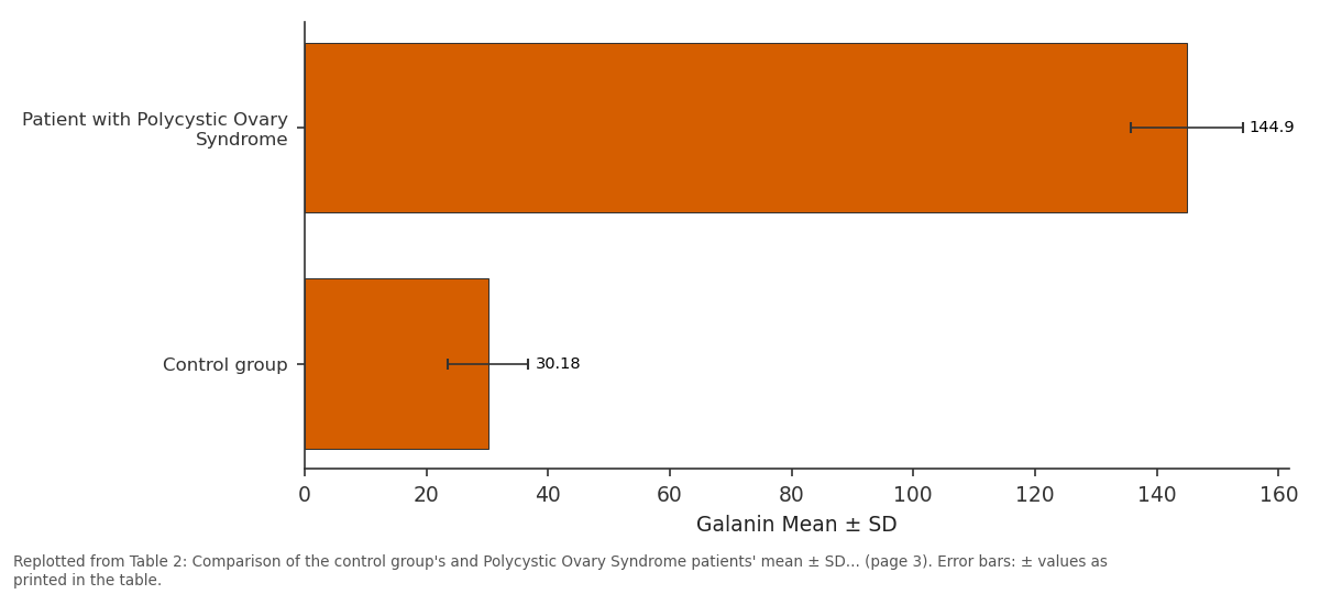
Galanin Mean ± SD: Patient with Polycystic... vs Control group
Replotted from Table 2 (page 3) of the original paper. Every bar is the paper's own table value.
View source evidence ↗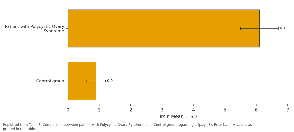
Irsin Mean ± SD: Patient with Polycystic... vs Control group
Replotted from Table 3 (page 3) of the original paper. Every bar is the paper's own table value.
View source evidence ↗Figures & tables
From the original paper Table 1
 Table 2
 Table 3
 Table 4
 Table 5
 Table 6
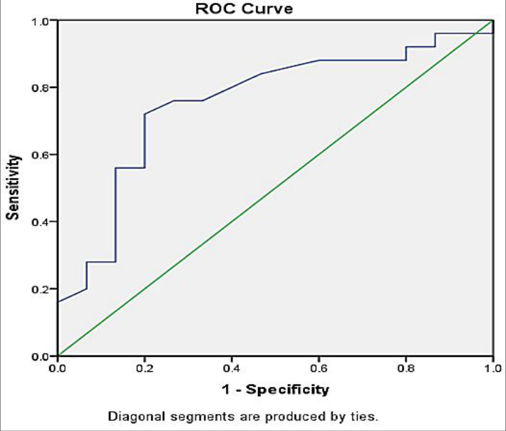 Figure 1
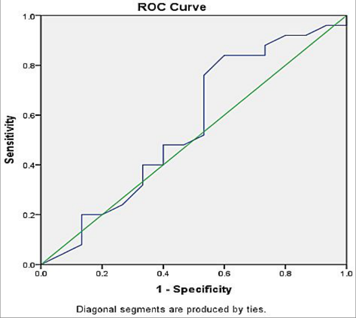 Figure 2
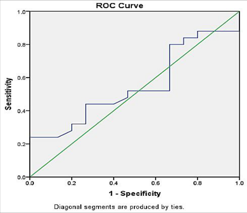 Figure 3
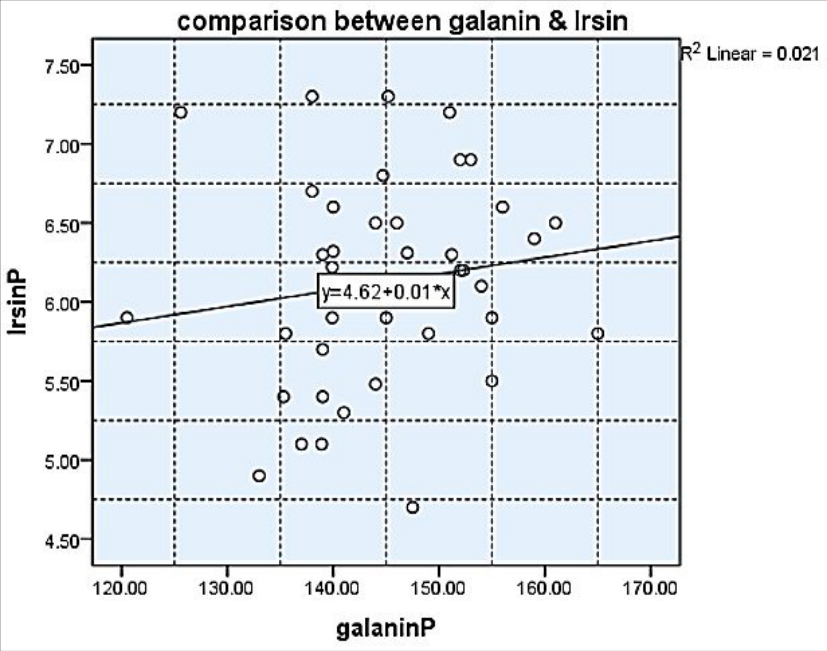 Figure 4
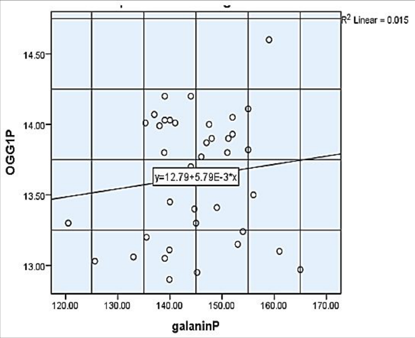 Figure 5
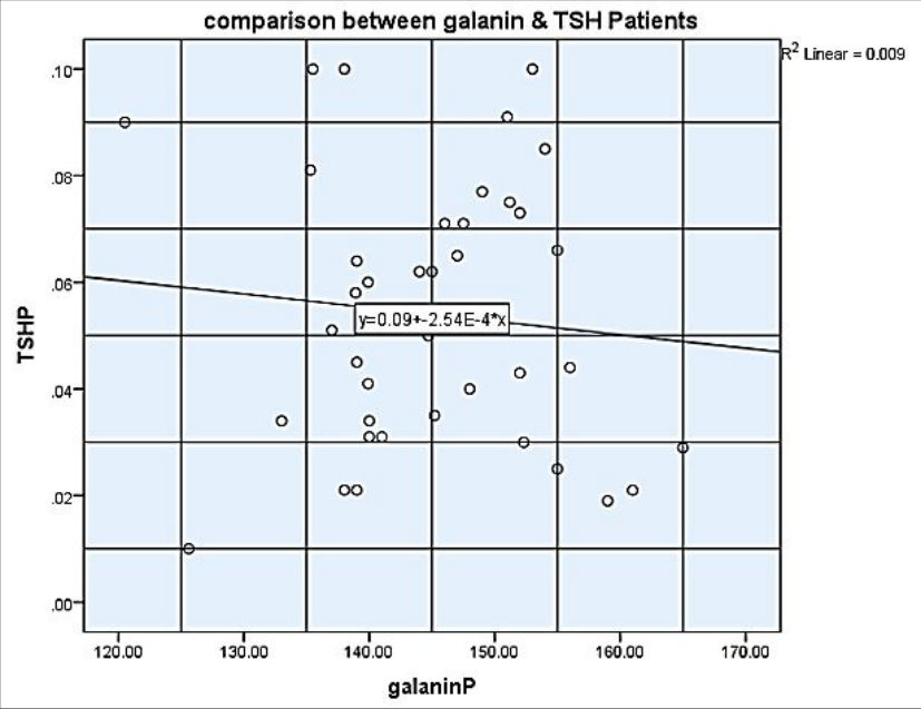 Figure 6
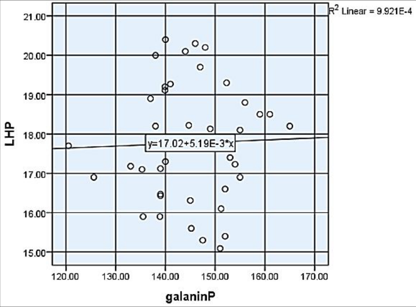 Figure 7
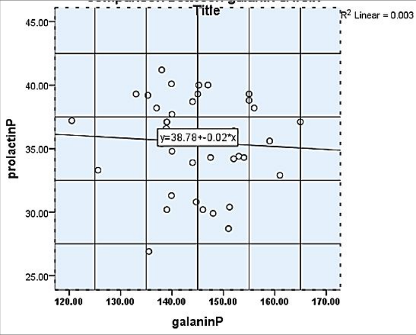 Figure 8
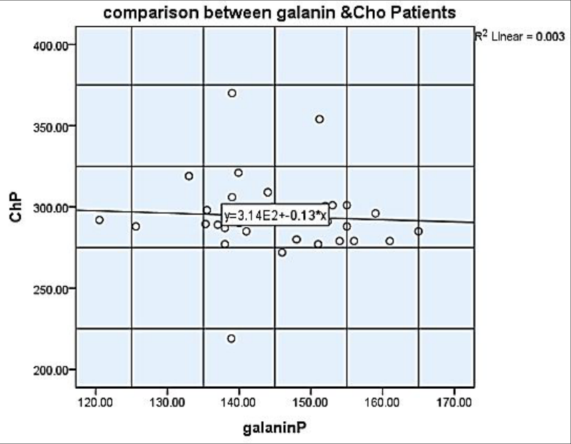 Figure 9
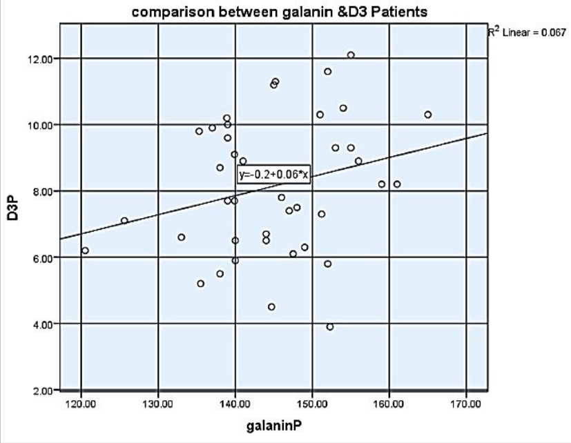 Figure 10
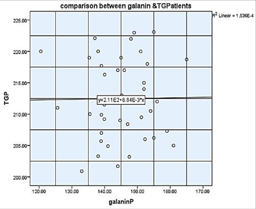 Figure 11
Introduction
The most prevalent endocrinopathy, polycystic ovarian syndrome (PCOS), affects seven to ten percent of women who are of reproductive age [1] . Hyperandrogenism, irregular menstrual periods, and polycystic ovaries are symptoms of this complex endocrine condition. Three criteria are used to diagnose PCOS: high androgen, ovulatory failure, or polycystic ovaries [2] . PCOS can be caused by a variety of hereditary and environmental factors. Family history, inactivity, diabetes mellitus, and obesity are risk factors for the development of PCOS. PCOS increases the risk of osteoporosis, cancer, high blood pressure, diabetes, lipid problems, mental health problems, and insulin resistance. PCOS is known to have a significant influence on female infertility, affecting between 0.6 and 3.4% of infertile couples. Recent advancements in lab equipment, sonography, and conventional laparoscopic testing for infertility have led to a large increase in the prevalence of PCOS. Women with PCOS need to be properly diagnosed and treated because of their elevated risk of diabetes, hypertension, cardiovascular disease, and cancers linked to hyperestrogen [3] . Both sexes' reproductive systems and the central and peripheral nervous systems have high concentrations of galanin, a neuropeptide composed of 29 amino acids [4] . Numerous physiological functions and illnesses, including feeding, hunger, inflammation, Alzheimer's disease (AD), epilepsy, neuroregeneration, convulsions, and pain, are linked to it. It belongs to the three members of the G-protein-coupled receptor (GPCR) superfamily (GAL1-3), each of which has unique signaling pathways and distribution patterns. Galanin, galanin-like peptide (GALP), galanin, alarin, and galanin-message associated peptide (GMAP) make up the so-called galaninergic system, which uses galanin as its primary signaling component [5] . In the hypothalamus, some of the neurons that produce GnRH also produce galanin. Estrogen significantly increases the synthesis of the galanin gene in these GnRH neurons. GAL2 has been shown to be present in the ovary. Galanin is believed to have a major role in the development of the gonadal secretory and preovulatory LH spike in animals. There may be more, as yet unidentified functions for galanin. It is thought that neurological influences over GnRH secretory neurons, especially galanin, are important in the development of polycystic ovarian syndrome since neuroendocrine abnormalities are frequently associated with unregulated gonadotropin production in patients with this illness. PCOS is one of the most prevalent conditions affecting women in their reproductive years. It is defined as metabolic and endocrine issues linked to modifications in the ovarian and pituitary-gonadal axis's function [6] . A myokine that muscles make called irisin was recently discovered to act as a messenger between skeletal muscle and other body parts. The Greek goddess Iris, a god-to-god mediator, is the source of the name Irisin [7] . When fibronectin type III domain containing 5 (FNDC5) is broken down by the circulating factor Irisin, white adipose tissue (WAT) experiences thermogenic responses, often known as browning. Increased oxygen demand and mitochondrial density are linked to weight loss and heat production caused by WAT browning. The pathogenesis of insulin-resistant conditions like type 2 diabetes and metabolic syndromes may involve irisin. Muscle mass, adipose tissue mass, and body mass index (BMI) have all been linked to higher irisin levels. Irisin plasma levels and body fat are correlated in both PCOS patients and healthy individuals [8] . This study sought to determine whether irisin and gelanin, two hormones, were associated with PCOs. To do this, we evaluated the relationship between the hormone levels of irisin and gelanin and another biological trait.
Materials and Methods
The patients included in this study were recruited during the period from January 2024 and March 2024.
Patients Group: Patients diagnosed with PCOs by upper abdomen ultra-sonogram were included in the study. This study investigated 40 patients, they ranged in age from 20 to 40. Submitted to the primary accommodations from Tikrit Teaching Hospital.
Controls Group:
For standardization and comparison, 20 seemingly healthy persons, neither smokers nor PCOs matched for age, and weight with the patient groups, were chosen as controls. The ages varied from 20 to 40 years. The control group was assessed to be free of sickness, not taking any drugs, and consisted primarily of medical staff and friends.
Data Collection:
The primary source of data was gathered directly from patients and control individuals through interviews and the use of a specifically designed questionnaire that recorded detailed information about each patient and control subject. It includes the following information: name, age (Years), mobile number, weight (kg), height (m), BMI, smoking history, family history of PCOs, length of sickness, kind of treatment, duration of treatment. Data received indirectly from patient records were also analyzed and documented.
Sample Collection
A sterile disposable syringe was used to extract approximately 8 milliliters of venous blood from each subject, which was then placed into gel tubes. After the blood in a gel tube was centrifuged for 10 minutes at 4000 rpm and allowed to clot at room temperature for a while, the serum was extracted and divided into Eppendorf tubes for every sample. After that, the samples were stored at -20 °C in a deep freezer until analysis time. Samples from control subjects were collected and processed using the same protocols. Even after adjusting for BMI, female PCOS patients had a 4.5-fold higher risk of Metabolic Syndrome (MBS), which rose by 3.8 times for each quartile increase in bioavailable testosterone, per a study by COVIELLO [9] . The study's PCOS patients had irisin levels that were abnormally high in comparison to what is typically seen in PCOS patients. Although its exact cause is uncertain, PCOS is associated with metabolic problems, as was previously indicated. Serum irisin levels were greater in patients with metabolic syndrome, per a study by Park et al. [10] . Serum fasting irisin levels may be higher in PCOS patients than in control subjects because of the metabolic syndrome, which is linked to PCOS. Another theory suggests that a high serum irisin level may provide protection during the prediabetic stage of PCOS. Furthermore, it has been suggested that irisin resistance may be linked to PCOS, as it is comparable to leptin resistance in people who are obese and/or have metabolic syndrome [11] . Gelanin levels were higher in PCOS patients than in controls, according to our findings. Slim PCOS patients exhibited considerably greater galanin levels than the BMI-matched controls, according to group analysis that further separated the subjects depending on their BMI; however, this difference was not present in the overweight/obese group [12] . Insulin resistance, diabetes, appetite, obesity, dyslipidemia, hypertension, metabolic syndrome, and reproduction are all impacted by galanin. Numerous investigations have discovered a link between galanin and PCOs [13] . This result is consistent with other research regarding the negative association between PCOs and TSH in the healthy control group. One study found that a drop in TSH levels was correlated with a decrease in the number of antral follicles generated [14] . AMH is released by both antral and pre-antral follicles, hence it seems logical that the two chemicals would be inversely correlated in healthy people [15] . According to a number of studies, women with PCOS had higher blood levels of the hormone LH. Ambiger et al. found that compared to healthy women, women with PCOS had higher levels of LH [16] . Elevated blood levels of LH are consistent with our investigation's findings. But keep in mind that a lot of obese women without PCOS also have elevated LH levels. This suggests that LH secretion control is altered by fat, which may cause PCOS-like symptoms in obese women. On the other hand, the blood LH levels of obese women with PCOS did not differ statistically significantly from those of women without the illness. Therefore, PCOS cannot be diagnosed by LH levels alone [17] . The primary glycosylase that catalyzes the removal of DNA oxidation products is 8-oxoguanine DNA glycosylase (OGG1). In this work, we looked into possible routes and OGG1's function in PCOS development. When we initially looked at OGG1 levels in PCOS patients' blood and follicular fluid (FF), we found that these patients had much higher OGG1 levels. In line with a study conducted in 2022 by Xia, Jing, et al. [18] , we likewise found a notable increase in OGG1 expression levels in the ovarian tissue of PCOS brought on by dehydroepiandrosterone (DHEA). 8oxoguanine DNA glycosylase (OGG1) is the main glycosylase that catalyzes the elimination of DNA oxidation products. In this study, we investigated potential pathways and the role of OGG1 in the development of PCOS. When we first examined OGG1 levels in the blood and follicular fluid (FF) of PCOS patients, we discovered that these patients had significantly higher OGG1 levels (Aldhalemi AA et al., 2024) [19] . We also discovered a significant rise in OGG1 expression levels in the ovarian tissue of PCOS caused by dehydroepiandrosterone (DHEA), which is consistent with a study done in 2022 by Xia, Jing, et al. [18] . This study found that vitamin D treatment did not stabilize the women's serum profiles or mitigate the effects of the condition. Drug administration may still be an important area of research, but it should not be overlooked in the fight against PCOS [22] . Rashidi et al. (2016) examined the relationship between serum 25-OH-Vit D3 levels and metabolic variables in women with polycystic ovarian syndrome who were not obese. A normal weight group and an overweight group (based on BMI) were established among non-fat women with polycystic ovarian syndrome. Serum levels of 25 (OH) D, FBS, CRP, TC, TG, LDL, HDL, INS, and IR were measured. 84.1% of women were found to be vitamin D deficient; yet, there was no discernible difference between the two groups. Their main finding was that there was a significant relationship between HDL serum levels, age, and 25 (OH) D. However, Rashidi et al. discovered that a decrease in 25 (OH) D levels was not statistically significantly associated with an increase in IR risk in non-obese women with polycystic ovarian syndrome [22] .
Exclusion criteria
Exclusion criteria
Conclusion
The information gathered suggests that irisin could be used as a PCO diagnostic biomarker. PCOS patients had significantly higher serum levels of galanin and irisin than the control groups, and obese PCOS patients had higher levels than participants of normal weight. PCO patients had higher levels of blood irisin, galanin, 8 oxoguanine DNA glycosylase, lipid profiles (TG and cholesterol), and D3.Elevated irisin levels in PCOS patients can lead to risk factors like insulin resistance and dyslipidemia. The polycystic ovarian syndrome can be identified by changes in blood levels of the irisin biomarker. . Ethical Approval On December 24, 2023, the Tikrit University College of Medicine's Ethical Committee approved this study under file number 68/240. Consent from participants or their guardians was obtained prior to study participation and blood collection.
Figures and tables from the paper
| Characteristic | PCOS No.40 | Control No.20 | P-Value |
|---|---|---|---|
| Mean ± SD | Mean ± SD | ||
| Age (Years) | 32.7±5.3 | 28.35±5.21NS | 0.06 |
| BMI (kg/m²) | 36.1±2.7 | 29.47±0.99 | 0.01 |
| Total | 40 | 20 |
Table text extracted from the original PDF; superscript letters are shown inline. The original table image is below.
View original table image
| Study groups | n | Galanin Mean ± SD | P. value |
|---|---|---|---|
| Patient with Polycystic Ovary Syndrome | 40 | 144.9±9.2 | 0.01 Significant difference |
| Control group | 20 | 30.18±6.6 |
Table text extracted from the original PDF; superscript letters are shown inline. The original table image is below.
View original table image
| Study groups | n | Irsin Mean ± SD | P. value |
|---|---|---|---|
| Patient with Polycystic Ovary Syndrome | 40 | 6.1±0.6 | 0.01 Significant difference |
| Control group | 20 | 0.9±0.3 |
Table text extracted from the original PDF; superscript letters are shown inline. The original table image is below.
View original table image
| Study groups | n | 8 Oxoguanine DNA glycosylase (OGG1) Mean ± SD | P. value |
|---|---|---|---|
| Patient with Polycystic Ovary Syndrome | 40 | 13.6±o.4 | 0.01 Significant difference |
| Control group | 20 | 2.3±0.3 |
Table text extracted from the original PDF; superscript letters are shown inline. The original table image is below.
View original table image
| Study groups | n | Mean ± SD | — | — | — |
|---|---|---|---|---|---|
| TSH | Prolactin | LH | FSH | ||
| Patient with Polycystic Ovary Syndrome | 40 | 0.05±0.02 | 35.5±3.5 | 17.7±1.5 | 28.01±3.8 |
| Control group | 20 | 2.29±0.8 | 11.2±3.8 | 9.07±2.7 | 9.6±1.5 |
| P. value | 0.01 | 0.01 | 0.01 | 0.01 |
Table text extracted from the original PDF; superscript letters are shown inline. The original table image is below.
View original table image
| Study groups | n | Mean ± SD | — | — |
|---|---|---|---|---|
| D3 | Cholesterol | TG | ||
| Patient with Polycystic Ovary Syndrome | 40 | 8.1±2.0 | 294.2±22.2 | 212.4±6.4 |
| Control group | 20 | 51.8±9.2 | 92.1±4.7 | 73.6±9.4 |
| P. value | 0.001 | 0.01 | 0.01 |
Table text extracted from the original PDF; superscript letters are shown inline. The original table image is below.
View original table image
References
22 in the original paper- Vaidya A, Yadav S, Vaidya A. A study on the clinical and hormonal profile of polycystic ovarian syndrome patients attending a tertiary care hospital: a descriptive cross-sectional study. JNMA J Nepal Med Assoc. 2020;58(231):875-888.
- Krishnan A, Muthusami S. Hormonal alterations in PCOS and its influence on bone metabolism. J Endocrinol. 2017;232(2):R99-R113.
- Rajbanshi I, Poudel P, Bhattarai S, Bhattarai S, Maharjan S, Pradhan P, et al. Metabolic and biochemical profile in women with polycystic ovarian syndrome attending tertiary care centre of central Nepal. BMC Womens Health. 2023;23(1):208.
- Azin F, Khazali H. Neuropeptide galanin and its effects on metabolic and reproductive disturbances in female rats with estradiol valerate (EV)-induced polycystic ovary syndrome (PCOS). Neuropeptides. 2020;80:102026.
- Šípková J, Křenová D, Klaschka J, Bubnová K, Hrabáková H, Pospíšilová M, et al. The galanin and galanin receptor subtypes, its regulatory role in the biological and pathological functions. Physiol Res. 2017;66(5):699-708.
- Mohd Zahir I, Ching YH, Jalaludin H, Zainalabidin S, Hussin AH. Spexin and galanin in metabolic functions and social behaviors with a focus on non-mammalian vertebrates. Front Endocrinol. 2022;13:882772.
- Pukajlo K, Kolackov K, Laczmanski L, KuliczkowskaPlaksej J, Lenarcik-Kabza A, Milewicz A, et al. Irisin plasma concentration in PCOS and healthy subjects is related to body adipose content. Endocr Abstr. 2014;35:758.
- Kruszewska J, Laudy-Wiaderny H, Kunicki M. Review of novel potential insulin resistance biomarkers in PCOS patients-The debate is still open. Int J Environ Res Public Health. 2022;19(4):2099.
- Coviello AD, Legro RS, Dunaif A. Adolescent girls with polycystic ovary syndrome have an increased risk of the metabolic syndrome associated with increasing androgen levels independent of obesity and insulin resistance. J Clin Endocrinol Metab. 2006;91(2):492497.
- Park KH, Zaichenko L, Brinkoetter M, Thakkar B, Sahin-Efe A, Joung KE, et al. Circulating irisin in relation to insulin resistance and the metabolic syndrome. J Clin Endocrinol Metab. 2013;98(11):48994907.
- Zhang Y, Scarpace PJ. The role of leptin in leptin resistance and obesity. Physiol Behav. 2006;88(3):24956.
- Altinkaya SO. Galanin and glypican-4 levels depending on metabolic and cardiovascular risk factors in patients with polycystic ovary syndrome. Arch Endocrinol Metab. 2021;65(4):479-487.
- Zhang Z, Fang P, Yu M, Wang Y, Li Y, Shi M, et al. Serum Galanin Concentration is increased in subjects with impaired glucose tolerance. Can J Diabetes. 2017;41(6):563-566.
- Liang Z, Xu Z, Liu J. Mendelian randomization study of thyroid function and anti-Müllerian hormone levels. Front Endocrinol. 2023;14:1188284.
- Jeppesen JV, Anderson RA, Kelsey TW, Christiansen SL, Kristensen SG, Jayaprakasan K, et al. Which follicles make the most anti-Müllerian hormone in humans? Evidence for an abrupt decline in AMH production at the time of follicle selection. Mol Hum Reprod. 2013;19(8):519-527.
- Ambiger S, Patil SB, Rekha M, Dhananjaya S. Role of luteinising hormone (LH) and insulin resistance in polycystic ovarian syndrome. Int. J Reprod Contracept Obstet Gynecol. 2017;6(9):3892-3898.
- Madikyzy M, Khaiboullina SF, Kassenova K, Abduvokhidov A, Muratbekova A. Evaluation of biochemical serum markers for the diagnosis of polycystic ovary syndrome (PCOS) in obese women in Kazakhstan: is anti-Müllerian hormone a potential marker? Biomedicines. 2024;12(10):2333.
- Xia J, Shi J, Ma L, Zhao Y, Zhou X. Inhibition of 8oxoguanine DNA glycosylase (OGG1) expression suppresses polycystic ovarian syndrome via the NF-κB signaling pathway. Reprod Biol. 2022;22(3):100679.
- Aldhalemi AA, Lahhob QR. The study of chemical reactions in the human body through the use of hormones. International Journal of Advanced Biochemistry Research. 2024;8(5):01-9.
- Rashidi H, Ghaderian SB, Moradi L. The effect of vitamin D3 on improving lipid profile, fasting glucose and insulin resistance in polycystic ovary syndrome women with vitamin D deficiency. Middle East Fertil Soc J. 2018;23(3):178-183.
- Selimoglu CD, Kiyici S, Ersoy C, Guclu M, Ozkaya G, Aydin H. The effect of vitamin D replacement therapy on insulin resistance and androgen levels in women with polycystic ovary syndrome. J Endocrinol Invest. 2014;37(6):1740-1746.
- Rashidi M, Toolabi M, Najafian E, Sadrian N, Safapoor P, Nazari P. The relationship of serum 25-dihydroxy vitamin D3 concentrations with metabolic parameters in non-obese women with polycystic ovarian syndrome. Middle East Fertil Soc J. 2016;21(4):264-268.