
Research article · 2025 · Full text
International Journal of Veterinary Sciences and Animal HusbandryNavigating the challenges of Pyothorax: The inspiring story of a cat's recovery
Volume 10(1) · Pages 153–155 · DOI: 10.22271/veterinary.2025.v10.i1c.2012 ↗
Research areasCongenital Diaphragmatic Hernia StudiesCerebrospinal fluid and hydrocephalus
Abstract
Author-writtenA six-month-old female domestic shorthaired cat was presented with a three-day history of respiratory distress. On clinical examination, the cat was severely tachypnoeic and lung auscultation revealed muffled sounds. Clinical examination revealed severe tachypnoea, and lung auscultation detected muffled sounds, indicating potential pleural effusion. Diagnostic imaging, including radiography and ultrasonography, revealed fluid accumulation in the thoracic cavity. To alleviate the cat's distress, emergency thoracocentesis was performed, removing approximately 150 ml of brown opaque fluid from the thoracic cavity. A 28-day course of Amoxicillin-Clavulonic acid was administered to address potential underlying infections. Follow-up thoracocentesis, performed two weeks later, removed an additional 70 ml of light brown fluid, and the cat's condition continued to improve. Ultimately, the cat made a full recovery, with no evidence of pleural effusion noted during a four-week follow-up examination.
Summary
Source-checkedPlain-language reading of the full paper, prepared with AI and checked against the text. Each point links to the authors' own words.
Why it matters
- Pyothorax, or thoracic empyema, is a life-threatening condition where septic purulent fluid builds up in the pleural space, causing inflammation and infection in surrounding tissues. View evidence ↗
- In cats, pyothorax is often caused by the spread of infection from the lungs (parapneumonic spread), where bacteria from the mouth and throat colonize lung tissue and move into the pleural space. View evidence ↗
- Because infected fluid accumulates in the pleural cavity, fast and accurate diagnosis is essential for effective treatment and positive outcomes. View evidence ↗
The question
- The study describes the diagnosis and treatment of a six-month-old male domestic shorthaired cat presenting with a three-day history of inappetence and respiratory distress. View evidence ↗
What they did
- Diagnosis was confirmed using radiography, which showed fluid accumulation and cardiac silhouette border effacement, and ultrasonography, which visualized low-echogenicity contents. View evidence ↗
- Blood analysis showed severe inflammation, indicated by a white blood cell count of 59,000/cmm (88% neutrophils), low protein (4.6 g/dL), low albumin (2.45 g/dL), and high lactate dehydrogenase (326.5 IU/L). View evidence ↗
- Under sedation with Butorphanol (0.2 mg/kg) and local Lignocaine, emergency thoracocentesis was used to remove approximately 150 ml of foul-smelling brown fluid. View evidence ↗
- Bacterial culture on MacConkey Agar identified the causative agent as gram-negative Klebsiella sp., which was found to be sensitive to Amoxicillin-Clavulonate. View evidence ↗
- Treatment consisted of intravenous Amoxicillin-Clavulonic acid and Ringer's Lactate, followed by a 28-day course of oral antibiotics. View evidence ↗
What they found
- Cytology of the pleural fluid revealed a high total nucleated cell count of 3,07,000/cmm, consisting of numerous degenerated neutrophils and bacteria. View evidence ↗
- The cat's appetite and hydration status returned to normal within four days of starting treatment. View evidence ↗
- A follow-up thoracocentesis in the second week removed 70 ml of light brown fluid, indicating the pleural effusion was gradually resolving. View evidence ↗
- After four weeks, the cat achieved a full recovery with normalized blood tests and no remaining fluid in the thoracic cavity. View evidence ↗
What it means
- The authors conclude that rapid recognition and intervention are critical to prevent life-threatening complications such as sepsis, organ failure, and death. View evidence ↗
- The authors recommend maintaining a high index of suspicion and using imaging (radiography/ultrasonography) and fluid cytology to guide targeted antibiotic therapy. View evidence ↗
Visual summary
1 visualsIllustrations explain the paper; they do not show study data.
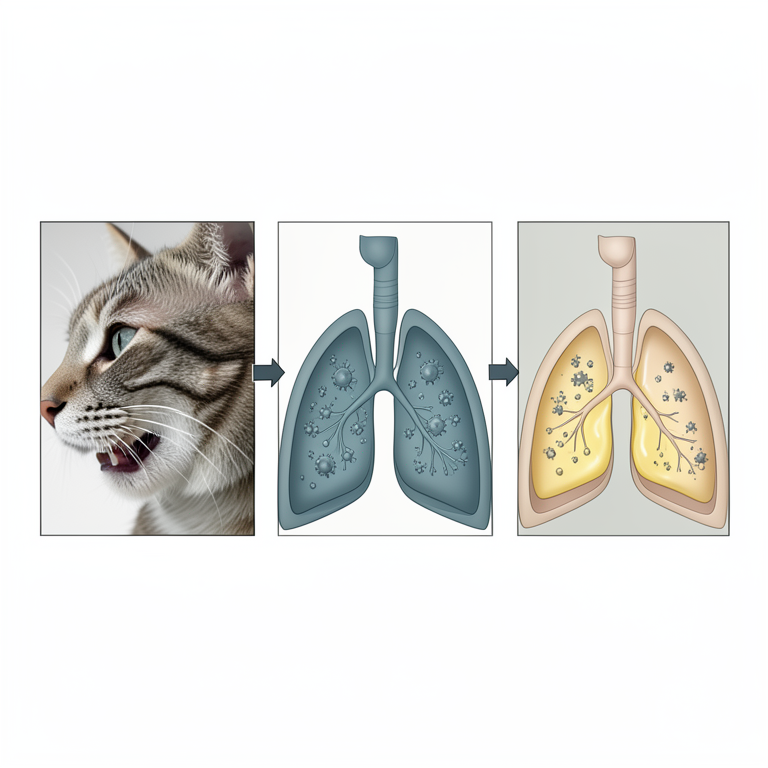
Pathogenesis of Feline Pyothorax
AI-generated conceptual illustration (domestic cat). It explains the idea and does not show study data.
View source evidence ↗Figures & tables
From the original paper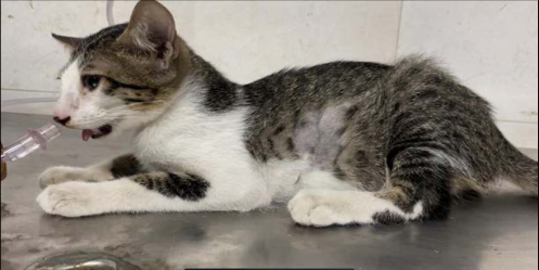 Figure 1
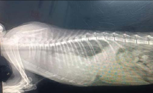 Figure 2
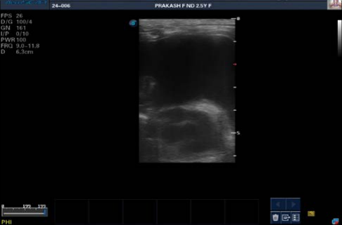 Figure 3
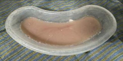 Figure 4
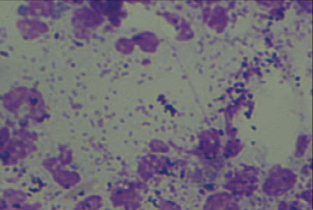 Figure 5
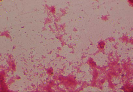 Figure 6
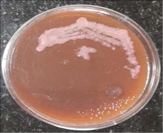 Figure 7
Introduction
Case History and Treatment A six-month-old male domestic shorthaired cat was presented with a three-day history of respiratory distress and inappetence, exhibiting severe clinical signs, including sternal recumbency, open-mouth breathing, and a slightly arched back, indicative of significant thoracic cavity compromise. Upon clinical examination, the cat's temperature was notably elevated at 103.8°F, with concurrent severe tachypnoea, pale mucous membranes, and evidence of severe dehydration and tachycardia, collectively suggesting a critical underlying condition. Radiographic examination revealed substantial fluid accumulation within the thoracic cavity, accompanied by border effacement of the cardiac silhouette. On Ultrasonographic examination, contents with less echogenicity in the thoracic cavity were visualized, thereby confirming the presence of severe pleural effusion.
Haemato-biochemical analysis revealed leucocytosis, characterized by an elevated WBC count of 59,000/cmm, with a predominance of neutrophils (88%), alongside hypoproteinaemia (4.6 g/dL) and hypoalbuminemia (2.45 g/dL), as well as an elevated lactate dehydrogenase level (326.5 IU/L), collectively indicating a severe underlying inflammatory process. Following sedation with Butorphanol (0.2 mg/kg intramuscular route) and local anaesthesia with Lignocaine, emergency thoracocentesis was performed, and approximately 150 ml of foul-smelling, brown fluid was removed, which was subsequently subjected to cytological examination, revealing numerous degenerated neutrophils (Total nucleated cell count - 3,07,000/cmm) with bacteria. The fluid was subjected to bacterial culture on MacConkey Agar, which showed pink mucoid growth, identifying gram-negative Klebsiella sp. as the causative agent. Antimicrobial sensitivity tests were also performed, demonstrating sensitivity of the organism to Amoxicillin-Clavulonate.
The cat was treated with intravenous Amoxicillin-Clavulonic acid and Ringer's Lactate, followed by oral antibiotics for 28 days (Sim et al., 2021) [6], resulting in normalization of hydration status and appetite within four days. VET 2025; 10(1): 153-155 David Karunyakaran H M.V.Sc Scholar, Department of Veterinary Clinical Medicine, Madras Veterinary College, Tamil Nadu Veterinary and Animal Sciences University, Chennai, Tamil Nadu, India Gowri B Professor, Department of Veterinary Clinical Medicine, Madras Veterinary College, Tamil Nadu Veterinary and Animal Sciences University, Chennai, Tamil Nadu, India Selvaraj P Professor and Head, Veterinary University Peripheral Hospital, Madhavaram Milk Colony, Tamil Nadu Veterinary and Animal Sciences University, Chennai, Tamil Nadu, India Tirumurugaan KG Professor and Project Director Incharge, Translational Platform for Veterinary Biologicals, Tamil Nadu Veterinary and Animal Sciences University, Tamil Nadu, India Sabareeswaran S Undergraduate Student, Madras Veterinary College, Tamil Nadu Veterinary and Animal Sciences University, Chennai, Tamil Nadu, India David Karunyakaran H M.V.Sc Scholar, Department of Veterinary Clinical Medicine, Madras Veterinary College, Tamil Nadu Veterinary and Animal Sciences University, Chennai, Tamil Nadu, India https://www.veterinarypaper.com Serial follow-up examinations, including thoracic ultrasonography and thoracocentesis on second week, revealed 70 ml of light brown fluid, signalling gradual resolution of pleural effusion. With complete normalization of blood test results and no evidence of fluid accumulation within the thoracic cavity after four weeks, the cat had an uneventful recovery. Fig 5: Pleural fluid cytology-Numerous neutrophils with bacteria Fig 1: Cat with open mouth breathing and slightly arched back Fig 6: Gram staining-Gram negative bacilli Fig 2: Thoracic radiography - Fluid accumulation in the thoracic cavity with border effacement of cardiac silhouette Fig 3: Thoracic ultrasonography-Anechoic contents in the thoracic cavity Fig 4: Pleural fluid-pyothorax Fig 7: MacConkey Agar-Pink mucoid colonies-Klebsiella sp
Discussion
Pyothorax, a severe and potentially life-threatening condition also known as thoracic empyema, is characterized by the accumulation of septic purulent fluid within the pleural space, precipitating a cascade of inflammation and infection that compromises the surrounding tissues (Beatty and Barrs, 2010) [1]. The primary etiological factor underlying feline pyothorax is thought to be parapneumonic spread of infection (Murphy, and Papasouliotis, 2011) [4], wherein oropharyngeal flora colonizes lung tissue, frequently as a complication of pneumonia, thereby facilitating the penetration of bacteria into the lung parenchyma and subsequent dissemination to the pleural space (Demetriou et al., 2002) [2]. This complex sequence of events culminates in the accumulation of infected fluid within the pleural cavity, necessitating prompt and accurate diagnosis to guide effective treatment and ensure successful outcomes (Epstein, 2014) [3]. A comprehensive diagnostic approach, incorporating clinical evaluation, laboratory analysis, and imaging studies, is crucial for identifying the underlying cause of pyothorax and informing https://www.veterinarypaper.com the development of a tailored treatment plan (Stillion and Letendre, 2015) [5].
Conclusion
The presentation of pyothorax in cats is often insidious and nonspecific, manifesting as vague and seemingly unrelated signs such as lethargy, anorexia, and respiratory distress, which can lead to a delayed diagnosis and initiation of treatment. As a result, prompt recognition and rapid intervention are critical to preventing severe and potentially life-threatening complications, including sepsis, organ failure, and death, which can arise from the proliferation of bacteria within the pleural space. The key to timely intervention lies in maintaining a high index of suspicion, combined with the strategic use of diagnostic imaging modalities, such as radiography and ultrasonography, and cytologic examination of pleural fluid to detect the presence of bacteria and guide targeted antimicrobial therapy. By adopting a proactive and multidisciplinary approach, a successful outcome can be ensured, thereby reducing the risk of complications, and improve the quality of life for affected animals Conflict of interest Authors have no conflict of interest in this study.
Financial Support: Not available Reference 1. Beatty JA, Barrs V. Pleural effusion in the cat. A practical approach to determining aetiology.
J Feline Med Surg. 2010;12:693-707. 2. Demetriou JL, Foale RD, Ladlow J. Canine and feline pyothorax: a retrospective study of 50 cases in the UK and Ireland. J Small Anim Pract. 2002;43(9):388-394. 3.
Epstein SE. Exudative pleural disease in small animals. Vet Clin North Am Small Anim Pract. 2014;44:161-180. 4. Murphy K, Papasouliotis K.
Pleural effusions in dogs and cats. Diagnostic investigation. In Pract. 2011;33:462-469. 5. Stillion JR, Letendre JA.
A clinical review of the pathophysiology, diagnosis, and treatment of pyothorax in dogs and cats. J Vet Emerg Crit Care. 2015;25(1):113- 129. 6. Sim JA, Lau SF, Omar S, Watanabe M, Aslam MW. A retrospective study on bacteriology, clinicopathologic and radiographic features in 28 cats diagnosed with pyothorax.
Animals. 2021;11(8):2286. How to Cite This Article Karunyakaran DH, Gowri B, Selvaraj P, Tirumurugaan KG, Sabareeswaran S. Navigating the challenges of Pyothorax: The inspiring story of a cat's recovery. International Journal of Veterinary Sciences and Animal Husbandry. 2025;10(1):153-155.
Creative Commons (CC) License This is an open-access journal, and articles are distributed under the terms of the Creative Commons Attribution-Non Commercial-Share Alike 4.0 International (CC BY-NC-SA 4.0) License, which allows others to remix, tweak, and build upon the work non-commercially, as long as appropriate credit is given and the new creations are licensed under the identical terms.