
Research article · 2025 · Full text
International Journal of Advanced Chemistry ResearchSynthesizing nanostructures of complex preparations by Staphysagria and cantharis mother tincture under influence of potentization
Volume 7(1) · Pages 18–21 · DOI: 10.33545/26646781.2025.v7.i1a.250 ↗
Research areasPhytochemical compounds biological activitiesBeetle Biology and Toxicology StudiesEssential Oils and Antimicrobial Activity
Abstract
Author-writtenThrough this research work synthesizing the nanostructures, a massless particles by complex mixture between Staphysagria and Cantharis mother tincture Afterwards Standardization done by Scanning electron microscope.
Summary
Source-checkedPlain-language reading of the full paper, prepared with AI and checked against the text. Each point links to the authors' own words.
Why it matters
- Nanoparticles are used in diagnostics, image contrast agents, and drug administration due to their unique biological, chemical, and physical properties. View evidence ↗
The question
- The study aimed to synthesize nanostructures (described as massless particles) using a complex mixture of Staphysagria and Cantharis mother tinctures and standardize them using a Scanning Electron Microscope (SEM). View evidence ↗
What they did
- The research was conducted at the Micro-Nano R & D Centre of the Parul Institute of Homoeopathy & Research (PIHR). View evidence ↗
- The mixture was prepared by combining 1 ml each of Cantharis Q and Staphysagria Q with 20 ml of distilled water and 1 gm of Potassium Ferricyanide. View evidence ↗
- The resulting complex heterogeneous mixture was heated in a hot water bath for 10 minutes. View evidence ↗
- The mixture underwent potentization using an electric potentizer machine with 10 downward strokes. View evidence ↗
- The final product was analyzed using a Scanning Electron Microscope (SEM) equipped with Energy Dispersive X-ray Spectroscopy (EDS). View evidence ↗
What they found
- SEM analysis was performed on the complex mixture at a scale of 1-50 microns. View evidence ↗
- SEM analysis of the complex mixture was conducted at a scale of 2-15 microns. View evidence ↗
- SEM analysis of the complex mixture was conducted at a scale of 3-15 microns. View evidence ↗
- SEM analysis of the complex mixture was conducted at a scale of 4-15 microns. View evidence ↗
- SEM analysis of the complex mixture was conducted at a scale of 9-1-1 microns. View evidence ↗
- SEM analysis of the complex mixture was conducted at a scale of 7-2-1 microns. View evidence ↗
- SEM analysis of the complex mixture was conducted at a scale of 3-5 microns. View evidence ↗
What it means
- The authors conclude that the SEM scanning of the heterogeneous complex structure created from Staphysagria Q and Cantharis Q was successfully completed. View evidence ↗
Visual summary
1 visualsIllustrations explain the paper; they do not show study data.
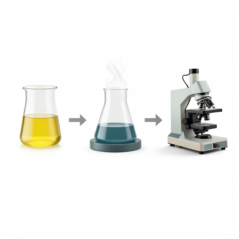
Synthesis Workflow
AI-generated conceptual illustration (Potassium ferricyanide solution with plant tinctures in a laboratory setting). It explains the idea and does not show study data.
View source evidence ↗Figures & tables
From the original paper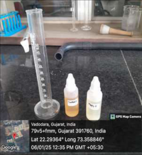 Figure 1
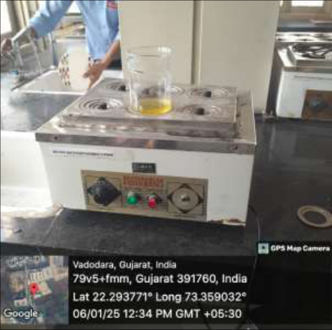 Figure 2
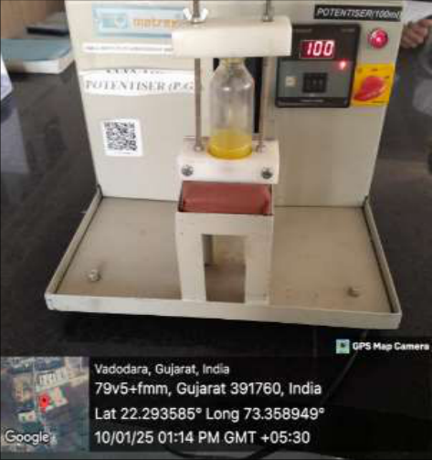 Figure 3
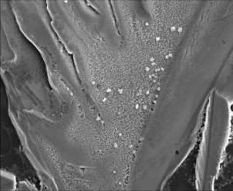 Figure 4
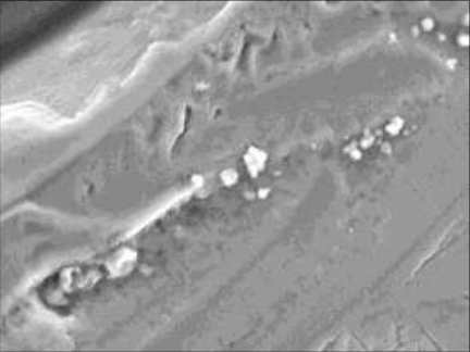 Figure 5
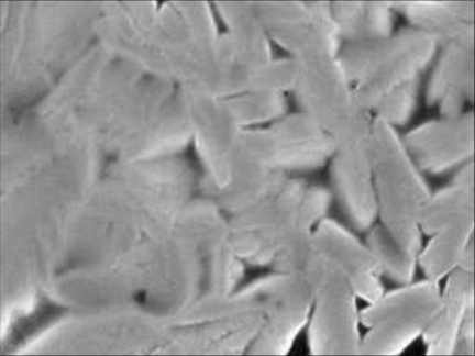 Figure 6
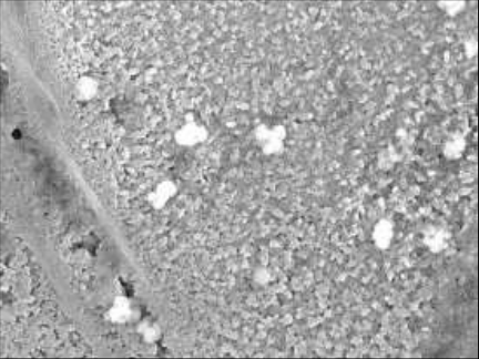 Figure 7
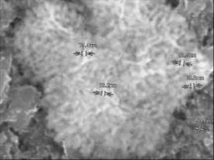 Figure 8
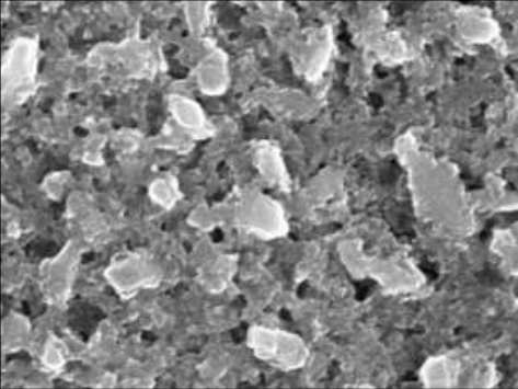 Figure 9
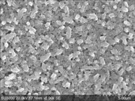 Figure 10
Introduction
As scientists utilize the special qualities of atomic and molecular assemblies constructed at the nanoscale scale, nanotechnology has emerged as one of the most important scientific initiatives of the early twenty-first century. Our capacity for manipulation Researchers can logically create and employ nanoparticles for drug administration, image contrast agents, and diagnostic applications thanks to their physical, chemical, and biological characteristics. The combination of these recently developed skills with developments in imaging, bioinformatics, and systems biology shows great potential for addressing some of the most difficult genetic and biochemical problems in biology. Even if there are many technical advancements in biology today, few could ever have the same revolutionary effect on fundamental research, medication development, and clinical medicine as nanotechnology.
Nanotechnology provides a vast array of tools and applications by functioning at the nanoscale level, at the very scale of biomolecules. Drug delivery platforms [1], improved image contrast agents [2], chip-based nanolabs that can monitor [3] and manipulate individual cells [4], and nanoscale probes that can follow the movements of individual molecules [6] and cells [5] as they move through their surroundings are some examples of near-term applications. The current toolkit for drug delivery and non-invasive drug monitoring is greatly expanded by this unparalleled capacity to observe and modify complex systems in vivo and in real time, which offers comprehensive insights into the underlying mechanisms and signalling pathways involved in the development of disease. By offering structures that can integrate several functions into a single nanoscale Moreover, nanotechnology provides the chance to track and identify cellular and molecular alterations linked to illness conditions [7].
With this multipurpose feature, it is possible to envision creating a nanoparticle that can target a particular tissue or cell type and deliver a therapeutic payload and a contrast agent that enables non-invasive imaging to the target. A reporter, like an apoptotic marker, may even be present in a nanoparticle to indicate that the payload has been delivered and is producing the intended therapeutic effect. By customizing drug delivery to each patient's reaction, such combinatorial nanostructures may eventually offer the way to "personalize medicine." Several groups have already developed multifunctional Nano devices and are evaluating them in in vitro and in vivo systems, despite the fact that this may appear futuristic [1, 8-18]. Materials & Methodology Type of study: Analytical work https://www.chemistryjournals.net Fig 2: Heating of Complex mixture under Hot water bath Site of Study: PIHR, Parul Institute of Homoeopathy & Research, Micro-Nano R & D Centre Association: Within Parul University Equipment Scanning Electron Microscope (SEM) with EDS, Electric Potentizer Machine Selected Services SEM Micrographs, Gold Coating for Non-conductive samples Medicinal Product: Staphysagria Q, Cantharis Q Procure from GMP Certified Pharmaceutical Company Preparations: For preparing heterogeneous mixtures for massless particles steps are given below; Step 1: Sterile all the Laboratory equipment’s with the help of Hot air oven Step 2: Mix 1 ml Cantharis Q and Staphysagria Q in 20 ml Distilled water with 1 gm Potassium Ferricynaide Step 3: After mixing the complex mixtures, placed under cool and dark place, away from sunlight in hard glass bottle.
Step 4: Complex Heterogeneous mixture undergoes Heating under Hot water bath for 10 minutes Step 5: Complex Heterogeneous mixture with Cantharis Q and Staphysagria Q undergoes into potentization, 10 downwards strokes by electric potentizer Machine Step 6: Analysis under Scanning Electron Microscope (SEM) with EDS.
Results
SEM Scanning of Heterogeneous complex mixture as Cantharis Q and Staphysagria Q are given below; Fig 3: Potentization of Complex mixture under Electric Potentizer Machine Fig 1: Sample of Staphysagria Q & Cantharis Q Fig 4: SEM Analysis of Complex mixture under 1-50 micron https://www.chemistryjournals.net Fig 5: SEM Analysis of Complex mixture under 2-15 micron Fig 9: SEM Analysis of Complex mixture under 7-2-1 micron Fig 6: SEM Analysis of Complex mixture under 3-15 micron Fig 7: SEM Analysis of Complex mixture under 4-15 micron Fig 8: SEM Analysis of Complex mixture under 9-1-1 micron Fig 10: SEM Analysis of Complex mixture under 3-5 micron
Conclusion
Scanning of heterogeneous complex structure by Staphysagria Q and Cantharis Q by SEM Scanning electron microscope had been completed. Conflict of interest No such
Acknowledgement
Authors would like to thanks Parul University and Faculty of Micro-Nano R & D Centre for completing this work.
References
18 in the original paper- Rose C, Parker AH, Jefferson B, Cartmell E. The characterization of feces and urine: A review of the literature to inform advanced treatment technology. Crit Rev Environ Sci Technol. 2015;45:1827-1879.
- Zaha DC, Bungau S, Aleya S, Tit DM, Vesa CM, Popa AR, et al. What antibiotics for what pathogens? The sensitivity spectrum of isolated strains in an intensive care unit. Sci Total Environ. 2019;687:118-127.
- Tamalli M, Bioprabhu S, Alghazal MA. Urinary tract infection during pregnancy at Al-Khoms. Int J Med Med Sci. 2013;3:455-459.
- Siddiq DM, Darouiche RO. New strategies to prevent catheter-associated urinary tract infections. Nat Rev Urol. 2012;9:305-314.
- Yassin MA, Elkhooly TA, Elsherbiny SM, Reicha FM, Shokeir AA. Facile coating of urinary catheter with bio- inspired antibacterial coating. Heliyon. 2019;5:e02986. https://www.chemistryjournals.net
- Syed MA, Manzoor U, Shah I, Bukhari SH. Antibacterial effects of tungsten nanoparticles on the Escherichia coli strains isolated from catheterized urinary tract infection (UTI) cases and Staphylococcus aureus. New Microbiol. 2010;33:329-335.
- Syed MA, Babar S, Bhatti AS, Bokhari H. Antibacterial effects of silver nanoparticles on the bacterial strains isolated from catheterized urinary tract infection cases. J Biomed Nanotechnol. 2009;5:209-214.
- Odoki M, Aliero AA, Tibyangye J, Maniga JN, Wampande E, Kato CD, et al. Prevalence of bacterial urinary tract infections and associated factors among patients attending hospitals in Bushenyi district, Uganda. Int J Microbiol. 2019;2019:1-8.
- Warkulwiz G, Hannon K, Cabano D, Mehta K. Urinary tract infections (UTIs) in rural Kenya: Screening challenges, treatment pathways, and technological solutions. 2017 IEEE Global Humanitarian Technology Conference (GHTC); 2017 Oct 19-22; San Jose, CA, USA. p. 1-6.
- Stamm WE, Norrby SR. Urinary tract infections: Disease panorama and challenges. J Infect Dis. 2001;183(1):S1-S4.
- Siakwa M, John ME, Kpikpitse D, Ankobil A, Hansen- Owoo E. Pregnancy outcomes: A comparison of women with symptomatic and asymptomatic bacteriuria in Cape Coast, Ghana. Afr J Pregnancy Childbirth. 2014;2:27-30.
- Tan CW, Chlebicki MP. Urinary tract infections in adults. Singap Med J. 2016;57:485-490.
- Rajivgandhi G, Vijayan R, Kannan M, Santhanakrishnan M, Manoharan N. Molecular characterization and antibacterial effect of endophytic actinomycetes Nocardiopsis sp. GRG1 (KT235640) from brown algae against MDR strains of uropathogens. Bioact Mater. 2016;1:140-150.
- Casertano M, Menna M, Imperatore C. The ascidian- derived metabolites with antimicrobial properties. Antibiotics. 2020;9:510.
- Ayuningrum D, Liu Y, Riyanti, Sibero MT, Kristiana R, Asagabaldan MA, et al. Tunicate-associated bacteria show a great potential for the discovery of antimicrobial compounds. PLoS ONE. 2019;14:e0213797.
- Tit DM, Pallag A, Iovan C, Furău G, Furău C, Bungau S. Somatic-vegetative symptoms evolution in postmenopausal women treated with phytoestrogens and hormone replacement therapy. Iran J Public Health. 2017;46:1528-1534.
- Kunin CM. Urinary tract infections in females. Clin Infect Dis. 1994;18:1-12.
- Kant S, Lohiya A, Kapil A, Gupta SK. Urinary tract infection among pregnant women at a secondary level hospital in Northern India. Indian J Public Health. 2017;61:118-123.