
Research article · 2025 · Full text
International Journal of Veterinary Sciences and Animal HusbandryConcurrent infections of theileriosis and amphistomiasis in a cross breed calve and its therapeutic management: A case study
Volume 10(1) · Pages 100–102 · DOI: 10.22271/veterinary.2025.v10.i1b.2001 ↗
Research areasHelminth infection and controlAnimal Behavior and Welfare Studies
Abstract
Author-writtenAn 8 month old crossbred female calve with a history of incomplete deworming, anorexia, weakness, fever, respiratory distress, deep yellow urine and diarrhoea was presented to the Veterinary Clinical Complex, CVSc. & A.H., RK Nagar, Tripura. On clinical examination, the calf revealed paper white conjunctival membrane, ruminal atony, soiled hind quarters and enlarged pre-scapular lymph node. Haematological examination revealed a lower level of Haemoglobin (4.8 g/dl), Total RBC (2.2 million/cmm) and PCV (11.4%). The blood smear and faecal examination revealed the presence of Theileria orientalis piroplasm and amphistome eggs. The animal was successfully treated with injection Buparvaquone @ 2.5 mg/kg BW I/M single dose, Injection Oxytetracycline-LA, deep I/M @ 20 mg/kg BW at every 48 hours interval on three occasions, Bolus oxyclozanide @ 15 mg/kg BW orally for 3 days along with supportive drugs.
Summary
Source-checkedPlain-language reading of the full paper, prepared with AI and checked against the text. Each point links to the authors' own words.
Why it matters
- Bovine tropical theileriosis is a significant tick-borne blood parasite disease in cattle that leads to high rates of illness and death. View evidence ↗
- While all cattle breeds can be affected, young calves and crossbred or exotic cattle are particularly susceptible to theileriosis. View evidence ↗
- Amphistomes, a type of trematode, have become a major source of economic loss for farmers because they reduce productivity and cause death in cattle. View evidence ↗
The question
- The study aims to describe a case of a calf concurrently infected with Theileria and amphistomiasis and evaluate the success of a specific therapeutic management plan. View evidence ↗
What they did
- The subject was an 8-month-old crossbred female calf presented at a veterinary clinic in Tripura with symptoms including fever, anorexia, and respiratory distress. View evidence ↗
- Blood samples were collected from the jugular vein using sterilized EDTA vials to perform hematological tests and detect blood parasites. View evidence ↗
- Diagnosis was confirmed using Giemsa-stained blood smears for Theileria orientalis piroplasms and a sedimentation technique for fecal examination to find amphistome eggs. View evidence ↗
- Treatment included a single dose of Buparvaquone (2.5 mg/kg), Meloxicam (0.2 mg/kg for 3 days), Pheniramine Maleate (0.5 mg/kg), and Oxytetracycline-LA (20 mg/kg every 48 hours for three doses). View evidence ↗
- The amphistomiasis infection was specifically treated with oral Bolus oxyclozanide at a dose of 15 mg/kg for three days. View evidence ↗
What they found
- Initial hematological tests showed severe anemia, with hemoglobin at 4.8 g/dl, total RBC at 2.2 million/cmm, and PCV at 11.4%. View evidence ↗
- Clinical signs at presentation included paper-white conjunctival membranes, enlarged pre-scapular lymph nodes, and ruminal atony. View evidence ↗
- Improvement began after the fourth day of treatment, characterized by the calf eating again, normalization of temperature and urine color, and the conjunctival membrane turning pink. View evidence ↗
- The calf appeared to have fully recovered and returned to normal health by the 10th day of the treatment regimen. View evidence ↗
What it means
- The authors conclude that the combination of Buparvaquone, Oxytetracycline, and Oxyclozanide is an effective treatment for concurrent theileriosis and amphistomiasis in calves. View evidence ↗
- The authors recommend implementing appropriate treatment regimens and increasing farmer awareness to control tick-borne infections in cattle. View evidence ↗
Visual summary
1 visualsIllustrations explain the paper; they do not show study data.
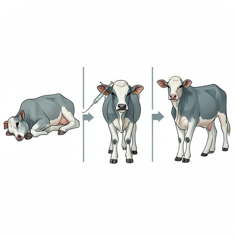
Therapeutic Management Flow
AI-generated conceptual illustration (crossbreed calf). It explains the idea and does not show study data.
View source evidence ↗Figures & tables
From the original paper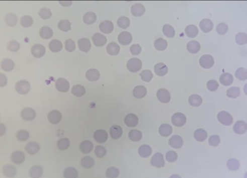 Figure 1
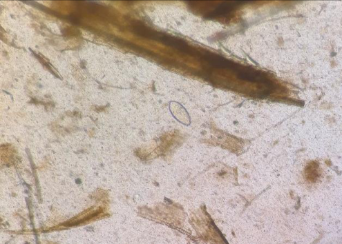 Figure 2
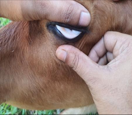 Figure 3
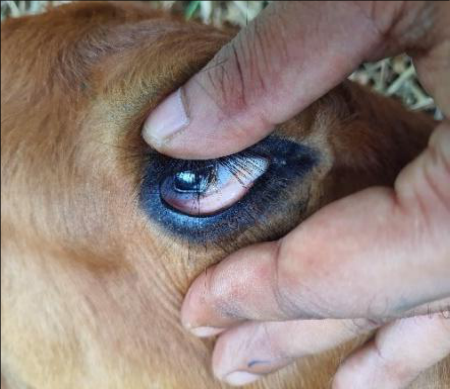 Figure 4
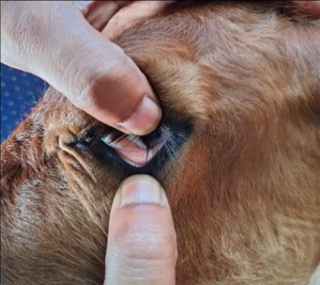 Figure 5
Introduction
Bovine tropical theileriosis is a major tick-borne haemo-parasitic disease of cattle and is associated with high morbidity and mortality. Though all breeds of cattle are equally susceptible to theileriosis but exotic, crossbred cattle as well as young calves are more affected by this disease. The disease is mainly caused by Theileria annulata, T. parva and T. orientalis in cattle in India and transmitted by ticks of genera Hyalomma and Rhipicephalus (Krishnamoorthy et al., 2021) [10] . The clinical signs include pyrexia (Up to 107°F), dullness, anorexia, enlarged superficial lymph nodes, ruminal atony, dehydration, salivation, lacrimation and discharge from nostrils. The clinically recovered animal may act as carriers with long-term persistent infections (Brown, 1990) [4] . The disease has a profound effect on the haematological values of the animals and is responsible for causing huge morbidity and mortality in the cattle population. Confirmatory diagnosis of theileriosis is mainly based on history of tick infestation, clinical signs and microscopic examination of Giemsa-stained thin blood smears of the suspected animal (Aktas et al., 2006) [1] or PCR. Among several trematodes affecting cattle, amphistomes have recently emerged as an important cause of economic loss to farmers due to mortality and low productivity (Kilani et al., 2003) [9] . In ruminants, clinical symptoms associated with amphistomiasis were diarrhoea, loss of body condition, rough hair coat, dullness, weakness, loss of appetite, intestinal haemorrhages, anaemia and intermandibular swelling (Blood et al., 1983) [3] . Diagnosis of amphistomiasis at field level was done mainly by faecal examination. This article describes a case of concurrent infection of Theleria and amphistomiasis along with its successful therapeutic management using Buparvaquone, Oxytetracyline and Oxyclozanide.
Case history and observations
An 8 month old crossbred female calves with approx. 52 kg was presented. to the Veterinary Clinical Complex, CVSc. & A.H., R.K. Nagar, Tripura with a history of incomplete deworming, anorexia, weakness, fever, respiratory distress, deep yellow urine and diarrhoea. Clinical examination of the animal revealed paper white conjunctival membrane (Figure 3), ruminal atony, soiled hind quarters and enlarged pre-scapular lymph node. Heavy tick infestation was also observed. On the basis of clinical examination, blood sample was collected aseptically from jugular vein in a sterilized EDTA vial for haematological examination as well as for haemo-parasite detection.
Haematological examination revealed Haemoglobin (4.8 g/dl), Total RBC (2.2 million/cmm), PCV (11.4%), Total WBC (7250/cmm), DLC (Neutrophil-71%, Lymphocyte-21%, Ecsinophil-5%, Monocyte-3% and Basophil-0%), Platelet count (6120/cmm). MCV, MCH and MCHC were 66.82 fL, 21.77 pg and 32.29 g/dl, respectively. The blood smear was subjected to Giemsa staining and observed under light microscopy, which revealed the presence of Theileria orientalis piroplasm (Fig. 1). Faecal examination by sedimentation technique revealed the presence of Amphistome eggs (Fig. 2). On the basis of history, clinical signs and laboratory findings the condition was diagnosed as concurrent infection of Theileriosis and Amphistomiasis.
Therapeutic management
The animal was treated with a single dose of injection of Buparvaquone @ 2.5 mg/kg BW I/M, injection of Meloxicam @ 0.2 mg/kg BW I/M for 3 days and injection of Pheniramine Maleate @ 0.5 mg/kg BW I/M,, injection of Oxytetracycline-LA, deep I/M @ 20 mg/kg BW at every 48 hours interval on three occasions along with supportive drugs eg. Injection Feritas @ 1.0 ml I/M every alternate day for ten occasions and Injection Tribivet 2ml I/M for 5 days. Amphistomiasis was treated by giving Bolus oxyclozanide @ 15 mg/kg BW orally for 3 days. Following the fourth day of treatment, the animal's health began to improve. The calve started eating, the temperature and urine colour became normal, the diarrhoea subsided and the conjunctival mucous membrane turned to pink in colour. The animal apparently became normal after the 10 th day of treatment.
Discussion
Theileriosis is one of the most significant arthropod-borne blood protozoan illnesses that cause morbidity and death, particularly in calves, those frequently afflicted during the prenatal period. In the present case, an eight months old calve was infected with Theileria orientalis. Various workers have reported that young calves are more susceptible to Theileriosis (Gupta et al., 2004) [6] . The haematology of the present case revealed that the total erythrocyte count, haemoglobin concentration and PCV mean values were significantly low as compared to normal levels suggestive of anaemia. These observations were in parallel with Hasanpour et al. (2008) [7] . Anemia in Theileriosis might be because of erythrophagocytosis which causes destruction of RBC's infected with theileria schizoints (Uilenberg, 1981) [12] . However, increased level of activated complement products (Omer et al., 2002) [11] and removal of piroplasm infected erythrocytes by macrophages in the RE system have also been suggested as a cause of anemia (Campbell et al., 1999) [5] . Several clinicians successfully used the combination of buparvaquone and oxytetracycline as a curative drug for the treatment of theileriosis in cattle (Bhatt et al., 2012; Kachhawa et al., 2016) [2,8] . Oxyclozanide is considered as the drug of choice for treating amphistomiasis in cattle and been successfully used by many workers (Yogeshpriya et al., 2017) [13] .
Conclusion
In conclusion, it was reported that a concurrent infection of theileriosis and amphistomasis in a calf was successfully treated with Buparvaquone, Oxytetracycline and Oxyclozanide. Since haemoprotozoan diseases are causing devastating losses to the livestock industry and pose major constraints to the dairy industry in India as well as throughout the world, therefore, following appropriate treatment regime along with awareness to the farmers are utmost important regarding controlling of these tick-borne infections in cattle.
Acknowledgement
AcknowledgmentsThe authors thank to all the staffs and students of Veterinary Clinical Complex, CVSc. & A.H., R.K. Nagar, Tripura for their assistance in the management of the case.
Figures and tables from the paper
References
13 in the original paper- Aktas M, Altay K, Dumanli N. A molecular survey of bovine Theileria parasites among apparently healthy cattle and with a note on the distribution of ticks in eastern Turkey. Vet Parasitol. 2006;138:179-185.
- Bhatt P, Kumar S, Sharma G, Shukla SK. Management of concurrent amphistomosis and theileriosis in a cross bred cattle. Vet Pract. 2012;13(1):109-110.
- Blood DC, Radostits OM, Henderson JA. Veterinary Medicine. 6th ed. ELBS, Bailliere and Tindall, UK, 1983, p. 906-907.
- Brown CG. Control of tropical theileriosis (Theileria annulata infection) of cattle. Parassitologia. 1990;32:2331.
- Campbell JD, Spooner RL, Campbell JD. Macrophages behaving badly: infected cells and subversion of immune responses to Theileria annulata. Parasitol Today. 1999;15(1):10-6.
- Gupta SK, Yadav A, Raina AK, Singh R. Theileriosis in a seven-day old bovine calf: A case report. Indian J Vet Med. 2004;24:55.
- Hasanpour A, Moghaddam GA, Namatollahi A. Biochemical, hematological and electrocardiographic changes in buffaloes naturally infected with Theileria annulata. Korean J Parasitol. 2008;46(4):223-227.
- Kachhawa JP, Kumar S, Sharma A, Singh AP, Ahuja A. Studies on alterations of clinical and hemato-biochemical parameters before and after treatment in calves naturally infected with theileriosis. Vet World. 2016;9(12):13811385.
- Kilani K, Guillot J, Chermett R. Amphistomes digestive. Principales maladies infectieuses et parasitaires du betail. 2003;1400-1410.
- Krishnamoorthy P, Akshata LG, Jacob SS, Suresh KP, Roy P. Theileriosis prevalence status in cattle and buffaloes in India established by systematic review and meta-analysis. Indian J Anim Sci. 2021;91(4):269-279.
- Omer OH, El-Malik KH, Mahmoud OM, Haroun EM, Hawas A, Sweeney D, Magzoub M. Haematological profiles in pure bred cattle naturally infected with Theileria annulata in Saudi Arabia. Vet Parasitol. 2002;107(1-2):161-168.
- Uilenberg G. Theileria infections other than east coast fever. Curr Top Vet Med Anim Sci. 1981;6:41.
- Yogeshpriya S, Saravanan M, Krishnakumar S, Veeraselvam M, Selvaraj P. Clinico-therapeutic management of amphistomiasis in cattle. Bull Environ Pharmacol Life Sci. 2017;6(6):92-94.